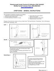
Gait Analysis Handout - painfreefeet.ca
Gait Analysis Handout - painfreefeet.ca
Gait Analysis Handout - painfreefeet.ca

Create successful ePaper yourself
Turn your PDF publications into a flip-book with our unique Google optimized e-Paper software.
PODIATRIC GAIT ANALYSIS<br />
Southerland stated that ‘no other group of health <strong>ca</strong>re professionals spends as much<br />
time in their professional edu<strong>ca</strong>tion learning “biomechanics.” Furthermore,<br />
‘clini<strong>ca</strong>l gait evaluation is a fundamental skill for podiatric physicians.’ 1<br />
<strong>Gait</strong> analysis forms part of the overall assessment of the locomotor system. The main<br />
objective of the overall assessment focuses on the position and alignment of the body<br />
and the relationship of the foot to the ground during gait. This involves discriminating<br />
between normal and abnormal gait indi<strong>ca</strong>tors using visual or computerized gait analysis.<br />
<strong>Gait</strong> depends on the repeated<br />
performance of the lower limbs of a<br />
sequence of motions that<br />
simultaneously advance the body<br />
along a desired line of progression<br />
while maintaining a stable weight<br />
bearing posture.<br />
In order for normal gait to occur a<br />
person must be able to accomplish<br />
the following four objectives:<br />
1. Each leg must be able to support<br />
the body’s weight without collapsing.<br />
2. Balance must be maintained,<br />
either stati<strong>ca</strong>lly or dynami<strong>ca</strong>lly,<br />
during the single support phase.<br />
3. The swinging leg must be able to<br />
advance to a position where it <strong>ca</strong>n<br />
take over the supporting role.<br />
4. Sufficient power must be provided to make the necessary limb movements and to<br />
advance the trunk.<br />
The effectiveness of normal gait depends on free joint mobility and muscle action that is<br />
selective in both timing and intensity. Therefore, pathologi<strong>ca</strong>l conditions of the muscles,<br />
bones, joints, sensory nervous system, central motor control, and the <strong>ca</strong>rdiopulmonary<br />
system will alter the mode and efficiency of gait. With a detailed knowledge of normal<br />
function of the foot and lower limb, the chiropodist/podiatrist becomes able to identify<br />
the signifi<strong>ca</strong>nt deficits of abnormal gait.<br />
THE FIVE COMPONENTS OF GAIT:<br />
1. Source of motion<br />
Muscles provide force for desired motion.<br />
Muscle dependent on the nerve supply.<br />
Therefore, the source of motion is the motor unit.<br />
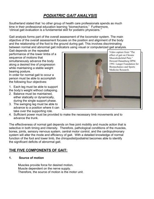
Video <strong>ca</strong>pture from “The<br />
effect of gait on Chronic<br />
Musculoskeletal Pain.”<br />
Howard Dananberg DPM.<br />
1992. Langer Foundation for<br />
Biomechanics and Sports<br />
Medicine Research.
2. Articulated levers<br />
The skeleton with its complex articulations and connecting ligaments provide the<br />
needed mobile lever system. Joint anatomy, dictates the directions motion <strong>ca</strong>n<br />
occur. Bone length proportionally magnifies the motor units’ actions. Fibrous<br />
tissue flexibility determines joint freedom of movement.<br />
3. Awareness of motion needed<br />
Information is needed as to the position (proprioception), velocity, force and<br />
direction of motion. The information is continually sensed by peripheral receptors<br />
and transmitted by sensory pathways to neural control centres.<br />
4. Control source to provide the desired motion<br />
Upper motor neurons arise from neural control centres that coordinate sensory<br />
input with anticipated motion. This results in muscles that contract with<br />
appropriate timing and intensity. There are numerous sites within the brain and<br />
spinal cord where a pathologi<strong>ca</strong>l process <strong>ca</strong>n introduce control deficits.<br />
5. Energy to move<br />
Muscles use oxygen to generate energy needed for force production.<br />
Availability of an adequate oxygen supply depends on the state of the<br />
<strong>ca</strong>rdiovascular system.<br />
Classifi<strong>ca</strong>tion of injury or disease of the structures essential for gait:<br />
1. Structural Impairment - biomechani<strong>ca</strong>l etiology<br />
2. Motor Unit Insufficiency<br />
3. Combined Motor and Sensory Impairment<br />
4. Central Control Dysfunction<br />
5. Energy Lack<br />
The focus of this outline is the structural impairments (biomechani<strong>ca</strong>l) of the foot and<br />
lower limb and their affect on gait.<br />
During a normal gait cycle, the joints should be free to move and possess optimal joint<br />
alignment. This depends on fibrous tissue mobility, architectural accuracy of the bones<br />
and articular <strong>ca</strong>rtilage smoothness. This should be evaluated during a static<br />
biomechani<strong>ca</strong>l examination and if possible examined utilizing objective gait analysis<br />
techniques.<br />
The four remaining classifi<strong>ca</strong>tions which are important for the chiropodist/podiatrist to<br />
realize, but may not be identified until further investigation by a family physician,<br />
psychiatrist, or orthopaedic surgeon. This paper assumes these injuries are not present
if the problem is of a biomechani<strong>ca</strong>l etiology. However, the discussion may point to<br />
these classifi<strong>ca</strong>tions as an alternative etiology for the gait problems observed.<br />
Power of <strong>Gait</strong><br />
The power of gait is generated by the interplay of the factors in the flowchart outlined<br />
below:<br />
Elastic Energy<br />
Elastic energy response<br />
There are many muscles in the lower extremity that use eccentric muscle contractions<br />
to store potential energy to be utilized during a subsequent concentric muscle<br />
contraction as kinetic energy.<br />
The table below outlines the timing of eccentric and concentric muscle contractions of<br />
the flexors and extensors of the hip, knee, and ankle during the gait cycle. For example,<br />
the hip flexors stretch (eccentric contraction) and store elastic energy during single<br />
support and use the stored energy to initiate the concentric contraction occurring during<br />
preswing that powers the swing leg forward.<br />
Late<br />
Swing<br />
Loading<br />
Response<br />
Hip Ext Ecc Conc<br />
SOURCE OF POWER FOR WALKING<br />
Swing phase action<br />
* Pendulum Action<br />
* Kinetic Energy<br />
Mid<br />
stance<br />
Terminal<br />
Stance<br />
Pre<br />
Swing<br />
Hip Flex Ecc Conc<br />
Knee Ext Ecc Conc<br />
Pull of swing limb<br />
Momentum Gravity<br />
Lever Effect of Stance Limb<br />
Heel, Ankle, and Forefoot Rocker<br />
Knee Flex Ecc Conc<br />
Ankle Pl. Ecc Conc<br />
Early<br />
Swing<br />
Ankle Dors. Ecc Conc Ecc Conc
Energy Transfer<br />
The verti<strong>ca</strong>l displacement of the pelvis during<br />
stance allows for the exchange between kinetic<br />
energy and potential energy. This is illustrated in<br />
the diagram to the right. The diagram below<br />
illustrates when the exchange between potential<br />
and kinetic energy occurs in relation to the<br />
verti<strong>ca</strong>l ground reaction force graph<br />
Force N/KG<br />
12.00<br />
10.00<br />
8.00<br />
6.00<br />
4.00<br />
2.00<br />
0.00<br />
-2.00<br />
Verti<strong>ca</strong>l Ground<br />
Reaction Force<br />
0<br />
6<br />
12<br />
18<br />
24<br />
30<br />
36<br />
42<br />
48<br />
54<br />
60<br />
66<br />
72<br />
78<br />
84<br />
90<br />
96<br />
Stride %<br />
Pull of swing limb and momentum<br />
The momentum of the<br />
swing limb helps with the<br />
progression of the stance<br />
leg and aids in the verti<strong>ca</strong>l<br />
displacement of the pelvis.<br />
Diagrams from Perry, J., “Normal<br />
and Pathologi<strong>ca</strong>l <strong>Gait</strong>, Disorders of<br />
the Foot Churchill and Livingstone,<br />
1985<br />
Highest point of<br />
centre of body mass<br />
High Potential Energy<br />
Lowest point of<br />
centre body mass<br />
High kinetic energy<br />
The diagram to the left depicts<br />
the muscle activity during loading<br />
response to terminal stance. The<br />
hip extensors store potential<br />
energy during late swing phase<br />
and release kinetic energy during<br />
a very powerful concentric during<br />
the loading response phase.<br />
Diagram from Perry, J., “Normal and Pathologi<strong>ca</strong>l <strong>Gait</strong>, Disorders of the<br />
Foot Churchill and Livingstone, 1985.<br />
C of Mass<br />
Forward Progression<br />
Forward progression of the stance<br />
leg is accomplished by the lever<br />
effect of the stance leg. The<br />
progression of the stance leg <strong>ca</strong>n<br />
be stopped by any problem with<br />
either the heel, or ankle, or forefoot<br />
rockers.<br />
Lever Effect of Stance Leg
Conservation of Momentum<br />
The power of gait is really about the conservation of momentum. There is a<br />
dissipation of kinetic energy if stance leg and foot functions normally. If the power of<br />
motion is blocked for some reason this energy must be dissipated. The dissipation of<br />
energy occurs at the weakest link.<br />
Excessive STJ and MTJ pronation is a visible form of power dissipation.<br />
Requirements to conduct a gait analysis<br />
Upon completion of a static biomechani<strong>ca</strong>l examination, the chiropodist/podiatrist should<br />
get the patient to walk to observe any obvious or subtle changes from normal gait<br />
patterns. The data acquired from both the static examination and the gait analysis<br />
should correlate to give the chiropodist a total clini<strong>ca</strong>l picture. If possible, you <strong>ca</strong>n even<br />
correlate the static and vidio gait data to an F S<strong>ca</strong>n/EMED Pedar test.<br />
The main problem while observing gait is to train your eye to focus in on problem areas<br />
within the whole kinetic chain from head to toe. Initially, you may need a video <strong>ca</strong>mera<br />
to detect these changes, but with repeated practice you <strong>ca</strong>n observe even the most<br />
subtle sagittal and frontal plane changes in gait with the naked eye.<br />
Most articles concerning gait analysis focus on the trunk, hips, knees and ankles, but<br />
fail to mention the area of the body the chiropodist is concerned with. This part of the<br />
outline will deal with the upper body, hips, knees and ankle, but will focus on the<br />
indi<strong>ca</strong>tors of abnormal gait within the foot. The outline assumes knowledge of how the<br />
trunk, hips, knees, ankles and foot normally function during gait.<br />
The walkway<br />
• Hard surface, straight & level<br />
• Good lighting<br />
• Minimum length 8-12 m<br />
• The ability to view in both the frontal and sagittal<br />
planes<br />
• Minimum width 1.1 m<br />
• Can use treadmill plus/minus <strong>ca</strong>mera<br />
The patient<br />
• Shod & unshod with patellae visible<br />
• Clothing should be worn to make observations easy and<br />
• Needs to walk back and forth several times looking straight ahead and letting<br />
arms swing naturally<br />
• A bisection of the posterior <strong>ca</strong>l<strong>ca</strong>neus and posterior aspect of leg is often helpful<br />
to aid visualization<br />
• A mark on the medial side of the navicular is also helpful
Patient walking speed<br />
• Many parameters of gait vary with walking speed (eg kinematics)<br />
• Controlling <strong>ca</strong>dence reduces variability but may reduce the validity of the<br />
observations<br />
• Use own judgment e.g. athlete versus non-athlete<br />
The analysis<br />
• Requires a systematic and logi<strong>ca</strong>l approach observing the gait patterns<br />
• Initially - gross review of gait to sense the ‘flow of action’<br />
• Followed by specific analysis in the frontal & sagittal planes to identify signifi<strong>ca</strong>nt<br />
gait deviations<br />
• Observe one segment at a time comparing R & L sides for symmetry of timing<br />
and joint positions<br />
• Start at the foot, and progressing sequentially upward (or vice-versa)<br />
• Separately observe events occurring during each of the periods of gait (e.g.<br />
contact, midstance, propulsion<br />
• Problem identifi<strong>ca</strong>tion and subsequent treatment.<br />
Problem identifi<strong>ca</strong>tion<br />
Finally, it may be helpful to remember that an abnormal or subtle change in gait may be<br />
performed for one of two reasons:<br />
1. The subject has no choice, since the movement is forced upon them due to<br />
weakness spasticity or deformity.<br />
2. The movement is a compensation which the subject is using to correct for some<br />
other problem which therefore needs to be identified.<br />
3. May be observing effects not <strong>ca</strong>uses<br />
4. The observed gait pattern is not the direct result of a pathologi<strong>ca</strong>l process, but<br />
the net result of a pathologi<strong>ca</strong>l process and the patient’s attempt to compensate<br />
for it’ (Whittle, 1996)<br />
INDICATORS OF ABNORMAL GAIT<br />
HEAD TILT AND SHOULDER DROP<br />
• head tilt suggests a leg length difference (LLD)<br />
• normally, the eyes should be level<br />
• the position of the head depends on the amount of compensation in the spine for<br />
the leg length difference<br />
• normally, the shoulders should be level<br />
• if shoulder drop is present, this is suggestive of a LLD<br />
there are many combinations of head tilt and shoulder drop (Figure 1)<br />
• lateral trunk bending may be observed.
FIGURE 1<br />
no compensation cervi<strong>ca</strong>l partial thoraco- complete<br />
compensation lumbar compensation compensation<br />
• make sure the side of the shoulder drop has the lower hand, since right or left<br />
handedness <strong>ca</strong>n <strong>ca</strong>use a rounding of the shoulder muscles producing an<br />
apparent shoulder drop.<br />
ARM SWING<br />
• arm swing reflects shoulder motion which reflects opposite pelvic rotation<br />
• increased arm swing on one side reflects, increased pelvic rotation on the other<br />
side<br />
• classi<strong>ca</strong>lly, if there is a LLD, the long leg will circumduct to prevent toe drag<br />
resulting in a greater arm swing on the side of the short leg.<br />
• the short leg has been observed to move excessively in keeping up with the step<br />
of the long leg, resulting in the arm swing being greater on the long side.<br />
ARM ABDUCTION AND ELBOW FLEXION<br />
• look for arm abduction, if present, this suggests an uncompensated or partially<br />
compensated LLD<br />
• the arm is held more abducted to increase inertia on the side of the long leg<br />
where the centre of gravity is deviated towards the short side<br />
• elbow flexion on one side decreases the moment of inertia of this side therefore<br />
increasing speed<br />
INCREASED LUMBAR LORDOSIS<br />
Many people have increased lumbar lordosis but it<br />
is only considered a gait abnormality if the lordosis<br />
is used to aid walking.<br />
Lordosis is usually observed from the side and<br />
generally peaks at the end of the stance phase<br />
on the affected side.<br />
http://www.seattlechildrens.org
Common <strong>ca</strong>uses of increased lordosis<br />
• flexion contracture of the hip<br />
• hip joint immobility preventing the femur from extending from its flexed position -><br />
extension of lumbar spine<br />
• imbalance between the hip extensors and abdominal muscles<br />
HIP HEIGHT<br />
• looking for signs of LLD or Trendelenberg’s gait<br />
• place pants on hips evenly using the ASIS as your reference<br />
• the ASIS may be uneven due to pelvic incongruities<br />
• in this <strong>ca</strong>se, use the iliac crest levels as your reference<br />
• when evaluating hips, look for a height difference from the front and back view<br />
• structural LLD will show the longer limb higher at the hips on both views<br />
• a functional LLD due to foot pronation on one side will show a drop in the anterior<br />
hip area and an elevation in the posterior hip area of the more pronated foot<br />
• this is due to an anterior-posterior tilt around the hip joint with midstance<br />
pronation<br />
• the combination of structural and functional is difficult to sort out visually<br />
TRENDELENBERG’S GAIT OR LATERAL TRUNK BENDING<br />
When standing on one leg, the centre of gravity (in front of S2 ) is<br />
brought over the weight bearing foot by the gluteal muscles on that<br />
side tilting the pelvis. The pelvic tilt results in elevation of the<br />
buttocks on the other side (Figure 2.A). The bending of the trunk<br />
towards the side of the supporting limb during stance phase is<br />
known as lateral trunk bending or Trendelenberg’s gait (Figure 2B).<br />
The purpose of this movement is to reduce the forces in the<br />
abductor muscles and the hip joint during single support stance.<br />
Trendelenberg’s gait <strong>ca</strong>n be applied to people with an antalgic gait<br />
or weak hip abductors.<br />
A positive Trendelenberg’s sign appears when the buttock on the non-<br />
Primal Pictures<br />
weight bearing side fails to rise (Figure 2C). Lateral trunk bending is best<br />
observed from the front or back. During double support, the trunk is upright, however<br />
when the swing leg leaves the ground, the trunk will lean across towards the side of the<br />
stance phase leg (Figure 2B). This lateral trunk bending will end at the next double<br />
support phase if the problem is unilateral.<br />
If bilateral, there is a waddling gait with excessive shoulder sway.
FIGURE 2<br />
A. SINGLE STANCE B. STABLE C. UNSTABLE STANCE<br />
Normal Lateral Trunk Bending Inactive hip abductors<br />
Positive Trendelenberg’s<br />
Causes of Trendelenberg’s <strong>Gait</strong><br />
1. Painful hip<br />
• amount of pain in hip depends on the force transmitted through the joint<br />
• lateral trunk bending will reduce the total joint forces<br />
2. Hip abductor weakness<br />
• hip abductors not able to contract with sufficient force to stabilize the pelvis<br />
during single support (positive Trendelenberg’s sign)<br />
• lateral trunk bending is employed to reduce the force that the hip adductors<br />
are attempting to oppose<br />
3. Abnormal hip joint<br />
• congenital hip dislo<strong>ca</strong>tion and coxa vara will lead to difficulties in stabilizing<br />
the pelvis<br />
• the effective length of the hip abductors is reduced be<strong>ca</strong>use the greater<br />
trochanter of the femur moves upward toward the pelvic brim -> hip abductors<br />
contract with a reduced tension<br />
• this combination of reduced muscle length and tension is a powerful incentive<br />
to walk with lateral trunk bending<br />
4. Unequal leg length<br />
• the pelvis tips downwards on the side of the shortened limb as the body<br />
weight is transferred to it<br />
• this exaggerated pelvic tilt is accompanied by a compensatory lateral bend of<br />
the trunk (Figure 1).
5. Wide walking base >100mm<br />
• a wide walking base may be <strong>ca</strong>used by an abducted hip, a valgus knee, or<br />
the fear of falling<br />
• lateral trunk bending <strong>ca</strong>n be used to keep the centre of gravity over the<br />
supporting leg<br />
• a waddling gait results from this lateral trunk bending.<br />
SHORT LEG GAIT<br />
In order for natural walking to occur the stance leg needs to be longer than the swing<br />
phase leg. It this does not occur, the swing leg will collide with the ground. Conversely,<br />
if the stance leg is too long, it <strong>ca</strong>use the swing leg to excessively reach before heel<br />
strike possibly <strong>ca</strong>using a greater impact at heel strike.<br />
There are three types of short leg gait, each of which is <strong>ca</strong>used by a different etiology:<br />
1. Structural leg length difference is <strong>ca</strong>used by a difference in bone length in either<br />
the femur or the tibia.<br />
2. Functional leg length difference is usually <strong>ca</strong>used by a neurologi<strong>ca</strong>l problem<br />
affecting the muscles of the hip, thigh, tibia and ankle. This results in the leg not<br />
being able to adjust to the appropriate length in either the stance phase or swing<br />
phase of gait. This occurs in a CVA patient.<br />
3. Unilateral excessive pronation at the STJ due to a compensation for another<br />
problem will <strong>ca</strong>use the leg to be shorter during its stance phase. This in turn<br />
<strong>ca</strong>uses the opposite swing leg to become much longer.<br />
Compensations for short leg gait:<br />
• head tilt<br />
• shoulder tilt<br />
• waist drop<br />
• lateral trunk bending<br />
• greater arm swing usually on short leg<br />
side<br />
• excessive internal rotation<br />
Swing phase adjustment (long leg)<br />
• knee flexion<br />
• hip flexion<br />
• steppage<br />
• circumduction<br />
Stance phase adjustment (short leg)<br />
• equinus<br />
• vaulting hip hiking
COXA VALGA / VARA<br />
• check out distal 1/3 of femur<br />
• normally 8° coxa vara<br />
• compare with static examination<br />
HIP ROTATION<br />
The hip is able to make large rotation in the transverse plane to compensate for<br />
problems which the knee and ankle <strong>ca</strong>nnot compensate.<br />
Transverse plane hip compensations or problems results in a in-toed or out-toed<br />
gait.<br />
There are three <strong>ca</strong>uses of abnormal hip rotation:<br />
1. Muscle weakness<br />
• spasticity or weakness of the muscles which rotate the femur about the hip joint<br />
• cerebral palsy may include an element of internal or external rotation since there<br />
<strong>ca</strong>n be weak medial or lateral hamstrings.<br />
2. Foot placement problems<br />
• foot problems such as pes <strong>ca</strong>vus or pes planus will produce abnormal rotation at<br />
the hip<br />
• weak peroneals <strong>ca</strong>use an inverted foot and a weak tibialis posterior <strong>ca</strong>use an<br />
everted foot. Both these problems <strong>ca</strong>use abnormal hip rotations.<br />
3. Compensatory hip rotation<br />
• external hip rotation <strong>ca</strong>n overcome weak quadriceps, this alters the line through<br />
the knee<br />
• external hip rotation <strong>ca</strong>n be used to facilitate hip flexion using the hip adductors<br />
as flexors<br />
• external rotation <strong>ca</strong>n be used to compensate for ankle equinus<br />
GENU VALGUM / VARUM<br />
• check proximal 1/3 of tibia<br />
• compare with static examination does middle of knee <strong>ca</strong>p line up with the 2 nd &<br />
3 rd toes during gait
TRANSVERSE ROTATION OF KNEE<br />
• Knee flexion at contact will <strong>ca</strong>use the tibia to internally<br />
rotate (remember the screw home mechanism).<br />
• Bisect the patella with big dot to visualize knee<br />
rotation.<br />
• At heel contact, the knees/patella should be slightly<br />
external to neutral on the transverse plane.<br />
• During stance, the knee should internally rotate 5°<br />
from the heel contact position of the knee.<br />
• Try to develop the knack of observing the knee right<br />
at heel contact.<br />
• Observe if the knee is functioning excessively internal<br />
or external, compared to your findings to the static hip<br />
rotation measurements.<br />
• If hip rotation is normal on static, but knees are excessively external, look for an<br />
internal malleolar position or a short leg where the hip will externally rotate to<br />
compensate.<br />
• Asymmetri<strong>ca</strong>l internal rotation signifies a functional or combined LLD where one<br />
foot is pronating more than the other.<br />
EXCESSIVE INTERNAL KNEE ROTATION<br />
• Draw line down anterior middle aspect of shin, this allows for visualization of tibial<br />
internal rotation.<br />
• Excessive pronation will hold the knee in its most internal position which results<br />
in delayed supination of the STJ and knee extension during terminal stance.<br />
EXCESSIVE KNEE FLEXION – EXTENSION<br />
Try to visualize sagittal knee motion at heel contact from a side view.<br />
At heel contact, the knee should be completely extended followed by 20° flexion to<br />
absorb shock of heel strike and to allow tibial internal rotation<br />
Effects of excessive knee flexion:<br />
• uses up the available ankle joint dorsiflexion too soon therefore, this disrupts the<br />
ankle rocker mechanism.<br />
• does not interfere with shock absorption<br />
• may prevent normal extension of the knee during terminal stance<br />
• with time you will observe excessive knee flexion by observing the lack of<br />
transverse skin creases just above the superior pole of the patella with<br />
incomplete knee extension
Causes of excessive knee flexion:<br />
• flexion contracture of the knee-hamstrings and gastrocnemius<br />
• flexion contracture of the hip-iliopsoas<br />
• spasticity of knee flexors, may <strong>ca</strong>use anterior trunk bending since the<br />
quadriceps will be weak<br />
• prevention of plantar flexion of the ankle during heel contact<br />
• compensation for a long leg during stance<br />
• compensation for functional hallux limitus<br />
Effects of excessive knee extension:<br />
• prevents shock absorption occurring with knee flexion at heel strike<br />
• the knee may rock into hyperextended state (Genu recurvatum)<br />
• may prevent ground clearance of the toes just after toe off<br />
Causes of excessive knee extension:<br />
• quadriceps weakness leading to anterior trunk compensation<br />
• compensation for an equinus state in children<br />
• quadriceps spasticity<br />
• weakness in knee flexors <strong>ca</strong>using excessive extension at heel lift<br />
• compensation for a boney equinus in children<br />
Re-extension of the knee and hip during terminal stance is a very important event<br />
for the progression of the stance leg just before opposite heel strike.<br />
Video <strong>ca</strong>pture from “The effect of gait on Chronic Musculoskeletal Pain.” Howard Dananberg DPM.<br />
1992. Langer Foundation for Biomechanics and Sports Medicine Research.
SIGNS OF EXCESSIVE SHOCK TRAVELLING UP THE LEGS:<br />
Your best bet is to listen for the sounds of the feet for sounds of excessive pounding<br />
and observing the back of the patients shirt or neck area for abnormal shock waves.<br />
TIBIAL VARUM / VALGUM<br />
• check distal 1/3 of tibia<br />
• compare with static examination<br />
HEEL POSITION / MOTION – CONTACT<br />
• if you see eversion of the <strong>ca</strong>l<strong>ca</strong>neus at contact, you are probably observing more<br />
than 5° of motion.<br />
• observe the position of the <strong>ca</strong>l<strong>ca</strong>neus just before heel strike.<br />
• evaluate if there is a high , low, or medially deviated STJ axis<br />
• if you see a lot of STJ motion evaluate if there s a low STJ axis<br />
• if there is a lot of talar bulge with hardly any STJ motion, evaluate if there is a<br />
high STJ axis<br />
• if there is talar bulge with a lot of STJ motion, evaluate if there is medially<br />
deviated axis<br />
HEEL POSITION - MIDSTANCE/PROPULSION<br />
• Normally, the <strong>ca</strong>l<strong>ca</strong>neus should invert during midstance.<br />
• Look for signs of resupination.<br />
BASE OF GAIT<br />
• Measured from medial heel to medial heel.<br />
• A wide base of gait is inefficient probably compensating for some instability.<br />
• A narrow base of gait <strong>ca</strong>n <strong>ca</strong>use an increase in rearfoot varus leading to shock<br />
absorption problems if the <strong>ca</strong>l<strong>ca</strong>neus <strong>ca</strong>nnot evert enough.<br />
• A wide base of gait sustains the pronatory torque at the STJ midstance since it<br />
takes longer to get the centre of body mass over the supporting leg.<br />
POSITION OF TOES<br />
• During contact, the proximal phalanges should be 5-10° dorsiflexed on the<br />
metatarsals.<br />
• At midstance, the toes and the metatarsals should be parallel.<br />
• If the proximal phalanges are dorsiflexed 5-10° or lesser, toe deformity is present<br />
from opposite side toe off to same side heel lift, this is a sign of forefoot<br />
instability.
ABDUCTORY TWIST<br />
• Abductory twist is actually a misnomer; it was originally thought to be an<br />
abduction of the forefoot on the rearfoot secondary to STJ and MTJ pronation.<br />
• In reality, this abductory twist is an adduction of the rearfoot after the heel rises.<br />
• To understand the concept of abductory twist involves an understanding of pelvic<br />
rotation during midstance.<br />
• During midstance, the pelvis begins to externally rotate from its most internal<br />
position.<br />
• At the end of midstance (30% of stride), the foot should begin to supinate.<br />
• If the foot remains pronated, it holds the tibia internally rotated while the femur<br />
externally rotates around the pelvis.<br />
• Horizontal torque is produced at the knee.<br />
• As the heel lifts, the pronated STJ <strong>ca</strong>n no longer hold the tibia in the extremely<br />
internally rotated.<br />
• Only the friction in the area of the plantar forefoot is trying to prevent the external<br />
rotation.<br />
• Therefore, the tibia and foot are forcefully and externally rotated around the<br />
metatarsal heads producing an abductory twist to the forefoot and adductory<br />
twist to the rearfoot.<br />
• The STJ must be maximally pronated before an abductory twist occurs.<br />
• This motion is exaggerated in uncompensated or partially compensated forefoot<br />
varus which requires abductory twist to allow the hallux to contact the ground in<br />
propulsion.<br />
• Look for heel adduction at heel lift as the patient walks away from you.<br />
• Does one foot have more abductory twist<br />
• Watch the total motion of the whole twist not the end result.<br />
H. Dananberg has postulated that abductory twist is as a result of a sagittal plane<br />
blockade in the 1 st MPJ. The avoidance of the 1 st MPJ will result in the abductory<br />
twist as the person will follow the line of least resistance. This will result in the<br />
person using the low gear axis for toe off.<br />
ANGLE OF GAIT<br />
• Normally, the angle of gait is 7-10° abducted.<br />
• Angle of gait is governed by the hip motion available, the amount of femoral or<br />
malleolar torsion present plus or minus the adduction or abduction of the whole<br />
foot on the body of the talus.<br />
• The angle reflects the summation of these hip, femur, tibia and foot transverse<br />
plane measurements.<br />
• If this does not add up to the angle of gait, look for errors in the summation or the<br />
body is increasing the angle of gait for some other deformity, i.e. short leg.<br />
• During the swing phase of gait, the patella should remain within the sagittal<br />
plane.
• If the patella during swing functions:<br />
1. EXTERNALLY - external torsion of femur or an external positioned<br />
problem of the hip joint is present.<br />
2. INTERNALLY - internal torsion of femur of an internal positioned problem<br />
of the hip joint is present.<br />
• The angle of gait during stance is primarily due to an external torsion of the tibia.<br />
If the knee, during swing, faces straight ahead but the:<br />
1) angle of gait is increased, then an increased malleolar torsion is present.<br />
2) angle of gait is decreased, then a decreased malleolar torsion is present.<br />
An abducted angle of gait (>10°), <strong>ca</strong>uses a pronatory torque at the moment of heel<br />
strike and is sustained through until propulsion.<br />
LATERAL ARCH<br />
• Excessive abduction of the forefoot on the rearfoot at the <strong>ca</strong>l<strong>ca</strong>neo- cuboid joint.<br />
• Make sure a prominent styloid does not mislead you.<br />
• Lateral side of the foot should lift up in one motion.<br />
• In a badly subluxed <strong>ca</strong>l<strong>ca</strong>neo-cuboid due to an equinus state, the heel will rise<br />
first, then the cuboid/5 th metatarsal next.<br />
• This is suggestive of a rocker bottom foot.<br />
MECHANICS OF HEEL LIFT<br />
Prior to visual heel lift (42% of stride):<br />
1. The knee becomes extended; the gastrocnemius muscle maintains flexor tension<br />
to prevent hyperextension.<br />
2. The tibia accelerates in an anterior direction as the gastrocnemius, soleus and<br />
deep posterior decelerate the tibia.<br />
3. The trunk moves forward over the stance foot shifting weight from heel to forefoot<br />
allowing the heel to gradually become non weight bearing until visible heel lift<br />
occurs.<br />
4. The gastrocnemius, soleus, and deep posterior muscle group begin to decelerate<br />
ankle joint dorsiflexion as weight is relieved from heel.<br />
5. The gastrocnemius contraction flexes the knee at opposite heel strike.<br />
6. The tibia continues its forward momentum and ankle joint dorsiflexion is stopped.<br />
7. Visible heel lift occurs at the point at which the eccentri<strong>ca</strong>lly contracting<br />
gastrocnemius and soleus, overcomes the ankle dorsiflexion moment <strong>ca</strong>used by<br />
forward tibial momentum and the centre of gravity posterior to the 1 st MPJ.
8. Heel lift is dependent on 1 st MPJ dorsiflexion<br />
Observation of Visual Heel Lift<br />
Visual heel lift is easier to observe relative to the swing leg, therefore, heel lift is<br />
early, normal or late as compared to the opposite foot.<br />
Normal heel lift should occur prior to heel contact of the opposite foot.<br />
Late heel lift occurs if there is a forefoot instability, therefore, heel lift on the stance<br />
side will be delayed until after contact of the opposite foot.<br />
Early heel lift generally occurs around the time when the opposite swing leg is<br />
passing the stance foot<br />
Early heel lift <strong>ca</strong>n be <strong>ca</strong>used by an equinus state.<br />
Early heel lift <strong>ca</strong>n be:<br />
1. double - severe gastrocnemius equinus<br />
2. abrupt - classi<strong>ca</strong>l<br />
3. rolling - rocker bottom foot<br />
If an equinus state is not present<br />
Check to see if the following are present as they all lead to an early heel lift:<br />
1. Cavus foot may limit ankle joint dorsiflexion since a high <strong>ca</strong>l<strong>ca</strong>neal inclination<br />
angle places the talar neck up against the anterior aspect of the tibial ankle<br />
mortise as the STJ supinates.<br />
2. Neuromuscular disease may <strong>ca</strong>use hyperinnervation of the <strong>ca</strong>lf muscles <strong>ca</strong>using<br />
the heel to pop up earlier.<br />
3. Osseous limitation at the ankle joint.<br />
4. STJ axis deviated off sagittal plane if STJ axis becomes more perpendicular in<br />
rotation to the sagittal plane, more sagittal plane motion will be available.<br />
As STJ supination proceeds, more talar dorsiflexion will occur <strong>ca</strong>using bone to<br />
bone contact.<br />
5. Calf/Hamstring/Iliopsoas tightness<br />
• <strong>ca</strong>lf tightness limits ankle joint dorsiflexion<br />
• hamstring tightness <strong>ca</strong>uses knee flexion which <strong>ca</strong>uses the tibia to tilt forward<br />
and uses up available ankle joint dorsiflexion<br />
• iliopsoas tightness holds knee in flexed position tilts tibia forward and uses up<br />
available ankle joint dorsiflexion
SIGNS OF RESUPINATION<br />
1. Heel inversion<br />
• difficult to observe with naked eye<br />
• using video slow motion, not difficult to observe<br />
2. Arch height elevation use navicular tuberosity as a point of reference<br />
3. Tibial rotation<br />
• tibial rotation easiest sign to observe<br />
• if the tibia stays internally rotated throughout midstance, look for arch height<br />
elevation in propulsion<br />
4. Plantar prominence of the first metatarsal head in propulsion<br />
• normally, the 1 st metatarsal will plantarflex in terminal stance and preswing<br />
with STJ supination<br />
• look for evidence of functional hallux limitus such as avoidance or low gear<br />
toeoff<br />
• this must be distinguished from plantar flexion of the 1 st metatarsal on the<br />
hallux<br />
If no resupination occurs during propulsion, combined with a verti<strong>ca</strong>l or low gear toe<br />
off, the gait is deemed appropulsive.<br />
Watch for resupination in swing phase, if no resupination is observed in midstance<br />
or propulsion but the <strong>ca</strong>l<strong>ca</strong>neus everts during contact.<br />
Check if one side stayed pronated longer.<br />
PATHOLOGICAL GAITS<br />
Earlier, I stressed that this outline would focus on the structural impairment,<br />
which may affect foot during gait. Motor unit insufficiency, combined motor and<br />
sensory impairment central control dysfunction and energy lack were not<br />
discussed. The following types of pathologi<strong>ca</strong>l gaits <strong>ca</strong>n be classified into one of<br />
the above 5 classifi<strong>ca</strong>tions.<br />
ANTALGIC GAIT<br />
• avoidance of pain from ambulation<br />
• quick soft steps<br />
SPASTIC GAIT<br />
• unbalanced muscle action leading to deformity<br />
• scissor like gait where there is adduction and internal rotation of hips<br />
• with an equinus of the feet and flexion at the knees
ATAXIC GAIT<br />
Spinal:<br />
• proprioceptive pathways of spine or brainstems are interrupted<br />
• loss of position and motion sense<br />
• wide base of gait<br />
• may slap feet at heel contact<br />
• must watch feet<br />
Cerebellar:<br />
• coordinating functions are interfered with<br />
• wide base of gait<br />
• unsteady irregular gait<br />
PARKINSONIAN GAIT<br />
• trunk bent forward<br />
• legs and arms are stiff<br />
• short steps with shuffling<br />
• centre of gravity is being chased<br />
PARALYTIC GAIT<br />
• muscle paralysis or weakness due to nerve, muscle, osseous pathology body<br />
weight used to provide the pull or stabilization needed<br />
Gluteus medius lurch<br />
• Trendelenberg’s gait in which the truck shifts over to the side of weak muscle<br />
when in stance<br />
• minimizes fall of swing phase side of pelvis<br />
Gluteus maximus lurch<br />
• trunk lurches back on stance side hyperextending<br />
• the purpose of the posterior truck bending is to compensate for ineffective hip<br />
extensors during stance. Posterior trunk bending produces an external<br />
extension moment on the hip<br />
Hip flexors<br />
• trunk moves back on the affected side as that side leaves stance<br />
• gives momentum to swing phase<br />
• also used if the knee is unable to flex (anterior trunk bending)<br />
Quadriceps<br />
• trunk moves forward on the stance phase side in an effort to keep the knee in<br />
extension for weight bearing<br />
• anterior trunk bending produces an external extension moment on the knee
Ankle dorsiflexors<br />
• drop foot gait, steppage gait<br />
• knee must be brought higher during swing to allow for ground clearance<br />
Triceps<br />
• no propulsion for toe off<br />
STIFF KNEE GAIT<br />
Limb must swing outward in semicircular movement for foot to clear the ground.<br />
Stance limb may go up on toes spastic hemiplegia.<br />
PSYCHOGENIC GAIT<br />
Astasia abasia unable to stand or walk without assistance, but have<br />
normal use of legs in supine position.<br />
Comptocormia functional bent back syndrome.<br />
Both are termed conversion hysterias. There is some underlying psychologi<strong>ca</strong>l problem<br />
which <strong>ca</strong>uses gait disturbance. Usually dramatic results once the psychologi<strong>ca</strong>l<br />
problems have been resolved.<br />
REFERENCES<br />
1. Blake, R.L., “Evaluation of <strong>Gait</strong>”, J. Amer. Pod. Med. Assoc., 1981 71:6, 341.<br />
2. Blake, R.L., “Positives in Biomechani<strong>ca</strong>l <strong>Gait</strong> Evaluation”, 3 rd year Clini<strong>ca</strong>l Notes,<br />
California College of Podiatric Medicine, 1985.<br />
3. Levy, L.A., Heatherington, V.J., “Principles and Practice of Podiatric Medicine”,<br />
Churchill Livingstone, 1990.<br />
4. McRea, R., “Clini<strong>ca</strong>l Orthopaedic Examinations”, 2 nd ed., Churchill Livingstone,<br />
1983.<br />
5. Morris, J. and Scherer, P., “Pathomechanics Syllabus”, California College of<br />
Podiatric Medicine, 1991.<br />
6. Perry, J., “Normal and Pathologi<strong>ca</strong>l <strong>Gait</strong>, Disorders of the Foot”, Chapter 5,<br />
Churchill and Livingstone, 1985.<br />
7. Robins, J., “Clini<strong>ca</strong>l Handbook of Podiatric Medicine”, 2 nd ed., Ohio College of<br />
Podiatric Medicine, 1983.<br />
8. Orien, W.P., Root, M.L., Weed, J.H., Clini<strong>ca</strong>l Biomechanics Volume II, “Normal<br />
and Abnormal Function of the Foot”, Clini<strong>ca</strong>l Biomechanics Corporation, 1977.<br />
9. Seibel, M.O., “Foot Function”, William and Wilkins, 1988.<br />
10. Turlik, M., 2 nd year Biomechanics Notes, Ohio College of Podiatric Medicine,<br />
1990.<br />
11. Whittle, M., “<strong>Gait</strong> <strong>Analysis</strong>, An Introduction”, Butterworth Henemann, 1991.<br />
1 Valmassy R.L : Clini<strong>ca</strong>l Biomechanics of the Lower Extremities , St. Louis, 1996, Mosby, Chapter 7, pp. 149.<br />
<strong>Gait</strong> evaluation in clini<strong>ca</strong>l biomechanics, by C.C. Southerland, Jr.