Adenoid cystic carcinoma of the salivary giands - Tel-Aviv Sourasky ...
Adenoid cystic carcinoma of the salivary giands - Tel-Aviv Sourasky ...
Adenoid cystic carcinoma of the salivary giands - Tel-Aviv Sourasky ...

You also want an ePaper? Increase the reach of your titles
YUMPU automatically turns print PDFs into web optimized ePapers that Google loves.
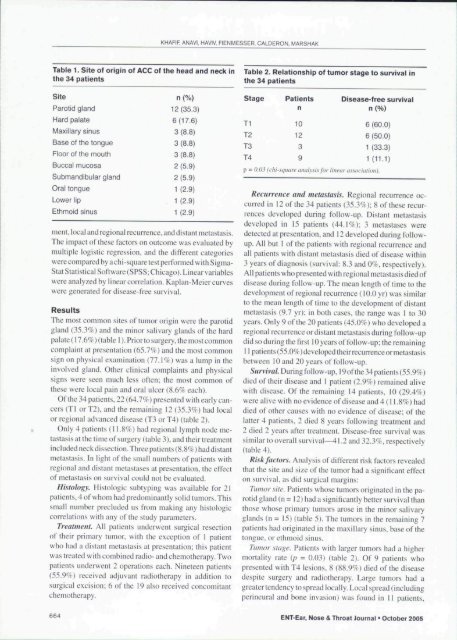
Table 1. Site <strong>of</strong> origin <strong>of</strong> ACC <strong>of</strong> <strong>the</strong> head and neck in<br />
<strong>the</strong> 34 patients<br />
Site<br />
Parotid gland<br />
Hard palate<br />
Maxillary sinus<br />
Base <strong>of</strong> tbe tongue<br />
Floor <strong>of</strong> <strong>the</strong> mouth<br />
Buccal mucosa<br />
Submandibular gland<br />
Oral tongue<br />
Lower lip<br />
Ethmoid sinus<br />
KHAFIF, ANAVI, HAVIV, FIENMESSER. CALDERON, MARSHAK<br />
n (%)<br />
12 (35.3)<br />
6(17.6)<br />
3 (8.8)<br />
3 (8.8)<br />
3 (8.8)<br />
2 (5.9)<br />
2(5.9)<br />
1 (2.9)<br />
1 (2.9)<br />
1 (2.9)<br />
ment, local and regional recurrence, and distant metastasis.<br />
The impact <strong>of</strong>" <strong>the</strong>se factors on outcome was evaluated by<br />
multiple logistic regression, and <strong>the</strong> different categories<br />
were compared by a chi-square test performed with Sigma-<br />
Stat Statistical S<strong>of</strong>tware (SPSS: Chicago). Linear variables<br />
were analyzed by linear correlation. Kaplan-Meier curves<br />
were generated for disease-free survival.<br />
Results<br />
The most common sites <strong>of</strong> tumor origin were <strong>the</strong> parotid<br />
gland I35.y/r) and <strong>the</strong> minor <strong>salivary</strong> glands <strong>of</strong><strong>the</strong> hard<br />
palate (17.6%) (table 1). Prior to surgery, <strong>the</strong> mo.st common<br />
complaint at presentation {b5.7^/c) and <strong>the</strong> most common<br />
sign on physical examination (77.1%) was a lump in <strong>the</strong><br />
involved gland. O<strong>the</strong>r clinical complaints and physical<br />
signs were seen much less <strong>of</strong>ten; <strong>the</strong> most common <strong>of</strong><br />
<strong>the</strong>se were local pain and oral ulcer (8.69f each).<br />
Of<strong>the</strong> 34 patients, 22 (64.7%) presented with early cancers<br />
(Tl orT2). and <strong>the</strong> remaining 12 (35.3%) had local<br />
or regional advanced disease (T3 or T4) (table 2).<br />
Only 4 patients (11.8%) had regional lymph node metastasis<br />
at <strong>the</strong> time <strong>of</strong> surgery (table 3). and <strong>the</strong>ir treatment<br />
included neckdissection.Threepatients (8.8%) had distant<br />
metastasis. In light <strong>of</strong><strong>the</strong> small numbers <strong>of</strong> patients with<br />
regional and distant metastases al presentation, <strong>the</strong> effect<br />
<strong>of</strong> metastasis on survival could not be evaluated.<br />
Histology. Histologic subtyping was available for 21<br />
patients, 4 <strong>of</strong> whom had predominantly solid tumors. This<br />
small number precluded us from making any histologic<br />
correlations with any <strong>of</strong><strong>the</strong> study parameters.<br />
Treatment. All patients underwent surgical resection<br />
<strong>of</strong> <strong>the</strong>ir primary tumor, with <strong>the</strong> exception <strong>of</strong> 1 patient<br />
who had a distant metastasis at presentation; this patient<br />
was treated with combined radio- and chemo<strong>the</strong>rapy. Two<br />
patients underwent 2 operations each. Nineteen patients<br />
(55.9%) received adjuvant radio<strong>the</strong>rapy in addition to<br />
surgical excision; 6 <strong>of</strong> <strong>the</strong> 19 also received concomitant<br />
chemo<strong>the</strong>rapy.<br />
Table 2. Relationship <strong>of</strong> tumor stage to survival in<br />
<strong>the</strong> 34 patients<br />
Stage Patients Disease-free survival<br />
n n (%)<br />
Tl 10 6(60.0)<br />
T2 12 6 (50.0)<br />
T3 3 1 (33.3)<br />
T4 9 1(11.1)<br />
p - 0.0.^ (ihi-sqimre imalysi\ for linear uswcicilioti).<br />
Recurrence and metastasis. Regional recurrence occurred<br />
in 12 <strong>of</strong> <strong>the</strong> 34 patients (35.3%); 8 <strong>of</strong> <strong>the</strong>se recurrences<br />
developed during follow-up. Distant metastasis<br />
developed in 15 patients (44.1%); 3 tnetastases were<br />
detected at presentation, and 12 developed during followup.<br />
All but 1 <strong>of</strong> <strong>the</strong> patients with regional recurrence and<br />
all patients with distant metastasis died <strong>of</strong> disease within<br />
3 years <strong>of</strong> diagnosis (survival: 8.3 and 0%, respectively).<br />
All patients who presented with regional metastasis died <strong>of</strong><br />
disease during follow-up. The mean length <strong>of</strong> time to <strong>the</strong><br />
development <strong>of</strong> regional recurrence (lO.Oyr) was simitar<br />
to <strong>the</strong> mean length <strong>of</strong> time to <strong>the</strong> development ol' distant<br />
metastasis (9.7 yr); in both cases, <strong>the</strong> range was 1 to 30<br />
years. Only 9 <strong>of</strong><strong>the</strong> 20 patients (45.0%) who developed a<br />
regional recurrence or distant metastasis during follow-up<br />
did so during <strong>the</strong> first 10 years <strong>of</strong> follow-up; <strong>the</strong> remaining<br />
11 patienls(55.0%)developed<strong>the</strong>irrecurrenceormetastasis<br />
between 10 and 20 years <strong>of</strong> follow-up.<br />
Survival. Duringfollow-up, 19<strong>of</strong><strong>the</strong>34patients(55.9%)<br />
died <strong>of</strong> <strong>the</strong>ir disease and 1 patient {2.97c) remained alive<br />
with disease. Of<strong>the</strong> remaining 14 patients. 10 (29.4%)<br />
were alive with no evidence <strong>of</strong> disease and 4 (11.8%) had<br />
died <strong>of</strong> o<strong>the</strong>r causes with no evidence <strong>of</strong> disease; <strong>of</strong> <strong>the</strong><br />
latter 4 patients, 2 died 8 years following treatment and<br />
2 died 2 years after treatment. Disease-free survival was<br />
similar to overall survival—41.2 and 32.3%, respectively<br />
(table 4).<br />
Risk factors. Analysis <strong>of</strong> different risk factors revealed<br />
that <strong>the</strong> site and size <strong>of</strong><strong>the</strong> tumor had a significant effect<br />
on survival, as did surgical margins:<br />
Tumor site. Patients whose tumors originated in <strong>the</strong> parotid<br />
gland (n = 12) had a significantly better survival than<br />
those whose primary tumors arose in <strong>the</strong> minor <strong>salivary</strong><br />
glands (n = 15) (table 5). The tumors in <strong>the</strong> remaining 7<br />
patients had originated in <strong>the</strong> maxillary sinus, base <strong>of</strong><strong>the</strong><br />
tongue, or ethmoid sinus.<br />
Ttwtor .stage. Patients with larger tumors had a higher<br />
mortality rate (p = 0.03) (table 2). Of 9 patients who<br />
presented with T4 lesions. 8 (88.9%) died <strong>of</strong><strong>the</strong> disease<br />
despite surgery and radio<strong>the</strong>rapy. Large tumors had a<br />
greatertendency to spread locally. Local spread (including<br />
pcrineural and bone invasion) was found in II patients.<br />
664 ENT-Ear, Nose & Throat Journal - October 2005