Gore RM, Ghahremani GG et al. Anomalies and - Department of ...
Gore RM, Ghahremani GG et al. Anomalies and - Department of ...
Gore RM, Ghahremani GG et al. Anomalies and - Department of ...

Create successful ePaper yourself
Turn your PDF publications into a flip-book with our unique Google optimized e-Paper software.
"<br />
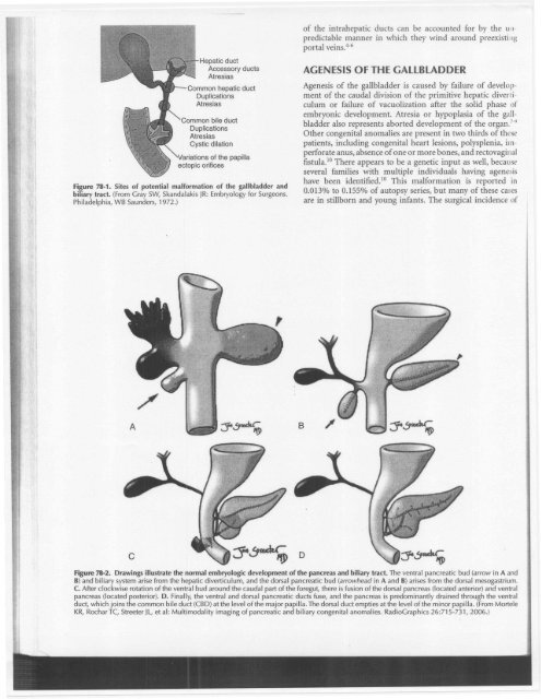
Hepatic duct<br />
Accessory ducts<br />
Atresias<br />
Common hepatic duct<br />
Duplications<br />
Atresias<br />
Common bile duct<br />
Duplications<br />
Atresias<br />
Cystic dilation<br />
Figure 78-1. Sites <strong>of</strong> potenti<strong>al</strong> m<strong>al</strong>formation <strong>of</strong> the g<strong>al</strong>lbladder <strong>and</strong><br />
biliary tract. (From Gray SW, Sk<strong>and</strong><strong>al</strong>akis JR:Embryology for Surgeons.<br />
Philadelphia, WB Saunders, 1972.)<br />
/<br />
<strong>of</strong> the intrahepatic ducts can be accounted for by the unpredictable<br />
manner in which they wind around preexisting<br />
port<strong>al</strong> veins.4-6<br />
AGENESIS OF THE GALLBLADDER<br />
Agenesis <strong>of</strong> the g<strong>al</strong>lbladder is caused by failure <strong>of</strong> development<br />
<strong>of</strong> the caud<strong>al</strong> division <strong>of</strong> the primitive hepatic diverticulum<br />
or failure <strong>of</strong> vacuolization after the solid phase <strong>of</strong><br />
embryonic development. Atresia or hypoplasia <strong>of</strong> the g<strong>al</strong>lbladder<br />
<strong>al</strong>so represents aborted development <strong>of</strong> the organ.7-"<br />
Other congenit<strong>al</strong> anom<strong>al</strong>ies are present in two thirds <strong>of</strong> these<br />
patients, including congenit<strong>al</strong> heart lesions, polysplenia, imperforate<br />
anus, absence <strong>of</strong> one or more bones, <strong>and</strong> rectovagin<strong>al</strong><br />
fistula.1O There appears to be a gen<strong>et</strong>ic input as well, because<br />
sever<strong>al</strong> families with multiple individu<strong>al</strong>s having agenesis<br />
have been identified.1O This m<strong>al</strong>formation is reported in<br />
0.013% to 0.155% <strong>of</strong> autopsy series, but many <strong>of</strong> these cases<br />
are in stillborn <strong>and</strong> young infants. The surgic<strong>al</strong> incidence <strong>of</strong><br />
Figure 78-2. Drawings illustrate the norm<strong>al</strong> embryologic development <strong>of</strong> the pancreas <strong>and</strong> biliary tract. The ventr<strong>al</strong> pancreatic bud (arrow in A <strong>and</strong><br />
B) <strong>and</strong> biliary system arise from the hepatic diverticulum, <strong>and</strong> the dors<strong>al</strong> pancreatic bud (arrowhead in A <strong>and</strong> B) arises from the dors<strong>al</strong> mesogastrium.<br />
C. After clockwise rotation <strong>of</strong> the ventr<strong>al</strong> bud around the caud<strong>al</strong> part <strong>of</strong> the foregut, there is fusion <strong>of</strong> the dors<strong>al</strong> pancreas (located anterior) <strong>and</strong> ventr<strong>al</strong><br />
pancreas (located posterior). D. Fin<strong>al</strong>ly, the ventr<strong>al</strong> <strong>and</strong> dors<strong>al</strong> pancreatic ducts fuse, <strong>and</strong> the pancreas is predominantly drained through the ventr<strong>al</strong><br />
duct, which joins the common bile duct (CBD) at the level <strong>of</strong> the major papilla. The dors<strong>al</strong> duct empties at the level <strong>of</strong> the minor papilla. (From Mortele<br />
KR, Rochar TC, Stre<strong>et</strong>er JL, <strong>et</strong> <strong>al</strong>: Multimod<strong>al</strong>ity imaging <strong>of</strong> pancreatic <strong>and</strong> biliary congenit<strong>al</strong> anom<strong>al</strong>ies. RadioGraphies 26:715-731, 2006.)