

Amoeboid - Thierry Karsenti
Amoeboid - Thierry Karsenti
Amoeboid - Thierry Karsenti

You also want an ePaper? Increase the reach of your titles
YUMPU automatically turns print PDFs into web optimized ePapers that Google loves.
available, bacterial classification remains a changing and expanding field. [4][115] For<br />
example, a few biologists argue that the Archaea and Eukaryotes evolved from Grampositive<br />
bacteria. [116]<br />
Identification of bacteria in the laboratory is particularly relevant in medicine, where<br />
the correct treatment is determined by the bacterial species causing an infection.<br />
Consequently, the need to identify human pathogens was a major impetus for the<br />
development of techniques to identify bacteria.<br />
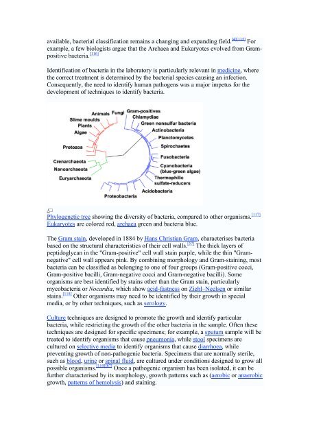
Phylogenetic tree showing the diversity of bacteria, compared to other organisms. [117]<br />
Eukaryotes are colored red, archaea green and bacteria blue.<br />
The Gram stain, developed in 1884 by Hans Christian Gram, characterises bacteria<br />
based on the structural characteristics of their cell walls. [57] The thick layers of<br />
peptidoglycan in the "Gram-positive" cell wall stain purple, while the thin "Gramnegative"<br />
cell wall appears pink. By combining morphology and Gram-staining, most<br />
bacteria can be classified as belonging to one of four groups (Gram-positive cocci,<br />
Gram-positive bacilli, Gram-negative cocci and Gram-negative bacilli). Some<br />
organisms are best identified by stains other than the Gram stain, particularly<br />
mycobacteria or Nocardia, which show acid-fastness on Ziehl–Neelsen or similar<br />
stains. [118] Other organisms may need to be identified by their growth in special<br />
media, or by other techniques, such as serology.<br />
Culture techniques are designed to promote the growth and identify particular<br />
bacteria, while restricting the growth of the other bacteria in the sample. Often these<br />
techniques are designed for specific specimens; for example, a sputum sample will be<br />
treated to identify organisms that cause pneumonia, while stool specimens are<br />
cultured on selective media to identify organisms that cause diarrhoea, while<br />
preventing growth of non-pathogenic bacteria. Specimens that are normally sterile,<br />
such as blood, urine or spinal fluid, are cultured under conditions designed to grow all<br />
possible organisms. [119][87] Once a pathogenic organism has been isolated, it can be<br />
further characterised by its morphology, growth patterns such as (aerobic or anaerobic<br />
growth, patterns of hemolysis) and staining.