Guide to identification of Lumbrineridae - NMBAQC
Guide to identification of Lumbrineridae - NMBAQC
Guide to identification of Lumbrineridae - NMBAQC

Create successful ePaper yourself
Turn your PDF publications into a flip-book with our unique Google optimized e-Paper software.
4<br />
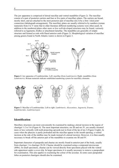
The jaw apparatus is comprised <strong>of</strong> dorsal maxillae and ventral mandibles (Figure 2). The maxillae<br />
consist <strong>of</strong> a pair <strong>of</strong> posterior carriers and four or five pairs <strong>of</strong> maxillary plates. The carriers are broad,<br />
mostly short, and are attached <strong>to</strong> the most posterior pair <strong>of</strong> maxillae (mx I) by a firm ’click-joint’<br />
connection (labidognath arrangement). The maxillary plates are usually referred <strong>to</strong> by numbers (roman<br />
numerals) from I <strong>to</strong> V (note that in older literature different numbering systems were <strong>of</strong>ten used). In<br />
addition <strong>to</strong> the numbered plates other more or less well-developed structures may be found, variously<br />
referred <strong>to</strong> as ligaments, bridles or attachment lamellae. The mandibles are generally <strong>of</strong> simple<br />
structure and formed as rods with flared anterior ends (Figure 2). Morphological variation <strong>of</strong> maxillae<br />
among genera found in North Atlantic waters is shown in Figure 3.<br />
Figure 2. Jaw apparatus <strong>of</strong> <strong>Lumbrineridae</strong>. Left: maxillae (from Lumbrineris). Right: mandibles (from<br />
Lumbrineris). Roman numerals indicate established numbering system for maxillary elements.<br />
Figure 3. Maxillae <strong>of</strong> <strong>Lumbrineridae</strong>. Left <strong>to</strong> right: Lumbrineris, Abyssoninoe, Augeneria, Eranno,<br />
Lumbrinerides, Lumbrineriopsis<br />
Identification<br />
Maxillary structures can most conveniently be examined by making a dorsal incision in the region <strong>of</strong><br />
chaetiger 1-2 <strong>to</strong> 5-6 (Figure 4). The most important structures, mx III and mx IV, are usually oriented<br />
more or less vertically with teeth projecting upwards just in front <strong>of</strong> the tip <strong>of</strong> mx I (Figure 4 right). In<br />
cases when the pharynx is partly protruded and the maxillae appear in the mouth opening, a ventral<br />
incision at the side <strong>of</strong> the midline may be made instead <strong>of</strong> a dorsal incision. However, it is then usually<br />
necessary <strong>to</strong> break <strong>of</strong>f the anterior part <strong>of</strong> the mandibles <strong>to</strong> examine mx III and IV.<br />
Important characters <strong>of</strong> parapodia and chaetae are mostly found in anterior part <strong>of</strong> the body, usually<br />
from chaetiger 1 <strong>to</strong> chaetiger 20-30. Chaetae should be examined using a compound microscope<br />
(400x). In small specimens, chaetae can be viewed directly from specimens placed with the ventral<br />
side uppermost under a cover-slip. In larger specimens it is usually necessary <strong>to</strong> remove a parapodium<br />
for examination. This also applies <strong>to</strong> examining the colour <strong>of</strong> the aciculae. In some cases parapodial<br />
lobes on posterior chaetigers should also be examined.