Abdi, Camillo Ricordi, Mohamed H. Sayegh, Antonello Pileggi and ...
Abdi, Camillo Ricordi, Mohamed H. Sayegh, Antonello Pileggi and ...
Abdi, Camillo Ricordi, Mohamed H. Sayegh, Antonello Pileggi and ...
You also want an ePaper? Increase the reach of your titles
YUMPU automatically turns print PDFs into web optimized ePapers that Google loves.
11<br />
DOI: 10.1161/CIRCULATIONAHA.112.123653<br />
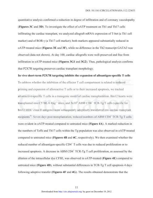
quantitative analysis confirmed a reduction in degree of infiltration <strong>and</strong> of coronary vasculopathy<br />
(Figures 3C <strong>and</strong> 3D). To investigate the effect of oATP treatment on Th1 <strong>and</strong> Th17 cells<br />
infiltrating the cardiac transplant, we analyzed allograft mRNA expression of T-bet (a Th1 cell<br />
marker) <strong>and</strong> of ROR- (a Th17 cell marker); both markers appeared substantially reduced in<br />
oATP-treated mice (Figures 3E <strong>and</strong> 3F), while no difference in the Th2 transcript GATA3 was<br />
observed (data not shown). At day 100, cardiac allografts were well-preserved <strong>and</strong> free from<br />
infiltration in oATP-treated mice (Figures 3G1 <strong>and</strong> 3G2). Thus, pathological analysis confirms<br />
that P2X7R targeting preserves cardiac transplant morphology.<br />
In vivo short-term P2X7R targeting inhibits the expansion of alloantigen-specific T cells<br />
To address whether the inhibition of the effector T cell compartment is related to redu reduced du duce ced d<br />
priming <strong>and</strong> expansion of alloreactive T cells or to their increased apoptosis, we tracked<br />
allo alloreactive-specific lore re reac acti tive ive<br />
ve-s -speci ci cifi fic T cells in a transgenic model el e of cardiac transp transplantation. sp splant ntat at ation. Bm12 hearts were<br />
Rag -/- mic<br />
transplanted into C57BL/6 Rag -/- mice, <strong>and</strong> 3x10 6 ABM CD4 + ransplanted int nto C5 C57B 7BL/ L/6 Ra ice, <strong>and</strong> 3x10 TCR-Tg T cells (specific for<br />
6 ABM CD4 D4 + TCR-Tg g T ce cells s (s (spe peci ci c fic fo for r<br />
bm12 bm bm12 12 MHC HC class cla lass II antigens) an antige ge g ns) ) were we were re subsequently su subs bseq eq eque uent nt ntly ly ado adoptively dopt pt ptiv ivel el ely y tr tran transferred an ansf sf sfer fer<br />
erre red in into<br />
to car cardiac ar ardi di diac ac tra transplant rans nspl pl p ant t<br />
recipients 31 . Seven days post-transplantation, reduced numbers of ABM CD4 + ecipients TCR-Tg T cells<br />
31 . Se Seve ve ven da days ys pos os osttt-tr<br />
tran an ansp sp spla la l nt ntat at ation on on, re redu du duce ce ced d nu numb mb mber er ers s of o ABM BM CD4 D4 D + TCR CR CR-T -T -Tg T cells<br />
were evident in oATP-treated compared to untreated mice (Figure 4A). A marked reduction in<br />
the numbers of Teffs <strong>and</strong> Th17 cells within the Tg population was also observed in oATP-treated<br />
compared to untreated mice (Figures 4B <strong>and</strong> 4C, respectively). We then examined whether the<br />
reduced number of alloantigen-specific CD4 + T cells was due to reduced proliferation or to<br />
increased apoptosis. A decrease in ABM CD4 + TCR-Tg T cell proliferation, as assessed by the<br />
dilution of the intracellular dye CFSE, was observed in oATP-treated (Figure 4E) compared to<br />
untreated mice (Figure 4D), without substantial differences in TCR-Tg T cell apoptosis 4 days<br />
following adoptive transfer (Figures 4F <strong>and</strong> 4G). The results obtained demonstrate that the<br />
Downloaded from<br />
http://circ.ahajournals.org/ by guest on December 19, 2012


