The study on the isolation and culture of the protoplast from ...
The study on the isolation and culture of the protoplast from ...
The study on the isolation and culture of the protoplast from ...

Create successful ePaper yourself
Turn your PDF publications into a flip-book with our unique Google optimized e-Paper software.
Protoplasts <strong>from</strong> Closterium<br />
were observed even in <strong>the</strong> same preparati<strong>on</strong>. <str<strong>on</strong>g>The</str<strong>on</strong>g> cell wall regenerati<strong>on</strong><br />
during 4 days <strong>of</strong> <strong>culture</strong> proceeded as follows.<br />
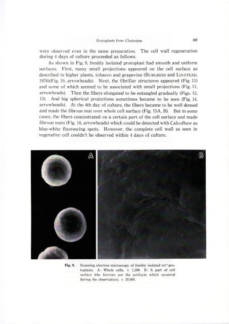
As shown in Fig. 9, freshly isolated <strong>protoplast</strong> had smooth <strong>and</strong> uniform<br />
surfaces. First, many small projecti<strong>on</strong>s appeared <strong>on</strong> <strong>the</strong> cell surface as<br />
described in higher plants, tobacco <strong>and</strong> grapevine (BURGRESS <strong>and</strong> LiNSTEAD,<br />
1976)(Fig. 10, arrowheads). Next, <strong>the</strong> fibrillar structures appeared (Fig. 11)<br />
<strong>and</strong> some <strong>of</strong> which seemed to be associated with small projecti<strong>on</strong>s (Fig. 11,<br />
arrowheads). <str<strong>on</strong>g>The</str<strong>on</strong>g>n <strong>the</strong> fibers el<strong>on</strong>gated to be entangled gradually (Figs. 12,<br />
13). And big spherical projecti<strong>on</strong>s sometimes became to be seen (Fig. 14,<br />
arrowheads). At <strong>the</strong> 4th day <strong>of</strong> <strong>culture</strong>, <strong>the</strong> fibers became to be well den sed<br />
<strong>and</strong> made <strong>the</strong> fibrous mat over whole cell surface (Fig. 15A, B). But in some<br />
cases, <strong>the</strong> fibers c<strong>on</strong>centrated <strong>on</strong> a certain part <strong>of</strong> <strong>the</strong> cell surface <strong>and</strong> made<br />
fibrous mats (Fig. 16, arrowheads) which could be detected with Calc<strong>of</strong>luor as<br />
blue-white fluorescing spots. However, <strong>the</strong> complete cell wall as seen In<br />
vegetative cell couldn't be observed within 4 days <strong>of</strong> <strong>culture</strong>.<br />
Fig_ 9_ Scanning electr<strong>on</strong> microscopy <strong>of</strong> freshly isolated mt+-<strong>protoplast</strong>s.<br />
A: Whole cells, X 1,300. B: A part <strong>of</strong> cell<br />
surface (<strong>the</strong> furrows are <strong>the</strong> artifacts which occurred<br />
during <strong>the</strong> observati<strong>on</strong>), x 20,000.<br />
107