A bibliography of Podospora and Schizothecium, a key to the ...
A bibliography of Podospora and Schizothecium, a key to the ...
A bibliography of Podospora and Schizothecium, a key to the ...
You also want an ePaper? Increase the reach of your titles
YUMPU automatically turns print PDFs into web optimized ePapers that Google loves.
108<br />
A.M.B. Centro Studi Micologici<br />
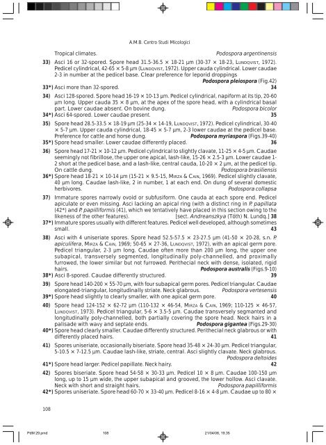
Tropical climates. <strong>Podospora</strong> argentinensis<br />
33) Asci 16 or 32-spored. Spore head 31.5-36.5 × 18-21 µm (30-37 × 18-23, LUNDQVIST, 1972).<br />
Pedicel cylindrical, 42-65 × 5-8 µm (LUNDQVIST, 1972). Upper cauda cylindrical. Lower caudae<br />
2-3 in number at <strong>the</strong> pedicel base. Clear preference for leporid droppings<br />
<strong>Podospora</strong> pleiospora (Fig.42)<br />
33*) Asci more than 32-spored. 34<br />
34) Asci 128-spored. Spore head 16-19 × 10-13 µm. Pedicel cylindrical, napiform at its tip, 20-60<br />
µm long. Upper cauda 35 × 8 µm, at <strong>the</strong> apex <strong>of</strong> <strong>the</strong> spore head, with a cylindrical basal<br />
part. Lower caudae absent. On bovine dung. <strong>Podospora</strong> bicolor<br />
34*) Asci 64-spored. Lower caudae present. 35<br />
35) Spore head 28.5-33.5 × 18-19 µm (25-34 × 14-19, LUNDQVIST, 1972). Pedicel cylindrical, 30-40<br />
× 5-7 µm. Upper cauda cylindrical, 18-45 × 5-7 µm, 2-3 lower caudae at <strong>the</strong> pedicel base.<br />
Preference for cattle <strong>and</strong> horse dung. <strong>Podospora</strong> myriaspora (Figs.39-40)<br />
35*) Spore head smaller. Lower caudae differently placed. 36<br />
36) Spore head 17-21 × 10-12 µm. Pedicel cylindrical <strong>to</strong> slightly clavate, 11-25 × 4-5 µm. Caudae<br />
seemingly not fibrillose, <strong>the</strong> upper one apical, lash-like, 15-26 × 2.5-3 µm. Lower caudae 1-<br />
2 short at <strong>the</strong> pedicel base, <strong>and</strong> a lash-like, central cauda, 10-20 × 2 µm, at <strong>the</strong> pedicel tip.<br />
On cattle dung. <strong>Podospora</strong> brasiliensis<br />
36*) Spore head 18-21 × 10-14 µm (15-21 × 9.5-15, MIRZA & CAIN, 1969). Pedicel slightly clavate,<br />
40 µm long. Caudae lash-like, 2 in number, 1 at each end. On dung <strong>of</strong> several domestic<br />
herbivores. <strong>Podospora</strong> collapsa<br />
37) Immature spores narrowly ovoid or subfusiform. One cauda at each spore end. Pedicel<br />
apiculate or even missing. Asci lacking an apical ring (with a distinct ring in P. papillata<br />
(42*) <strong>and</strong> P. papilliformis (41), which we tentatively have placed in this section owing <strong>to</strong> <strong>the</strong><br />
likeness <strong>of</strong> <strong>the</strong> o<strong>the</strong>r features). [sect. Andreanszkya (Tóth) N. Lundq.] 38<br />
37*) Immature spores usually with different features. Pedicel well developed, although sometimes<br />
small. 43<br />
38) Asci with 4 uniseriate spores. Spore head 52.5-57.5 × 23-27.5 µm (41-50 × 20-28, s.n. P.<br />
apiculifera, MIRZA & CAIN, 1969; 50-65 × 27-36, LUNDQVIST, 1972), with an apical germ pore.<br />
Pedicel triangular, 2-3 µm long. Caudae <strong>of</strong>ten more than 200 µm long, <strong>the</strong> upper one<br />
subapical, transversely segmented, longitudinally poly-channelled, <strong>and</strong> proximally<br />
furrowed, <strong>the</strong> lower similar but not furrowed. Peri<strong>the</strong>cial neck with dense, isolated, rigid<br />
hairs. <strong>Podospora</strong> australis (Figs.9-10)<br />
38*) Asci 8-spored. Caudae differently structured. 39<br />
39) Spore head 140-200 × 55-70 µm, with four subapical germ pores. Pedicel triangular. Caudae<br />
elongated-triangular, longitudinally striate. Neck glabrous. <strong>Podospora</strong> vertesensis<br />
39*) Spore head slightly <strong>to</strong> clearly smaller, with one apical germ pore. 40<br />
40) Spore head 124-152 × 62-72 µm (110-132 × 46-54, MIRZA & CAIN, 1969; 110-125 × 46-57,<br />
LUNDQVIST, 1973). Pedicel triangular, 5-6 × 3.5-5 µm. Caudae transversely segmanted <strong>and</strong><br />
longitudinally poly-channelled, both partially covering <strong>the</strong> spore head. Neck hairs in a<br />
palisade with wavy <strong>and</strong> septate ends. <strong>Podospora</strong> gigantea (Figs.29-30)<br />
40*) Spore head clearly smaller. Caudae differently structured. Peri<strong>the</strong>cial neck glabrous or with<br />
differently placed hairs. 41<br />
41) Spores uniseriate, occasionally biseriate. Spore head 35-48 × 24-30 µm. Pedicel triangular,<br />
5-10.5 × 7-12.5 µm. Caudae lash-like, striate, central. Asci slightly clavate. Neck glabrous.<br />
<strong>Podospora</strong> del<strong>to</strong>ides<br />
41*) Spore head larger. Pedicel papillate. Neck hairy. 42<br />
42) Spores biseriate. Spore head 54-58 × 30-33 µm. Pedicel 10 × 8 µm. Caudae 100-150 µm<br />
long, up <strong>to</strong> 15 µm wide, <strong>the</strong> upper subapical <strong>and</strong> grooved, <strong>the</strong> lower hollow. Asci clavate.<br />
Neck with short <strong>and</strong> straight hairs. <strong>Podospora</strong> papilliformis<br />
42*) Spores uniseriate. Spore head 60-70 × 33-40 µm. Pedicel 8-16 × 4-8 µm. Caudae up <strong>to</strong> 80 ×<br />
PdM 29.pmd 108<br />
21/04/08, 19.35


