Phylogenetic lineages in the Capnodiales - Cbs - KNAW
Phylogenetic lineages in the Capnodiales - Cbs - KNAW
Phylogenetic lineages in the Capnodiales - Cbs - KNAW

Create successful ePaper yourself
Turn your PDF publications into a flip-book with our unique Google optimized e-Paper software.
(Morris 1989). The fungus causes partial defoliation of mature<br />
plants (Dodd 1961, Auld 1969), though <strong>the</strong> impact depends on<br />
environmental conditions (Dodd 1961). Seedl<strong>in</strong>gs are however<br />
killed rapidly (Wang et al. 1997).<br />
This fungus, which has hi<strong>the</strong>rto been known simply as<br />
“Phaeoramularia” sp., still lacks a name and proper description.<br />
The genus Phaeoramularia is treated as a synonym of Passalora<br />
(Crous & Braun 2003), and hence <strong>the</strong> species is named <strong>in</strong> <strong>the</strong> latter<br />
genus as P. agerat<strong>in</strong>ae. Interest<strong>in</strong>gly, this species appears to be<br />
closely related to Passalora fulva, which is a serious pathogen of<br />
tomato (Solanaceae) (Thomma et al. 2005).<br />
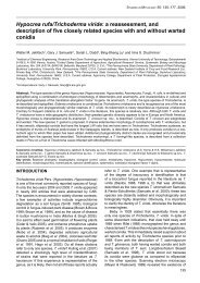
Passalora armatae Crous & A.R. Wood, sp. nov. MycoBank<br />
MB514698. Fig. 6.<br />
Etymology: Named after <strong>the</strong> host on which it occurs, Dalbergia<br />
armata.<br />
Passaloraea dalbergiicolae similis, sed conidiophoris <strong>in</strong> synnematibus densis,<br />
conidiis ad basim obconice truncatis, apice rostrato.<br />
Leaf spots amphigenous, on upper surface visible as red-brown,<br />
irregular to subcircular spots with <strong>in</strong>dist<strong>in</strong>ct marg<strong>in</strong>s, 0.5–2 mm<br />
diam; <strong>in</strong> reverse <strong>in</strong>dist<strong>in</strong>ct, chlorotic to medium or red-brown.<br />
Mycelium <strong>in</strong>ternal, consist<strong>in</strong>g of smooth, branched, pale brown,<br />
2–3 µm wide hyphae. Caespituli hypophyllous, fasciculate to<br />
synnematous, up to 200 µm high and 250 µm wide, situated on<br />
a prom<strong>in</strong>ently erumpent, pale brown stroma, up to 100 µm high<br />
and wide. Conidiophores subcyl<strong>in</strong>drical, unbranched, flexuous,<br />
guttulate, pale to medium brown, smooth, 120–180 × 4–6 µm,<br />
2–6-septate. Conidiogenous cells term<strong>in</strong>al, subcyl<strong>in</strong>drical,<br />
www.studies<strong>in</strong>mycology.org<br />
<strong>Phylogenetic</strong> l<strong>in</strong>eageS <strong>in</strong> <strong>the</strong> <strong>Capnodiales</strong><br />
guttulate, pale to medium brown, f<strong>in</strong>ely verruculose, becom<strong>in</strong>g<br />
somewhat swollen, appear<strong>in</strong>g slightly clavate, 25–70 × 6–8 µm;<br />
conidiogenous loci 4–20 per conidiogenous cell, sympodial, round,<br />
darkened, thickened, refractive, prom<strong>in</strong>ent, 2–3 µm wide, up to<br />
1 µm high. Conidia (27–)30–40(–45) × 9–10(–12) µm, pale to<br />
medium brown, smooth to f<strong>in</strong>ely verruculose, granular to guttulate,<br />
th<strong>in</strong>-walled, ellipsoidal to obovoid, transversely 2–4-euseptate,<br />
widest <strong>in</strong> middle of basal cell, or middle of conidium, taper<strong>in</strong>g to an<br />
obconically truncate base; hilum thickened, darkened and refractive;<br />
apical cell conical, elongat<strong>in</strong>g to an apical beak up to 20 µm long.<br />
When cultivated conidia rema<strong>in</strong> attached to conidiogenous cells,<br />
giv<strong>in</strong>g conidiophores <strong>the</strong> appearance of small tufts which is very<br />
characteristic, and not commonly observed <strong>in</strong> Passalora.<br />
Culture characteristics: On MEA slow grow<strong>in</strong>g, erumpent, with<br />
dense white aerial mycelium, which becomes mouse-grey, reach<strong>in</strong>g<br />
5 mm diam after 1 wk; on PDA mouse-grey (surface), iron-grey<br />
(reverse), with diffuse red pigment <strong>in</strong> agar; on OA similar to PDA,<br />
also with diffuse red pigment <strong>in</strong> agar.<br />
Host range and geographic distribution: Dalbergia armata, South<br />
Africa.<br />
Specimen exam<strong>in</strong>ed: south Africa, KwaZulu-Natal Prov<strong>in</strong>ce, South Coast, Mpenjati<br />
Nature Reserve, between Ramsgate and Port Edward, on leaves of Dalbergia<br />
armata, 28 May 2008, A.R. Wood, CBS H-20337 holotype, cultures ex-type CPC<br />
15419 = CBS 125420, CPC 15420, 15421.<br />
Notes: Passalora dalbergiae, which occurs on Dalbergia sissoo<br />
(Fabaceae) <strong>in</strong> India, is dist<strong>in</strong>ct from P. armatae <strong>in</strong> hav<strong>in</strong>g superficial<br />
mycelium and solitary conidiophores (Hernández-Gutiérrez &<br />
Fig. 6. Passalora armatae. A. Fruit<strong>in</strong>g <strong>in</strong> vivo. B–C. Caespituli with prom<strong>in</strong>ent basal stroma. D. Sporulation on MEA. E. Conidiogenous cells giv<strong>in</strong>g rise to conidia. F–G. Conidia.<br />
Scale bars: B = 125 µm, C–E = 10 µm.<br />
35