Determination of ergosterol in cellular fungi by HPLC - Asociación ...
Determination of ergosterol in cellular fungi by HPLC - Asociación ...
Determination of ergosterol in cellular fungi by HPLC - Asociación ...

You also want an ePaper? Increase the reach of your titles
YUMPU automatically turns print PDFs into web optimized ePapers that Google loves.
The Journal <strong>of</strong> the Argent<strong>in</strong>e Chemical Society Vol. 98, 10-15 (2011) 10<br />
Journal <strong>of</strong> the<br />
Argent<strong>in</strong>e Chemical<br />
Society<br />
DETERMINATION OF ERGOSTEROL IN CELLULAR FUNGI BY<br />
<strong>HPLC</strong>. A MODIFIED TECHNIQUE.<br />
V. M. Chiocchio 1,♥ and L. Matković 2<br />
1<br />
Cátedra de Microbiología Agrícola. Dto. Biología Aplicada y Alimentos. FAUBA- INBA –<br />
CONICET. Av. San Martín 4453. (1427DSE) CABA Argent<strong>in</strong>a.<br />
2<br />
Laboratorio de Esteroides, Departamento de Química Biológica Facultad de Ciencias Exactas y<br />
Naturales, UBA. Ciudad Universitaria Pabellón II 4to Piso Laboratorio de Esteroides (1428EHA)<br />
CABA Argent<strong>in</strong>a<br />
Received Mars 30, 2011. In f<strong>in</strong>al form July 18, 2011.<br />
Abstract<br />
Ergosterol is an important membrane sterol <strong>in</strong> almost all eumycotic <strong>fungi</strong> and has been postulated to<br />
be strongly associated with liv<strong>in</strong>g cytoplasmic <strong>fungi</strong> <strong>in</strong> the soil. However, <strong>ergosterol</strong> is not produced<br />
<strong>by</strong> all <strong>fungi</strong> and the <strong>ergosterol</strong> concentrations are known to vary between the same species<br />
depend<strong>in</strong>g on the physiological state <strong>of</strong> the fungus. In this research we studied a modification to an<br />
<strong>ergosterol</strong> extraction technique which has the advantage <strong>of</strong> hav<strong>in</strong>g a protocol with few steps <strong>of</strong><br />
purification. This technique also <strong>in</strong>cludes an <strong>in</strong>ternal loss marker to evaluate the efficiency <strong>of</strong><br />
extraction.<br />
Key words: <strong>ergosterol</strong>; <strong>HPLC</strong> quantification; soil <strong>fungi</strong><br />
♥ Correspond<strong>in</strong>g author. E-mail: chiocchi@agro.uba.ar, Tel. 005411-4524-8061<br />
J. Argent. Chem. Soc., 2011, 98, 10-15
11 V. M. Chiocchio and L. Matković<br />
Resumen<br />
El <strong>ergosterol</strong> es un lípido de la familia de los esteroles componente importante de la membrana<br />
celular, presente en casi todos los hongos pertenecientes a los llamados “hongos verdaderos”<br />
(eumycotas). Se ha postulado que se halla asociado fuertemente con el citoplasma fúngico en el<br />
suelo, dando idea de la subsistencia del mismo. S<strong>in</strong> embargo, el <strong>ergosterol</strong> no es producido por todos<br />
los hongos y las concentraciones de <strong>ergosterol</strong> se sabe que varían entre la misma especie en función<br />
del estado fisiológico del hongo. En este trabajo de <strong>in</strong>vestigación, se ha realizado una modificación a<br />
las técnicas de extracción de <strong>ergosterol</strong> ya conocidas, las cuales entre otras cosas, no tienen en<br />
cuenta las pérdidas de masa del esteroide a lo largo de los pasos de purificación. Esta <strong>in</strong>novación<br />
posee además la ventaja de tener un protocolo con pocos y sencillos pasos de purificación. Además,<br />
agrega la utilización de un marcador de pérdida para poder evaluar de esta manera la eficiencia de la<br />
extracción y así tener un resultado más cercano al real.<br />
Palabras clave: <strong>ergosterol</strong>; cunatificación en <strong>HPLC</strong>; hongos del suelo<br />
Introduction<br />
Ergosterol is an important membrane sterol <strong>in</strong> almost all eumycotic <strong>fungi</strong> [1]. A number <strong>of</strong><br />
techniques have been used to estimate biomass fungal: direct microscopic count<strong>in</strong>g <strong>of</strong> hyphae [2,4]<br />
(the most widely accepted and commonly used method), fluorescence microscopy [5], leaf clear<strong>in</strong>g<br />
and sta<strong>in</strong><strong>in</strong>g [5,6] and ATP assay [7] . Various serious sources <strong>of</strong> error exist <strong>in</strong> all <strong>of</strong> these<br />
techniques. A disadvantage <strong>of</strong> all these techniques is that they use conversion factors <strong>by</strong> means <strong>of</strong><br />
which biomass either is <strong>of</strong>ten under or overestimated [8,9]. Biochemical markers <strong>of</strong> biomass,<br />
pr<strong>in</strong>cipally glucosam<strong>in</strong>e and <strong>ergosterol</strong>, have also been used [10,11].<br />
The glucosam<strong>in</strong>e assays has been questioned, as this compound is not restricted to fungal<br />
cells and it is refractory thus persist<strong>in</strong>g after the death <strong>of</strong> the mycelium [10 and 12]. Therefore, at<br />
best, glucosam<strong>in</strong>e can only be an <strong>in</strong>dicator <strong>of</strong> both liv<strong>in</strong>g and dead hyphae.<br />
Ergosterol is an important membrane sterol <strong>in</strong> almost all eumycotic <strong>fungi</strong> [13] and has been<br />
postulated to be strongly associated with liv<strong>in</strong>g cytoplasmic <strong>fungi</strong> <strong>in</strong> the soil [14]. However,<br />
<strong>ergosterol</strong> is not produced <strong>by</strong> all <strong>fungi</strong> and the <strong>ergosterol</strong> concentrations are known to vary<br />
between the same species depend<strong>in</strong>g on the physiological state <strong>of</strong> the fungus [15,17].<br />
Ergosterol has been widely used to quantify biomass <strong>in</strong> several studies <strong>of</strong> soils and<br />
mycorrhizal systems [18,19,7] and to determ<strong>in</strong>e biomass associated with decay<strong>in</strong>g leaves <strong>in</strong><br />
freshwaters [15,16,20].<br />
Sung et al. (1995) [21] found good correlations between mycelial dry weight and <strong>ergosterol</strong><br />
content for some species <strong>of</strong> ectomycorrhizal <strong>fungi</strong>, but also that <strong>in</strong> other species, <strong>ergosterol</strong><br />
decreased as the mycelial culture aged.<br />
Age <strong>of</strong> mycelium used, extraction procedures, <strong>in</strong>cubation temperatures have been shown to<br />
<strong>in</strong>fluence <strong>ergosterol</strong> content [22]. However, we believe that these studies support the use <strong>of</strong><br />
<strong>ergosterol</strong> as a measure <strong>of</strong> fungal biomass <strong>in</strong> the soil. We found soil <strong>ergosterol</strong> concentration to be<br />
positively correlated to both total and liv<strong>in</strong>g hyphal lenghts <strong>in</strong> soil with a high degree <strong>of</strong><br />
significance, but only a moderate degree <strong>of</strong> l<strong>in</strong>ear correlation. Therefore, a moderate degree <strong>of</strong><br />
error will be <strong>in</strong>volved <strong>in</strong> estimation <strong>of</strong> liv<strong>in</strong>g fungal biomass from soil <strong>ergosterol</strong> content alone.<br />
Our method is a modification to an extraction <strong>ergosterol</strong> technique [18,23] which has the<br />
advantage <strong>of</strong> hav<strong>in</strong>g a protocol with few steps <strong>of</strong> purification. For example, <strong>in</strong> one <strong>of</strong> the most<br />
recent publications [24], we appreciate that the authors considered a lot <strong>of</strong> steps, which were carried<br />
out with several errors and greater extraction times. As a fundamental po<strong>in</strong>t <strong>of</strong> our method we<br />
developed a procedure <strong>of</strong> extraction <strong>of</strong> <strong>ergosterol</strong> different from those already published, tak<strong>in</strong>g <strong>in</strong>to<br />
account the losses caused <strong>by</strong> the extraction. In addition, we had <strong>in</strong> m<strong>in</strong>d the possible oxidation <strong>of</strong>
<strong>Determ<strong>in</strong>ation</strong> <strong>of</strong> <strong>ergosterol</strong> <strong>in</strong> <strong>cellular</strong> <strong>fungi</strong> <strong>by</strong> <strong>HPLC</strong>… 12<br />
<strong>ergosterol</strong>, so we added from the beg<strong>in</strong>n<strong>in</strong>g <strong>of</strong> the technic an antioxidant such as BHT thus avoid<strong>in</strong>g<br />
the use <strong>of</strong> nitrogen.<br />
The basic procedures <strong>in</strong>volve sample extraction, saponification, partition<strong>in</strong>g, purification,<br />
determ<strong>in</strong>ation and quantification <strong>of</strong> <strong>ergosterol</strong>.<br />
Material and Methods<br />
We collected different samples <strong>of</strong> wet soil from San Luis (Potrero de los Funes and<br />
Quebrada de la Bolsa), Argent<strong>in</strong>a, with vegetation cover <strong>of</strong> ferns (Bryopteris sp., Cheilantes sp.,<br />
and Anemia sp.). The samples (2 g <strong>of</strong> each; with 24% humidity) were saponificated with 2g KOH<br />
<strong>in</strong> 25 mL <strong>of</strong> methanol (<strong>HPLC</strong> grade), <strong>in</strong> the presence <strong>of</strong> 100 µL <strong>of</strong> corticosterone (loss marker) and<br />
100 µL <strong>of</strong> butylated hydroxytoluene (BHT) as antioxidant. After the saponification (1 h reflux to<br />
70°C), they were left to cool. The supernatant was placed <strong>in</strong> a funnel and 20 mL <strong>of</strong> methanol was<br />
added twice over the precipitate. We jo<strong>in</strong>ed the three aliquots <strong>in</strong> the funnel with 30 mL <strong>of</strong> dry nhexane<br />
(previously filtered the 60mL <strong>of</strong> methanol though Millex-HV 0.45 µm- Millipore S.A.,<br />
Molsheim, France). This extraction was repeated twice. Fractions were jo<strong>in</strong>ed and taken to dryness<br />
<strong>in</strong> a Rotavapor. The samples were stored (4°C <strong>in</strong> darkness) until <strong>HPLC</strong> analysis. Each sample was<br />
re-dissolved <strong>in</strong> 200 µL methanol. For <strong>HPLC</strong> analysis we used a P100 isocratic pump (Spectra<br />
Physics), a UV-100 detector (Spectra Physics) and a Data Jet <strong>in</strong>tegrator (Spectra Physics), a flow<br />
rate <strong>of</strong> 1.0 mL/m<strong>in</strong>. The wavelength was set at 290 nm for determ<strong>in</strong>ate <strong>ergosterol</strong> and 254nm for<br />
corticosterone. Thus, each sample was <strong>in</strong>jected twice <strong>in</strong> the <strong>HPLC</strong>. The column used was a<br />
Supelcosil LC18 (Supelco; 5 µm, 250 x 4.6 mm).<br />
Methanol:acetonitrile, 80:20 v/v (<strong>HPLC</strong>-grade) was the mobile phase. Under these conditions the<br />
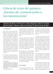
retention time was 13.4±0.2 m<strong>in</strong>. (Figure 1).<br />
13.4 m<strong>in</strong><br />
Figure 1. Ergosterol pr<strong>of</strong>ile isolated from <strong>cellular</strong> <strong>fungi</strong>.<br />
Spectra Physics <strong>HPLC</strong> equipment was used with an<br />
isocratic Pump P100 <strong>in</strong>corporated. The absorbance<br />
spectrum was plotted at 290nm with a UV 100 Spectra<br />
Physics detector, chart speed was 0.5 cm.m<strong>in</strong> -1 <strong>in</strong>tegrated<br />
with a Spectra Physics Data Jet Integrator. A Rheodyne<br />
<strong>in</strong>jector supplied with a 20μL loop was used. The column<br />
used was a Supelcosil C18 (Supelco; 5 µm, 250 x 4.6<br />
mm) and, as mobile phase, <strong>HPLC</strong>-grade methanol :<br />
acetonitrile (80:20 v/v). The retention time <strong>in</strong> these<br />
conditions was 13.4 m<strong>in</strong> ± 0.2 m<strong>in</strong> at a flow rate <strong>of</strong> 1.0<br />
mL.m<strong>in</strong> -1 and an attenuation <strong>of</strong> 128. A similar pr<strong>of</strong>ile was<br />
obta<strong>in</strong>ed for the steroid used to calculate the losses but,<br />
<strong>in</strong> this case the absorbance spectrum was plotted at<br />
254nm.
13 V. M. Chiocchio and L. Matković<br />
Stock calibration solutions <strong>of</strong> corticosterone and BHT were prepared <strong>in</strong> methanol.<br />
Ergosterol was recalculated from peak areas <strong>of</strong> <strong>in</strong>ternal standard (IS). The relative response<br />
obta<strong>in</strong>ed from the ratio <strong>of</strong> IS to <strong>ergosterol</strong> peak areas was 0.99, whereas the relative recovery (the<br />
ratio <strong>of</strong> absolute recoveries <strong>of</strong> IS and <strong>ergosterol</strong>) was 0.88.<br />
The l<strong>in</strong>earity <strong>of</strong> the method was tested for corticosterone and <strong>ergosterol</strong>. Increas<strong>in</strong>g<br />
amounts (5 to 20 µg) <strong>of</strong> analytes were <strong>in</strong>jected for each determ<strong>in</strong>ation.<br />
The regression equations were calculated <strong>by</strong> the method <strong>of</strong> standardized pr<strong>in</strong>cipal<br />
component, and the coefficient <strong>of</strong> correlation was determ<strong>in</strong>ed <strong>by</strong> l<strong>in</strong>ear regression.<br />
Results and discussion<br />
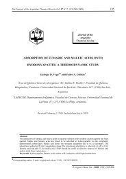
Calibration curves for peak areas vs. quantity <strong>of</strong> <strong>ergosterol</strong> were l<strong>in</strong>ear from 5 to 20 µg<br />
(Figure 2). The m<strong>in</strong>imum detectable amount <strong>of</strong> <strong>ergosterol</strong> and IS was 2 µg. By <strong>in</strong>ject<strong>in</strong>g 20µL <strong>of</strong><br />
the reconstituted extract we obta<strong>in</strong>ed 0.1 µg.g -1 <strong>ergosterol</strong>. A relative response factor <strong>of</strong> IS<br />
compared with that <strong>of</strong> <strong>ergosterol</strong> was calculated and found to be 0.99 for the entire range. Absolute<br />
recoveries <strong>of</strong> <strong>ergosterol</strong> were calculated and were different depend<strong>in</strong>g on the sample considered.<br />
Thus, we <strong>in</strong>corporated a relative recovery accord<strong>in</strong>g to the sample studied.<br />
Area<br />
20000<br />
16000<br />
12000<br />
8000<br />
4000<br />
0<br />
y = 791,44x<br />
R 2 = 0,9701<br />
0 5 10 15 20<br />
ug de <strong>ergosterol</strong><br />
Figure 2. Calibration plot <strong>of</strong> µg <strong>ergosterol</strong> (Std 1.015 mg/mL). Ergosterol was<br />
determ<strong>in</strong>ed <strong>by</strong> <strong>HPLC</strong> Spectra Physics equipment with UV 100 detector and a Supelcosil<br />
C18 column at room temperature. The mobile phase was <strong>HPLC</strong> grade methanol :<br />
acetonitrile (80:20 v/v). The elution time was 13.4 m<strong>in</strong> ± 0.2 m<strong>in</strong>. at a flow rate <strong>of</strong> 1.0<br />
mL m<strong>in</strong> -1 . The chat speed was 0.5 cm.m<strong>in</strong> -1 .<br />
The correlation <strong>of</strong> the losses measured with the standard <strong>of</strong> <strong>ergosterol</strong> and the standard <strong>of</strong><br />
corticosterone was 98.9% with 0.1% SD.<br />
These determ<strong>in</strong>ations were conducted on pooled <strong>ergosterol</strong> samples and thus reflect the<br />
entire process <strong>in</strong>clud<strong>in</strong>g the solid-phase extraction.<br />
Our <strong>HPLC</strong> <strong>ergosterol</strong> method was optimized tak<strong>in</strong>g <strong>in</strong>to account the losses produced <strong>in</strong> the<br />
various extraction steps. We used corticosterone as an <strong>in</strong>ternal standard, s<strong>in</strong>ce it allowed us to<br />
correct the values obta<strong>in</strong>ed for <strong>ergosterol</strong>. Various different treatments were used for <strong>ergosterol</strong><br />
determ<strong>in</strong>ation. The best result <strong>in</strong> the recovery <strong>of</strong> <strong>ergosterol</strong> was 100% methanol <strong>in</strong> the
<strong>Determ<strong>in</strong>ation</strong> <strong>of</strong> <strong>ergosterol</strong> <strong>in</strong> <strong>cellular</strong> <strong>fungi</strong> <strong>by</strong> <strong>HPLC</strong>… 14<br />
saponification process and n-hexane <strong>in</strong> the extraction process. A change <strong>of</strong> solvent <strong>in</strong> the process<br />
<strong>of</strong> saponification (to ethanol for example) gave a lower percentage <strong>of</strong> recovery <strong>in</strong> the first<br />
extraction (67%) and a lower percentage (9%) <strong>in</strong> a second extraction with the same solvent. The<br />
efficiency <strong>of</strong> the method proposed was already 80% <strong>in</strong> the first extraction (Table 1). Us<strong>in</strong>g this<br />
technique, we quantified <strong>ergosterol</strong> <strong>in</strong> soil samples (with Bryopteris) and showed the presence <strong>of</strong><br />
fungal mycelium, as an <strong>in</strong>direct measure <strong>of</strong> fungal biomass.<br />
We determ<strong>in</strong>ed <strong>ergosterol</strong> <strong>in</strong> a range (0.18-0.43) µg /g dry soil depend<strong>in</strong>g on the sample.<br />
So, our method to quantify <strong>ergosterol</strong> showed to be efficient and able to be carried out <strong>in</strong> only a<br />
few steps, also implemented, correction <strong>by</strong> loss. It should be noted that there were no overlapped<br />
peaks <strong>in</strong> the <strong>HPLC</strong> procedure.<br />
Table 1. Percentages <strong>of</strong> mass recovered from a total <strong>of</strong> 50.75 μg <strong>of</strong> standard <strong>ergosterol</strong>.<br />
Treatment with either ethanol or methanol <strong>in</strong>volves saponification. Retention times were<br />
13.4 m<strong>in</strong> ± 0.2 m<strong>in</strong> at a flow rate <strong>of</strong> 1.0 mL.m<strong>in</strong> -1 for all the determ<strong>in</strong>ations at the same<br />
conditions specified <strong>in</strong> Figure 2. Values are mean ± SDM <strong>of</strong> three <strong>in</strong>dependent experiments<br />
performed <strong>in</strong> duplicate.<br />
Sample<br />
Ergosterol<br />
+<br />
methanol<br />
Ergosterol<br />
+<br />
methanol<br />
Ergosterol<br />
+<br />
methanol<br />
Solvent<br />
Extraction<br />
First<br />
extraction<br />
Second<br />
extraction<br />
Total<br />
recovery<br />
n-hexane 80 ± 7 % 8 ± 1 % 88 ± 8 %<br />
n-heptane 15 ± 3 % 1.3 ± 0,9 % 16 ± 4 %<br />
etherethilic<br />
53 ± 2 % 4,8 ± 0,5 % 58 ± 3 %<br />
Ergosterol<br />
+ ethanol n-heptane 67 ± 3 % 9 ± 1 % 76 ± 4 %<br />
Acknowledgments. This work was supported <strong>by</strong> grants provided <strong>by</strong> the University <strong>of</strong> Buenos<br />
Aires and CONICET (Argent<strong>in</strong>a). The authors wish to thank Dr. Carlos Lantos and Dr. Alicia<br />
Godeas for their suggested revisions <strong>of</strong> the manuscript.<br />
References<br />
[1] D.S. Hibbett, M. B<strong>in</strong>der, J.F. Bisch<strong>of</strong>f, M. Blackwell, P.F. Cannon, O.E. Eriksson, S.<br />
Huhndorf, T. James, P.M. Kirk, R. Lück<strong>in</strong>g, T. Lumbsch, F. Lutzoni, P.B. Matheny, D.J.<br />
Mclaughl<strong>in</strong>, M.J. Powell, S. Redhead, C.L. Schoch, J.W. Spatafora, J.A. Stalpers, R.<br />
Vilgalys, M.C. Aime, A. Aptroot, R. Bauer, D. Begerow, G.L. Benny, L.A. Castlebury, P.W.<br />
Crous, Y.-C. Dai, W. Gams, D.M. Geiser, G.W. Griffith, C. Gueidan, D.L. Hawksworth, G.<br />
Hestmark, K. Hosaka, R.A. Humber, K. Hyde, J.E. Ironside, U. Kõljalg, C.P. Kurtzman, K.-<br />
H. Larsson, R. Lichtwardt, J. Longcore, J. Miądlikowska, A. Miller, J.M. Moncalvo, S.
15 V. M. Chiocchio and L. Matković<br />
Standridge Mozley, F. Oberw<strong>in</strong>kler, E. Parmasto, V. Reeb, J.D. Rogers, C. Roux, L.<br />
Ryvarden, J.P. Sampaio, A. Schüßler, J. Sugiyama, R.G. Thorn, L. Tibell, W.A. Untere<strong>in</strong>er,<br />
C. Walker, Z. Wang, A. Weir, M. Weiss, M.M. Blanco, K. W<strong>in</strong>ka, Y.-J. Yao, N. Zhang, La<br />
<strong>in</strong>vestigación micológica. 2007, 111, 509-547.<br />
[2] D. Park<strong>in</strong>son, Filamentous <strong>fungi</strong>, <strong>in</strong>: Methods <strong>of</strong> Soil Analysis, Part 2 (A. L. Page, R. H.<br />
Miller and D. R. Keeney, Eds). American Society <strong>of</strong> Agronomy - Soil Science Society <strong>of</strong><br />
America, Madison, 1982a, pp. 945-968.<br />
[3] K.H. Domsch, T.H. Beck, J.P.E. Anderson, B. Södeerström, D. Park<strong>in</strong>son, G. Trolldenier, Z.<br />
Pflanzen. Bodenkd, 1979, 142, 520-533.<br />
[4] P.D. Stahl, T.B. Park<strong>in</strong>, N.S. Eash, Soil Biol. Biochem., 1995, 27 (8), 1091-1097.<br />
[5] M. Bölter, J. Bloem, K. Me<strong>in</strong>ers, R. Möller, Biol. Fertil. Soils, 2002, 36, 249–259.<br />
[6] F. Bärlocher, B. Kendrick, J. Ecol., 1974, 62 (3), 761-791.<br />
[7] T. Larsen, J. Axelsen, H.W. Ravn, J. Chromatogr. A, 2004, 1026, 301-304.<br />
[8] M. Klamer, E. Bääth, Soil Biol. Biochem., 2004, 36, 57-65.<br />
[9] J. Rousk, E. Bääth, Soil Biol. Biochem., 2007, 39, 2173-2177.<br />
[10] S.Y. Newell, Estimat<strong>in</strong>g fungal biomass and productivity <strong>in</strong> decompos<strong>in</strong>g litter. In The<br />
Fungal Community: Its Organization and Role <strong>in</strong> the Ecosystem (G.C.Carroll and D. T.<br />
Wicklow. Eds.), Dekker, New York, 1992, pp. 521-561.<br />
[11] J. Whipps, D. Lewis, Trans. Brit. Mycol. Soc., 1980, 79, 178–179.<br />
[12] P.D. Sharma, P.J. Fisher, J. Webster, Trans. Brit. Mycol. Soc., 1977, 69 (3), 479-483.<br />
[13] B. Axelsson, A. Saraf, L. Larsson, J. Chromatogr. B, 1995, 666, 77–84.<br />
[14] S.Y. Newell, Fungal biomass and productivity, <strong>in</strong>: Methods <strong>in</strong> Microbiology, 2001, 30, 357–<br />
372.<br />
[15] M.O. Gessner, E. Chauvet, Appl. Environ. Microbiol., 1993, 59 (2), 502-507.<br />
[16] K. Suberkropp, M.O. Gessner, E. Chauvet, Appl. Environ. Microbiol., 1993, 59 (10), 3367-<br />
3372.<br />
[17] M.M. Müller, R. Kantola, V. Kitunen, Mycol. Res., 1994, 98, 593–603.<br />
[18] H. Wallander, H.B. Massicotte, J.E. Nylund, Soil Biol. Biochem., 1997, 29, 45-53.<br />
[19] X.R. Zhao, Q. L<strong>in</strong>, P.C. Brookes, Soil Biol. Biochem., 2005, 37, 311-317.<br />
[20] M.O. Gessner, J. Schwoerbel, Oecologia, 1991, 87 (4), 602-603.<br />
[21] S.S. Sung, L.M. White, D.H. Marx, W.J. Otros<strong>in</strong>a, Mycorrhiza, 1995, 5, 439-447.<br />
[22] S. Berm<strong>in</strong>gham, L. Malt<strong>by</strong>, R.C. Cooke, Mycol. Res., 1995, 9(4), 479-484.<br />
[23] S. Ruzicka, M.D. Norman, J.A. Harris, Soil Biol. Biochem., 1995, 27, (9), 1215-1217.<br />
[24] D. Tardieu, J.D. Bailly, G. Benard, P. Guerre, Revue Mèd. Vèt, 2007, 158 (8-9), 442-446.