BACTERIAL SEPSIS AND MENINGITIS - Nizet Laboratory at UCSD
BACTERIAL SEPSIS AND MENINGITIS - Nizet Laboratory at UCSD
BACTERIAL SEPSIS AND MENINGITIS - Nizet Laboratory at UCSD

You also want an ePaper? Increase the reach of your titles
YUMPU automatically turns print PDFs into web optimized ePapers that Google loves.
number of newborn children developing fever sank from<br />
45% to 11.3%.”[396]<br />
Closure of the umbilical vessels and the subsequent<br />
aseptic necrosis of the cord begins soon after the infant<br />
takes the first bre<strong>at</strong>h. The umbilical arteries contract;<br />
the blood flow is interrupted; and the cord tissues,<br />
deprived of a blood supply, undergo aseptic necrosis.<br />
The umbilical stump acquires a rich flora of microorganisms.<br />
Within hours, the umbilical stump is colonized with<br />
large numbers of gram-positive cocci, particularly Staphylococcus<br />
species, and shortly thereafter with fecal organisms<br />
[396,397]. These bacteria can invade the open umbilical<br />
wound, causing a localized infection with purulent discharge<br />
and, as a result of delayed obliter<strong>at</strong>ion of the<br />
umbilical vessels, bleeding from the umbilical stump.<br />
From this site, infection can proceed into the umbilical<br />
vessels, along the fascial planes of the abdominal wall, or<br />
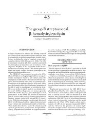
into the peritoneum (Fig. 6–1) [396,398,399].<br />
Although umbilical discharge or an “oozing” cord is<br />
the most common manifest<strong>at</strong>ion of omphalitis, periumbilical<br />
cellulitis and fasciitis are the conditions most often<br />
associ<strong>at</strong>ed with hospitaliz<strong>at</strong>ion [398]. Infants presenting<br />
with fasciitis have a high incidence of bacteremia, intravascular<br />
coagulop<strong>at</strong>hy, shock, and de<strong>at</strong>h [398]. Edema of<br />
the umbilicus and peau d’orange appearance of the surrounding<br />
abdominal skin, signaling obstruction of the<br />
underlying lymph<strong>at</strong>ics, can be an early warning sign,<br />
whereas the p<strong>at</strong>hognomonic purplish blue discolor<strong>at</strong>ion<br />
FIGURE 6–1 After birth, the necrotic tissue of the umbilical stump<br />
separ<strong>at</strong>es. This provokes some inflamm<strong>at</strong>ion, which is limited by a<br />
fibroblastic reaction extending to the inner margin of the coarsely stippled<br />
area. The inner half of the media and the intima of the umbilical arteries<br />
become necrotic, but this does not stimul<strong>at</strong>e an inflamm<strong>at</strong>ory reaction.<br />
Arrows indic<strong>at</strong>e routes by which infection may spread beyond the<br />
granul<strong>at</strong>ion tissue barriers. Organisms invading the thrombus in the vein<br />
may dissemin<strong>at</strong>e by emboli. (From Morison JE. Foetal and Neon<strong>at</strong>al<br />
P<strong>at</strong>hology, 3rd ed. Washington, DC, Butterworth, 1970.)<br />
CHAPTER 6 Bacterial Sepsis and Meningitis<br />
245<br />
implies advanced necrotizing fasciitis [393]. Septic emboliz<strong>at</strong>ion<br />
arising from the infected umbilical vessels is<br />
uncommon, but can produce metast<strong>at</strong>ic spread to various<br />
organs, including the lungs, pancreas, kidneys, and skin<br />
[394]. Such emboli can arise from the umbilical arteries<br />
and from the umbilical vein, because final closure of the<br />
ductus venosus and separ<strong>at</strong>ion of the portal circul<strong>at</strong>ion<br />
from the inferior vena cava and the systemic circul<strong>at</strong>ion<br />
are generally delayed until day 15 to 30 of life [400].<br />
Omphalitis is now a rare infection in developed<br />
countries because of modern umbilical cord care. Complic<strong>at</strong>ions<br />
of omphalitis include various infections, such<br />
as septic umbilical arteritis [394,401], suppur<strong>at</strong>ive thrombophlebitis<br />
of the umbilical or portal veins or the ductus<br />
venosus [401–403], peritonitis [399,401,402,404], intestinal<br />
gangrene [399], pyourachus (infection of the urachal<br />
remnant) [405], liver abscess, endocarditis, pyelophlebitis<br />
[399,406], and subacute necrotizing funisitis [407]. Some<br />
of these infections can occur in the absence of signs of<br />
omphalitis [394,401].<br />
ADMINISTRATION OF DRUGS TO THE<br />
MOTHER BEFORE DELIVERY<br />
Almost all antimicrobial agents cross the placenta. Antimicrobial<br />
drugs administered to the mother <strong>at</strong> term can<br />
alter the initial microflora of the neon<strong>at</strong>e and can complic<strong>at</strong>e<br />
the diagnosis of infection in the neon<strong>at</strong>e. Chapter 37<br />
reviews the clinical pharmacology of antimicrobial agents<br />
administered to the mother.<br />
Studies have shown th<strong>at</strong> corticosteroid administr<strong>at</strong>ion to<br />
mothers in preterm labor to enhance pulmonary m<strong>at</strong>ur<strong>at</strong>ion<br />
in the fetus resulted in a significant decrease in the incidence<br />
and severity of neon<strong>at</strong>al respir<strong>at</strong>ory distress syndrome, but<br />
an increase in m<strong>at</strong>ernal infection, particularly endometritis,<br />
compared with placebo [408]; however, the impacts of this<br />
practice on the risk of neon<strong>at</strong>al infection differed among<br />
early studies [408,409]. Roberts and Dalziel [410] more<br />
recently performed a large meta-analysis of 21 randomized<br />
controlled studies from the Cochrane Pregnancy and Childbirth<br />
Group Trials register, comprising 3885 pregnant<br />
women and 4269 infants, and concluded th<strong>at</strong> anten<strong>at</strong>al<br />
corticosteroid administr<strong>at</strong>ion (betamethasone, dexamethasone,<br />
or hydrocortisone) given to women expected to deliver<br />
singleton or multiple pregnancies, whether labor was spontaneous,<br />
induced by membrane rupture, or electively<br />
induced, was associ<strong>at</strong>ed with multiple favorable outcomes,<br />
including reduced neon<strong>at</strong>al de<strong>at</strong>h (rel<strong>at</strong>ive risk 0.69), intensive<br />
care admissions (rel<strong>at</strong>ive risk 0.80) and systemic infections<br />
in the first 48 hours of life (rel<strong>at</strong>ive risk 0.56).<br />
Substance abuse during pregnancy can affect immune<br />
function in the neon<strong>at</strong>e. Significant abnormalities in<br />
T-cell function and an apparent increased incidence of<br />
infections have been found during the first year of life<br />
among infants born to alcohol-addicted [411–413] and<br />
heroin-addicted [414,415] mothers. The adverse effects<br />
of cocaine and opi<strong>at</strong>es on placental function, fetal growth<br />
and development, and prem<strong>at</strong>urity also may predispose to<br />
a gre<strong>at</strong>er likelihood of neon<strong>at</strong>al infection [415,416]. Drug<br />
abuse is a multifactorial problem; it is virtually impossible<br />
to separ<strong>at</strong>e the consequences of direct pharmacologic<br />
effects on the fetus from the consequences secondary to