Folliculitis decalvans - Cicatricial Alopecia Research Foundation
Folliculitis decalvans - Cicatricial Alopecia Research Foundation
Folliculitis decalvans - Cicatricial Alopecia Research Foundation

Create successful ePaper yourself
Turn your PDF publications into a flip-book with our unique Google optimized e-Paper software.
The development of FD after scalp injury is<br />
occasionally reported by patients and can be<br />
found in literature (16). However, its pathogenetic<br />
significance is not clear.<br />
Seborrhea as a pathogenetic factor of FD has<br />
been emphasized by some authors in the past.<br />
Since scalp seborrhea is a common finding and<br />
FD is rare, the significance is doubtful (13).<br />
Clinical presentation<br />
<strong>Folliculitis</strong> <strong>decalvans</strong> predominantly involves the<br />
vertex and occipital area of the scalp. The initial<br />
lesion is an erythematous follicular papule. The<br />
hallmark of FD is the development of scarred areas<br />
and follicular pustules. Livid to bright erythema<br />
together with yellow-gray scales can be present<br />
especially around the follicles as well as follicular<br />
hyperkeratosis, erosions, and hemorrhagic crusts<br />
(6,17–19) (FIG. 1).<br />
Patients occasionally report spontaneous bleeding<br />
and frequently complain about pain, itching,<br />
and/or burning sensations. In the cause of the<br />
progressing disease, small to extensive, irregularly<br />
shaped, atrophic flesh-colored or ivory-white<br />
patches of cicatricial alopecia develop (18,20–22).<br />
In our experience, the scarred areas are often times<br />
thicker and indurated compared to the atrophic<br />
cicatricial patches of other inflammatory primary<br />
scarring scalp disorders. In older lesion pustules<br />
can be absent but progressive scarring may still<br />
continue (FIG. 2).<br />
Tufted folliculitis is a common finding in patients<br />
with FD. It is characterized by multiple hairs (5–20)<br />
emerging from one single dilated follicular orifice.<br />
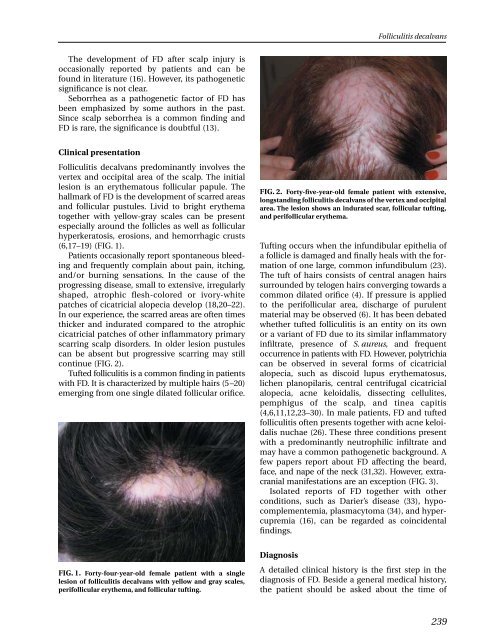
FIG. 1. Forty-four-year-old female patient with a single<br />
lesion of folliculitis <strong>decalvans</strong> with yellow and gray scales,<br />
perifollicular erythema, and follicular tufting.<br />
<strong>Folliculitis</strong> <strong>decalvans</strong><br />
FIG. 2. Forty-five-year-old female patient with extensive,<br />
longstanding folliculitis <strong>decalvans</strong> of the vertex and occipital<br />
area. The lesion shows an indurated scar, follicular tufting,<br />
and perifollicular erythema.<br />
Tufting occurs when the infundibular epithelia of<br />
a follicle is damaged and finally heals with the formation<br />
of one large, common infundibulum (23).<br />
The tuft of hairs consists of central anagen hairs<br />
surrounded by telogen hairs converging towards a<br />
common dilated orifice (4). If pressure is applied<br />
to the perifollicular area, discharge of purulent<br />
material may be observed (6). It has been debated<br />
whether tufted folliculitis is an entity on its own<br />
or a variant of FD due to its similar inflammatory<br />
infiltrate, presence of S. aureus,<br />
and frequent<br />
occurrence in patients with FD. However, polytrichia<br />
can be observed in several forms of cicatricial<br />
alopecia, such as discoid lupus erythematosus,<br />
lichen planopilaris, central centrifugal cicatricial<br />
alopecia, acne keloidalis, dissecting cellulites,<br />
pemphigus of the scalp, and tinea capitis<br />
(4,6,11,12,23–30). In male patients, FD and tufted<br />
folliculitis often presents together with acne keloidalis<br />
nuchae (26). These three conditions present<br />
with a predominantly neutrophilic infiltrate and<br />
may have a common pathogenetic background. A<br />
few papers report about FD affecting the beard,<br />
face, and nape of the neck (31,32). However, extracranial<br />
manifestations are an exception (FIG. 3).<br />
Isolated reports of FD together with other<br />
conditions, such as Darier’s disease (33), hypocomplementemia,<br />
plasmacytoma (34), and hypercupremia<br />
(16), can be regarded as coincidental<br />
findings.<br />
Diagnosis<br />
A detailed clinical history is the first step in the<br />
diagnosis of FD. Beside a general medical history,<br />
the patient should be asked about the time of<br />
239