

In partnership with : - CHU - Marrakech
In partnership with : - CHU - Marrakech
In partnership with : - CHU - Marrakech

Create successful ePaper yourself
Turn your PDF publications into a flip-book with our unique Google optimized e-Paper software.
parameters (behavior, school, performance and mood) in<br />
children and this has lead to increasing use of VNS in this<br />
population. This is in addition to the fact that it would limit<br />
the complications resulting from pharmacotherapy side<br />
effects [12].<br />
Mechanism of Action<br />
The exact mechanism of action of VNS is not well<br />
established. Recent neurophysiologic and neuro-imaging<br />
studies have pointed out some of the neural pathways<br />
activated by VNS and have given some proof that the<br />
alteration of the afferent fibers of the vagus nerve leads<br />
to an increase in seizure threshold [2]. 80% of the vagus<br />
nerve fibers are afferent fibers, somatic and visceral, which<br />
transmit input from the head, thorax and abdomen into<br />
nucleus tractus solitarius. The nucleus solitarius has 3<br />
major outputs: Autonomic feed up loop, direct projection to<br />
the reticular formation of the medulla, and the Ascending<br />
Projections into the parabracheal (PB) nucleus and Locus<br />
ceruleus (LC) [13].<br />
The LC is a major Norepinephrine (NE) nucleus in the CNS.<br />
The NE released upon VNS stimulation increases seizure<br />
threshold by releasing y-aminobutyric acid [14]. It also<br />
inhibits glutamate secretion in the regions that project<br />
afferent fibers into the LC. Rat studies have shown that<br />
destroying the LC makes VNS ineffective in seizure control<br />
[15]. <strong>In</strong> addition, both PB and LC nuclei project efferent<br />
fibers into the amygdala and the stria terminalis. This<br />
is possibly the reason for the antidepressant and mood<br />
alteration effect observed <strong>with</strong> VNS.<br />
Another suggested mechanism is that VNS, by altering<br />
heart rate and contractility, alters cerebral blood flow<br />
(CBF) to specific areas in the brain leading to a higher<br />
seizure threshold [16]. Certain studies have shown<br />
some evidence on this: Vonck et al., using single photon<br />
emission computed tomography scans showed that there<br />
was acute limbic hyperperfusion and chronic thalamic<br />
hypo-perfusion concomitant <strong>with</strong> VNS stimulation [17].<br />
These correlated <strong>with</strong> positive clinical efficacy.<br />
Recent studies have concentrated on the cortical<br />
neurophysiology [18]. <strong>In</strong>itially, it was widely thought that<br />
C-fiber activation <strong>with</strong> high current is needed to achieve<br />
an anti-epileptic effect. However, Krahl et al reported that<br />
destroying the C-fibers in rat models didn’t affect VNS<br />
efficacy [19]. Other investigators interestingly showed that<br />
low current intensities, which cause stimulation purely<br />
of the myelinated A or B fibers, achieve a longer lasting<br />
seizure control effect than high currents, which are also<br />
associated <strong>with</strong> more side-effects [18].<br />
The VNS Device<br />
Unlike Cardiac pacemakers, which are on-demand devices<br />
that interfere in case of abnormal cardiac currents, VNS<br />
batteries usually work inter-ictally in a continuous fashion<br />
causing long term changes in the brain and increasing<br />
the seizure threshold, and thus, decreasing the seizure<br />
frequency. VNS devices are produced by Cyberonics<br />
<strong>In</strong>corporation in Houston, Texas. Each device has three<br />
parts: a current pulse generator composed of lithium<br />
6<br />
cadmium battery, a lead wire that is placed subcutaneously,<br />
and a silicone rubber embedded platinum electrode. Each<br />
electrode has three helical coils, and each has three loops<br />
to ensure maximum contact when wrapped around the left<br />
vagus nerve. The first and the distal coils are the positive<br />
and negative leads. The middle helix is only for anchoring.<br />
Each patient is given a hand-held magnet to activate the<br />
neural stimulation when an aura occurs, thus, aborting or<br />
minimizing a seizure activity.<br />
Surgical Placement of VNS<br />
Under General anesthesia, a skin incision is made on<br />
the left at the anterior border of the sternocleidomastoid<br />
muscle at the level of the cricothyroid membrane. The<br />
platysma is opened and careful dissection anterior to the<br />
sternocleidomastoid is then done to expose and then open<br />
the carotid sheath which contains the internal jugular<br />
vein, the carotid artery and the left vagus nerve. We need<br />
to expose at least 3 cm of the vagus nerve for proper<br />
electrode attachment. After isolating the vagus nerve, a<br />
subcutaneous pocket to hide the generator is made above<br />
the pectoralis fascia through a left chest skin incision. Some<br />
authors perform the surgery through a single incision at<br />
a midpoint between the two usual incisions. A tunneler<br />
is used to pass the connection lead from the neck to the<br />
chest incision (figure1). Following this, the electrodes are<br />
wrapped around the exposed part of the vagus nerve, and<br />
the lead connected to the generator which is placed in the<br />
chest pocket. Before closure, a programmer is used to<br />
interrogate the generator and to verify that there is good<br />
lead impedance, and all connections are verified. The<br />
current practice is to put on the system at 2 weeks after<br />
implantation.<br />
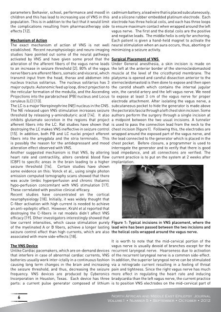
Figure 1: Typical incisions in VNS placement, where the<br />
lead wire has been passed between the two incisions and<br />
the helical coils wrapped around the vagus nerve.<br />
It is worth to note that the mid-cervical portion of the<br />
vagus nerve is usually devoid of branches except for the<br />
recurrent laryngeal nerve. Hoarseness due to activation<br />
of the recurrent laryngeal nerve is a common side-effect.<br />
<strong>In</strong> addition, the superior laryngeal nerve can be stimulated<br />
via a retrograde current resulting in a feeling of throat<br />
pain and tightness. Since the right vagus nerve has much<br />
more effect in regulating the heart rate and inducing<br />
bradycardia than the left vagus nerve, the current practice<br />
is to position VNS electrodes on the mid-cervical part of<br />
North African and Middle East Epilepsy Journal<br />
Volume1 • Number 5 • September • October • 2012