Number 4 - Jordan Journal of Biological Sciences
Number 4 - Jordan Journal of Biological Sciences
Number 4 - Jordan Journal of Biological Sciences

Create successful ePaper yourself
Turn your PDF publications into a flip-book with our unique Google optimized e-Paper software.
170<br />
© 2010 <strong>Jordan</strong> <strong>Journal</strong> <strong>of</strong> <strong>Biological</strong> <strong>Sciences</strong>. All rights reserved - Volume 3, <strong>Number</strong> 4<br />
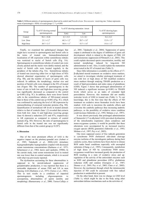
Table 2. Different patterns <strong>of</strong> spermatogenesis observed in control and N a n d ro lone Decan o ate -receiving rats. Values represents<br />
mean <strong>of</strong> percentages ± SEM, n in each group=7, *: p=0.001<br />
Group<br />
% <strong>of</strong> ST showing maturation Arrest<br />
% <strong>of</strong> ST showing normal<br />
spermatogenesis Early Late<br />
% <strong>of</strong> ST showing hypo-<br />
spermatogenesis<br />
Control 94.1 ± 0.26 4.3 ± 0.3 1.2 ± 0.1 0.4 ± 0.3<br />
Low Dose 21.1 ±1.2 * 44.3 ± 1.2 * 20.7 ± 1.8 * 13.9 ± 1.5 *<br />
High Dose 20.7 ± 2.1 * 30.0 ± 1.1 * 33.7 ± 1.8 * 15.6 ± 1.2 *<br />
Finally, we examined the pathological changes that<br />
might have occurred to spermatogonia and Sertoli cells in<br />
the testes <strong>of</strong> treated rats. Immunohistochemical<br />
localization <strong>of</strong> AR expression within seminiferous tubules<br />
was restricted to nuclei <strong>of</strong> Sertoli cells (Fig. 5A).<br />
Spermatogonia in seminiferous tubules <strong>of</strong> control rats were<br />
closely packed and the nuclei were darkly-stained, and the<br />
nuclei <strong>of</strong> Sertoli cells were located regularly in the<br />
periphery <strong>of</strong> the tubules (Fig. 5A). Seminiferous tubules<br />
<strong>of</strong> treated rats (receiving either low or high doses <strong>of</strong> ND)<br />
showed abnormal organization <strong>of</strong> spermatogenic cells<br />
(Fig. 5B), and the number <strong>of</strong> layers <strong>of</strong> germ cells was<br />
reduced. In addition, the morphology, nuclear size and<br />
position <strong>of</strong> Sertoli cells were changed in the STs <strong>of</strong> treated<br />
rats (Figure 5B). The number <strong>of</strong> spermatogonia in the<br />
testes <strong>of</strong> rats in both low and high-dose receiving groups<br />
was significantly decreased as compared to the control<br />
(p=0.001) (Fig. 5C). In addition, there were fewer Sertoli<br />
cells in the seminiferous tubules <strong>of</strong> ND-treated animals<br />
than in those <strong>of</strong> the control rats (p=0.001) (Fig. 5C), which<br />
was confirmed by analyzing the level <strong>of</strong> AR expression by<br />
immunoblotting <strong>of</strong> extracted testicular proteins (Fig. 5D).<br />
Quantification <strong>of</strong> normalized AR levels in treated animals<br />
relative to that <strong>of</strong> controls (lane 1,2) revealed that extracts<br />
from both low- (lane 3) and high-dose receiving groups<br />
(lane 4) showed a reduction (32% and 47%, respectively)<br />
in AR expression as compared to extracts <strong>of</strong> control<br />
animals (Fig. 5D). However, the ratio <strong>of</strong> spermatogonia to<br />
Sertoli cells in the treated rats was not significantly<br />
different from that <strong>of</strong> the control group (p=0.287).<br />
4. Discussion<br />
One <strong>of</strong> the most prominent effects <strong>of</strong> AAS is the<br />
negative impact on the pituitary-gonadal axis (Aubert et<br />
al., 1985; Takahashi et al., 2004).AAS stimulates<br />
hypogonadotrophic hypogonadism coupled with decreased<br />
serum testosterone concentrations (Harkness et al., 1975;<br />
Schurmeyer et al., 1984; Jarow and Lipshultz, 19908). In<br />
the current study, total serum testosterone level in treated<br />
rats was lower than in control animals, which is in accord<br />
with what was previously reported.<br />
The dysfunction accounting for these abnormalities is<br />
supposed to be steroid-induced suppression <strong>of</strong><br />
gonadotrophin production; it was reported that there is a<br />
noticeable depression <strong>of</strong> serum FSH and LH in men<br />
abusing AAS (Harkness et al., 1975; Clerico et al., 1981).<br />
This in turn results in a condition <strong>of</strong> impaired<br />
spermatogenesis including oligozoospermia to<br />
azoospermia, abnormalities <strong>of</strong> sperm motility and<br />
morphology (Ramaswamy et al., 2000; Torres-Calleja et<br />
al., 2001; Takahashi et al., 2004). Suppression <strong>of</strong> sperm<br />
output is attributed to the degree <strong>of</strong> inhibition <strong>of</strong> germ cell<br />
development which is related to the degree <strong>of</strong> FSH, LH<br />
and testosterone suppression (O'Donnell et al., 2001). This<br />
could explain decreased sperm concentration, motility and<br />
normal morphology induced by long-term ND<br />
administration to rats. Also, it could explain the maturation<br />
arrest noted in the ST <strong>of</strong> treated rats (Table 2).<br />
Since little information is available on the effects <strong>of</strong> 17β-alkylated<br />
steroid treatment on oxidative stress markers,<br />
we aimed to investigate whether prolonged treatment <strong>of</strong><br />
rats with low or high doses <strong>of</strong> ND modified oxidative<br />
stress markers through studying TBARS production as a<br />
result <strong>of</strong> lipid peroxidation and redox status <strong>of</strong> glutathione<br />
(GSH). Figure 3 shows that prolonged administration <strong>of</strong><br />
ND induced a significant increase (p=0.001) in TBARS<br />
levels which serves as an index <strong>of</strong> extended lipid<br />
peroxidation. However, this treatment did not modify<br />
testicular levels <strong>of</strong> GSH in treated rats (Table 1). To our<br />
knowledge, this is the first time that the effects <strong>of</strong> ND<br />
treatment on oxidative stress biomarker levels have been<br />
studied. AAS seek to maximize the anabolic effects and<br />
overcome the catabolic pathways thus increasing anabolic<br />
pathways, so the possibility <strong>of</strong> oxidative stress condition<br />
could increase (Saborido et al., 1993; Molano et al., 1997).<br />
It was shown previously that prolonged administration<br />
<strong>of</strong> Stanazolol (17-α-alkylated AAS) provoked dysfunction<br />
<strong>of</strong> the mitochondria respiratory chain complexes and<br />
mono-oxygenase systems; it would be possible that these<br />
alterations were accompanied by an increased reactive<br />
oxygen species (ROS) generation (Saborido et al., 1993;<br />
Molano et al., 1997; Pey et al., 2003).<br />
One more supposed source <strong>of</strong> free radicals generation<br />
is cytochrome P450 cholesterol side-chain cleavage<br />
enzyme is<strong>of</strong>orms <strong>of</strong> the steroidogenic pathway during their<br />
catalytic cycles, these is<strong>of</strong>orms have been shown to release<br />
ROS under basal conditions especially with uncoupled<br />
substrates (Chang et al., 1995). Consequently, metabolism<br />
<strong>of</strong> high doses <strong>of</strong> ND by cytochrome P450 monooxygenases<br />
would have increased greatly the production <strong>of</strong><br />
ROS resulting in a state <strong>of</strong> oxidative stress and upregulation<br />
<strong>of</strong> the activity <strong>of</strong> the antioxidant enzymes such<br />
as SOD and glutathione peroxidase (GPx), gluthatione<br />
reductase (GR) and catalase (Georgiou et al., 1987;<br />
Diemer et al., 2003; Pey et al., 2003; Chen et al., 2005).<br />
However, the mechanism through which the intramuscular<br />
injection <strong>of</strong> ND could be associated with free radical<br />
production is unidentified at present.<br />
On the other hand, there was no change on GSH level<br />
(Table 1), which might be due to the body using other<br />
antioxidant defense mechanisms such as the activities <strong>of</strong>