Lymphatic filariasis and Brugia timori - Rick Maizels' Group ...
Lymphatic filariasis and Brugia timori - Rick Maizels' Group ...
Lymphatic filariasis and Brugia timori - Rick Maizels' Group ...

Create successful ePaper yourself
Turn your PDF publications into a flip-book with our unique Google optimized e-Paper software.
352<br />
<strong>Brugia</strong> <strong>timori</strong> is a filarial nematode of humans, closely<br />
related to <strong>Brugia</strong> malayi [1,2], <strong>and</strong> is a causative agent<br />
of lymphatic <strong>filariasis</strong> in Timor <strong>and</strong> neighbouring isl<strong>and</strong>s<br />
of Indonesia. The successful control of B. <strong>timori</strong> in parts of<br />
Flores isl<strong>and</strong> using multiple doses of diethylcarbamazine<br />
(DEC) was reported in the 1980s [3]. However, this was not<br />
extended to other parts of Flores or to other isl<strong>and</strong>s. Today,<br />
B. <strong>timori</strong> is still abundant locally <strong>and</strong> is a frequent cause of<br />
morbidity. The target of the Global Program to Eliminate<br />
<strong>Lymphatic</strong> Filariasis (GPELF) is to eliminate disease<br />
caused by Wuchereria bancrofti, B. malayi <strong>and</strong> B. <strong>timori</strong><br />
worldwide, by the year 2020 [4]. To achieve this goal, an<br />
increased knowledge of B. <strong>timori</strong> <strong>and</strong> its differences <strong>and</strong><br />
similarities to the other lymphatic filarial parasites is<br />
needed.<br />
Distribution of B. <strong>timori</strong><br />
<strong>Brugia</strong> <strong>timori</strong> is restricted to the eastern isl<strong>and</strong>s of the<br />
lesser Sunda archipelago (Nusa Tenggara Timur), where it<br />
replaces B. malayi. Microfilariae (Mf) of the B. <strong>timori</strong> type<br />
were first described from East Timor [1]. The official<br />
description of the species was based on specimens collected<br />
in northeast Flores [2]. The species also occurs on Sumba,<br />
Lembata, Pantar, Alor <strong>and</strong> other small isl<strong>and</strong>s of the<br />
archipelago. The prevalence of microfilaraemia in the<br />
isl<strong>and</strong> inhabitants can reach up to 25%. In some areas,<br />
such as central Alor, 40% of the residents show signs of<br />
infection <strong>and</strong> .80% have antifilarial antibodies [5,6]. On<br />
Flores, B. <strong>timori</strong> was found co-endemic with W. bancrofti<br />
mainly in coastal lowl<strong>and</strong> areas with irrigated rice<br />
paddies. Therefore, B. <strong>timori</strong> was described to be a lowl<strong>and</strong><br />
species [7]. Recent studies from Alor showed that B. <strong>timori</strong><br />
occurs in rice-farming areas also in altitudes of ,800 m,<br />
whereas W. bancrofti was found in the coastal areas with<br />
dry l<strong>and</strong> agriculture [6]. Anopheles barbirostris is the only<br />
known natural vector of B. <strong>timori</strong>, but culicine mosquito<br />
species, such as Aedes togoi, were found to support<br />
development under experimental conditions [8]. On Alor<br />
<strong>and</strong> Flores, An. barbirostris breeds close to the rice fields in<br />
small water holes besides the rivers used for irrigation.<br />
On both isl<strong>and</strong>s, Anopheles subpictus, the vector of<br />
W. bancrofti, is found in the coastal areas, where it breeds<br />
in small ponds with brackish water. The global number of<br />
B. <strong>timori</strong> infections is not known, but it is assumed that<br />
,800 000 individuals are infected (i.e. extremely small<br />
when compared with infections with W. bancrofti <strong>and</strong><br />
B. malayi) [6].<br />
Diagnosis of B. <strong>timori</strong> infection<br />
The Mf of B. <strong>timori</strong> can be distinguished from those of<br />
B. malayi <strong>and</strong> W. bancrofti by their large size (,310 mm),<br />
the long nucleus-free anterior end <strong>and</strong> the larger number<br />
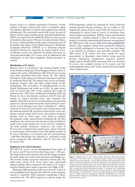
of small, single row nuclei in the posterior end (Figure 1a).<br />
The sheath usually cannot be stained by Giemsa [9]. The<br />
adult male worms of B. <strong>timori</strong> have more subventral<br />
adanal papillae when compared with those in B. malayi,<br />
<strong>and</strong> the females have larger ovejectors (see Table 1) [2].<br />
Molecular differentiation of B. <strong>timori</strong> from B. malayi is<br />
not straightforward. Around 10% of the B. malayi genome<br />
contain a repetitive DNA sequence containing a Hha I<br />
restriction site. The Hha I repeat is a proven target for<br />
www.sciencedirect.com<br />
Update TRENDS in Parasitology Vol.20 No.8 August 2004<br />
PCR diagnostics, which can separate B. malayi from the<br />
animal parasite <strong>Brugia</strong> pahangi, but is unable to distinguish<br />
B. malayi from B. <strong>timori</strong> [10]. The easiest way to<br />
distinguish B. malayi from B. <strong>timori</strong> is restriction fragment<br />
length polymorphism (RFLP) of the mitochondrial<br />
cytochrome c oxidase subunit 2. The B. malayi genome<br />
project is currently undertaking full genome sequencing of<br />
the TRS strain of B. malayi (http://www.tigr.org/tdb/e2k1/<br />
bma1/). This zoophilic isolate from peninsular Malaysia<br />
was initially pathogenic to humans, but was also found<br />
to be infective to monkeys, cats <strong>and</strong> other animals. The<br />
TRS strain of B. malayi has been reared for many<br />
generations in laboratory hosts before being used for<br />
genome sequencing. Comparative sequence analyses<br />
might help to identify DNA sequences that vary between<br />
B. <strong>timori</strong>, the zoophilic strains of B. malayi <strong>and</strong> the<br />
anthropophilic strains of B. malayi that do not successfully<br />
infect animal hosts.<br />
Figure 1. <strong>Brugia</strong> <strong>timori</strong> on Alor isl<strong>and</strong>, eastern Indonesia. (a) Giemsa-stained<br />
microfilaria of B. <strong>timori</strong> with an unstained sheath (arrows) <strong>and</strong> nucleus-free<br />
anterior end (arrow). Scale bar ¼ 50 mm. (b) Lymphedema <strong>and</strong> mossy lesion of the<br />
right foot caused by B. <strong>timori</strong>. (c) Typical lymphedema of the leg below the knee<br />
caused by B. <strong>timori</strong>. (d) Farmers in a B. <strong>timori</strong>-endemic area working in their rice<br />
paddies. (e) Acute adenolymphangitis (arrows) observed 6–24 h after treatment<br />
with a single dose diethylcarbamazine (6 mg kg 21 ) combined with albendazole<br />
(400 mg). (f) Filarial abscess caused by B. <strong>timori</strong> (arrow) next to urtical lesions. (g)<br />
Demonstration of local hygiene to reduce morbidity caused by B. <strong>timori</strong>. (h) Flipchart<br />
for health education developed by Deutsche Gesellschaft für Technische<br />
Zusammenarbeit (GTZ) for the lymphatic <strong>filariasis</strong> control programme on Alor<br />
(photo courtesy by GTZ).