View - Universitat de les Illes Balears
View - Universitat de les Illes Balears
View - Universitat de les Illes Balears

Create successful ePaper yourself
Turn your PDF publications into a flip-book with our unique Google optimized e-Paper software.
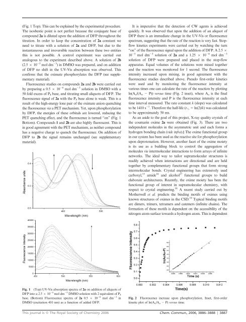
(Fig. 1 Top). This can be explained by the experimental procedure.<br />
The isosbestic point is not perfect because the conjugate base of<br />
compound 2a is diluted upon the addition of DFP throughout the<br />
titration. In or<strong>de</strong>r to keep the concentration of 2a constant we<br />
need to titrate with a solution of 2a and DFP, but due to the<br />
instantaneous and irreversible reaction between these two entities<br />
this is not possible. A control experiment was carried out<br />
analogous to the experiment <strong>de</strong>scribed above. A solution of 2b<br />
(2.5 6 10 25 mol dm 23 ) in DMSO was prepared, and on addition<br />
of DFP no shift in the UV-Vis absorption was observed. This<br />
confirms that the oximate phosphorylates the DFP (see supplementary<br />
material).<br />
Fluorescence studies on compounds 2a and 2b were carried out<br />
by preparing a 0.5 6 10 26 mol dm 23 solution in DMSO with a<br />
50 fold excess of P4 base, and titrating small aliquots of DFP. The<br />
fluorescence signal of 2a with the P4 base alone is weak. This is a<br />
result of the high-energy lone pair of the oximate anion quenching<br />
the fluorescence via a PET mechanism. Yet, upon phosphorylation<br />
by DFP, the energies of these orbitals are lowered, reducing the<br />
PET quenching effect, and the fluorescence is turned ‘‘on’’ (Fig. 1<br />
Bottom). Compounds 1 and 2b are also highly fluorescent. This is<br />
in good agreement with the PET mechanism, as neither compound<br />
has a negative charge to quench the fluorescence. On addition of<br />
DFP to 2b the signal remains unchanged (see supplementary<br />
material).<br />
Fig. 1 (Top) UV-Vis absorption spectra of 2a on addition of aliquots of<br />
DFP into a 2.5 6 10 25 mol dm 23 DMSO solution with 2 equivalent of P 4<br />
base. (Bottom) Fluorescence spectra of 2a 0.5 6 10 26 mol dm 23 in<br />
DMSO (excitation 410 nm) as a function of ad<strong>de</strong>d DFP.<br />
It is imperative that the <strong>de</strong>tection of CW agents is achieved<br />
quickly. It was observed that upon the addition of an aliquot of<br />
DFP there is an immediate change in the UV-Vis or fluorescence<br />
spectrum, suggesting that the rate of the reaction is very fast. Stopflow<br />
kinetics experiments were carried out by watching the turn<br />
‘‘on’’ of the fluorescence signal upon the addition of DFP. A 2.5 6<br />
10 25 mol dm 23 solution of 2a and a 1.25 6 10 24 mol dm 23<br />
solution of DFP were prepared and placed in the stop-flow<br />
apparatus. Equal volumes of the solutions were mixed together<br />
and the reaction was monitored for 1 second. The fluorescence<br />
intensity increased upon mixing, in good agreement with the<br />
fluorescence studies <strong>de</strong>scribed above. Pseudo first-or<strong>de</strong>r kinetics<br />
were used and by monitoring the fluorescence intensity at<br />
various times one can calculate the rate of the reaction by plotting<br />
ln(Ao/(Ao 2 P)) versus time (Fig. 2 inset), where Ao is the final<br />
fluorescence intensity and P is the fluorescence intensity at each<br />
time interval measured. The rate constant k (slope) was calculated<br />
to be 1410 s 21 . Therefore the half-life (tK 5 ln(2)/k) was calculated<br />
to be approximately 50 ms.<br />
As an asi<strong>de</strong> to the goal of this project, X-ray quality crystals of<br />
the coumarin oxime 2a were obtained (Fig. 3). There are two<br />
in<strong>de</strong>pen<strong>de</strong>nt molecu<strong>les</strong> in the asymmetric unit and each forms a<br />
hydrogen bonding chain (vi<strong>de</strong> infra).§ The oxime functional group<br />
in our system has been used as the reactive site for phosphorylation<br />
upon <strong>de</strong>protonation. However, another facet of the oxime moiety<br />
is its use as a building block to control the aggregation of<br />
molecu<strong>les</strong> via intermolecular interactions to form arrays of infinite<br />
networks. The i<strong>de</strong>al way to tailor supramolecular structures is<br />
readily achieved when interactions are directional and are held<br />
together by complementary functional groups that form strong<br />
intermolecular bonds. Crystal engineering has extensively used<br />
carboxyl, 15 ami<strong>de</strong> 16 and alcohol 17 functional groups to build<br />
elaborate architectures. Recently, the oxime moiety has been the<br />
functional group of interest in supramolecular chemistry, with<br />
respect to crystal engineering. 18 A recent study carried out by<br />
Motherwell et al. predicts the binding motifs of oximes using<br />
known structures of oximes in the CSD. 19 Typical binding motifs<br />
are dimers, trimers, tetramers and catemers (infinite chains). The<br />
formation of these motifs is <strong>de</strong>pen<strong>de</strong>nt on the accessibility of the<br />
nitrogen atom surface towards a hydrogen atom. This is <strong>de</strong>pen<strong>de</strong>nt<br />
Fig. 2 Fluorescence increase upon phosphorylation. Inset, first-or<strong>de</strong>r<br />
kinetic plot of ln(Ao/Ao 2 P) versus time.<br />
This journal is ß The Royal Society of Chemistry 2006 Chem. Commun., 2006, 3886–3888 | 3887