Enhanced ultraviolet sensitivity of zinc oxide nanoparticle ...
Enhanced ultraviolet sensitivity of zinc oxide nanoparticle ...
Enhanced ultraviolet sensitivity of zinc oxide nanoparticle ...

You also want an ePaper? Increase the reach of your titles
YUMPU automatically turns print PDFs into web optimized ePapers that Google loves.
360 L. Qin et al. / Optical Materials 33 (2011) 359–362<br />
to pure crystalline phases and high purity <strong>of</strong> the synthesized<br />
powders. Using the slow and controllable top-down chemical etching<br />
process described here, <strong>nanoparticle</strong>s as small as 10 nm sizes<br />
have been demonstrated. To start with, the micron size ZnO particles<br />
were suspended in deionized water using a magnetic stirrer.<br />
The solid weight percent <strong>of</strong> the ZnO particles in water was approximately<br />
2.5%. A separate mild acidic solution (pH 6) was prepared<br />
by adding acetic acid in water. The acidic solution was then added<br />
drop by drop (at a rate <strong>of</strong> 2–3 ml every minute) to the ZnO colloidal<br />
suspension while the liquid was vigorous stirred. The acetic acid<br />
etches ZnO by forming <strong>zinc</strong> acetate which dissolved in water. After<br />
2 h <strong>of</strong> etching, the solution was centrifuged and the ZnO particles<br />
were extracted. The extracted particles were suspended in deionized<br />
water and washed. The solution was then centrifuged and<br />
the particles were extracted again. The etching process is repeated<br />
several times as described above beginning with suspending the<br />
extracted ZnO particles in water and adding acetic acid solution<br />
for 2 h. After six etching cycles it was observed that ZnO <strong>nanoparticle</strong>s<br />
with size below 100 nm could be obtained. At the end <strong>of</strong> this<br />
etching process, the colloidal suspension was separated into two<br />
batches. One batch was surface treated by dissolving PVA<br />
(0.5% in weight in water). The PVA capping on the ZnO <strong>nanoparticle</strong>s<br />
occurred due to the reaction <strong>of</strong> excess Zn 2+ on the surface with<br />
the hydroxyl group <strong>of</strong> the PVA. The particles from both batches<br />
[uncoated (ZnO–A) and PVA-coated (ZnO–U)] were centrifuged<br />
and dispersed in ethanol to form 30 mg/ml suspension. These solutions<br />
were then spin-casted onto quartz plates and annealed in air<br />
at 150 °C for 5 min.<br />
Characterization methods used to determine the material quality<br />
and optical characteristics <strong>of</strong> the ZnO <strong>nanoparticle</strong>s are the following.<br />
X-ray diffraction (XRD) was performed on a Bruker D8<br />
Discover X-ray diffractometer with a high-star 2-d detector. The<br />
2-theta used in the measurement was from 30° to 100°. The size<br />
and morphology <strong>of</strong> the ZnO <strong>nanoparticle</strong>s were determined using<br />
a Carl Zeiss Ultra 1540 dual beam scanning electron microscope<br />
(SEM) set to 5 kV. The ZnO <strong>nanoparticle</strong>s were characterized by<br />
their excitation and emission spectra using a Spex Fluorolog<br />
Tau-3 spectr<strong>of</strong>luorimeter. UV–Vis absorption spectra were recorded<br />
using a Shimadzu UV–Vis 2550 spectrophotometer with a<br />
deuterium lamp (190–390 nm) and a halogen lamp (280–<br />
1100 nm). Auto switching between the lamps is synchronized to<br />
a wavelength and the switching range is selectable between 282<br />
and 393 nm. The scanning wavelength range used in the experiment<br />
is 240–800 nm with a switching wavelength <strong>of</strong> 330 nm.<br />
Photoconductors were made by depositing two irregular<br />
100 nm Al contacts on top <strong>of</strong> the ZnO <strong>nanoparticle</strong> film formed<br />
as stated above using E-beam via shadow mask. A schematic diagram<br />
<strong>of</strong> the detectors is presented in the inset <strong>of</strong> Fig. 1(b). Point<br />
contact current–voltage (I-V) characteristics <strong>of</strong> samples were measured<br />
using a HP4155B semiconductor parameter analyzer under<br />
darkness and under illumination by a 340 nm UV LED with an<br />
intensity <strong>of</strong> 45.58 mW/cm 2 . These measurements were performed<br />
at room temperature in air.<br />
3. Results and discussion<br />
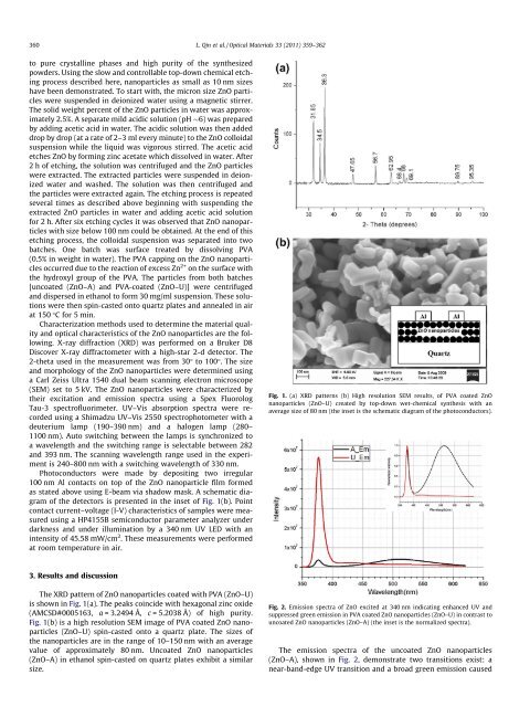
The XRD pattern <strong>of</strong> ZnO <strong>nanoparticle</strong>s coated with PVA (ZnO–U)<br />
is shown in Fig. 1(a). The peaks coincide with hexagonal <strong>zinc</strong> <strong>oxide</strong><br />
(AMCSD#0005163, a = 3.2494 Å, c = 5.2038 Å) <strong>of</strong> high purity.<br />
Fig. 1(b) is a high resolution SEM image <strong>of</strong> PVA coated ZnO <strong>nanoparticle</strong>s<br />
(ZnO–U) spin-casted onto a quartz plate. The sizes <strong>of</strong><br />
the <strong>nanoparticle</strong>s are in the range <strong>of</strong> 10–150 nm with an average<br />
value <strong>of</strong> approximately 80 nm. Uncoated ZnO <strong>nanoparticle</strong>s<br />
(ZnO–A) in ethanol spin-casted on quartz plates exhibit a similar<br />
size.<br />
Fig. 1. (a) XRD patterns (b) High resolution SEM results, <strong>of</strong> PVA coated ZnO<br />
<strong>nanoparticle</strong>s (ZnO–U) created by top-down wet-chemical synthesis with an<br />
average size <strong>of</strong> 80 nm (the inset is the schematic diagram <strong>of</strong> the photoconductors).<br />
Fig. 2. Emission spectra <strong>of</strong> ZnO excited at 340 nm indicating enhanced UV and<br />
suppressed green emission in PVA coated ZnO <strong>nanoparticle</strong>s (ZnO–U) in contrast to<br />
uncoated ZnO <strong>nanoparticle</strong>s (ZnO–A) (the inset is the normalized spectra).<br />
The emission spectra <strong>of</strong> the uncoated ZnO <strong>nanoparticle</strong>s<br />
(ZnO–A), shown in Fig. 2, demonstrate two transitions exist: a<br />
near-band-edge UV transition and a broad green emission caused