CUP2 binds in a bipartite manner to upstream activation sequence c ...
CUP2 binds in a bipartite manner to upstream activation sequence c ...
CUP2 binds in a bipartite manner to upstream activation sequence c ...

Create successful ePaper yourself
Turn your PDF publications into a flip-book with our unique Google optimized e-Paper software.
458<br />
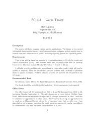
This difference is most clearly appreciated when the <strong>in</strong>tensities<br />
of bands at positions –128 and –127 are compared<br />
(Fig. 7B): for ace1, the <strong>in</strong>tensity of the band at<br />
position –128 is substantially reduced compared <strong>to</strong> the<br />
band at –127, while for <strong>CUP2</strong> the bands at –128 and<br />
–127 have almost equal <strong>in</strong>tensity. Further <strong>upstream</strong>,<br />
<strong>CUP2</strong> exhibits miss<strong>in</strong>g nucleoside signals at position<br />
–140 <strong>to</strong> –142, while ace1 does not. Although on the <strong>to</strong>p<br />
strand these differences are small, the comparison<br />
shown <strong>in</strong> Fig. 7A demonstrates that while at position<br />
–139 the two prote<strong>in</strong>s have similar signals, at position<br />
–140 the band <strong>in</strong>tensity for <strong>CUP2</strong> is significantly lower<br />
than that for ace1. Additional evidence for these contacts<br />
is provided by the scan of the cleavage pattern of<br />
the unbound fraction for <strong>CUP2</strong> (see Fig. 3), <strong>in</strong> which a<br />
peak of enhanced band <strong>in</strong>tensity is observed at position<br />
–141.<br />
The differences <strong>in</strong> miss<strong>in</strong>g nucleoside patterns are<br />
most pronounced on the bot<strong>to</strong>m strand. The most strik<strong>in</strong>g<br />
difference occurs at positions –139 <strong>to</strong> –141, where<br />
significant miss<strong>in</strong>g nucleoside signals are found for<br />
<strong>CUP2</strong> while ace1 shows none (Fig. 7B).<br />
These miss<strong>in</strong>g nucleoside results <strong>in</strong>dicate that <strong>CUP2</strong><br />
contacts four additional base pairs <strong>in</strong> the <strong>upstream</strong> halfsite,<br />
with greater <strong>in</strong>teraction with the bot<strong>to</strong>m strand<br />
than is the case for ace1. This conclusion is consistent<br />
with previous methylation <strong>in</strong>terference results [14],<br />
which showed that three of the guan<strong>in</strong>es with<strong>in</strong> this region<br />
<strong>in</strong>terfere with the formation of the <strong>CUP2</strong>-DNA<br />
complex when methylated, while formation of the ace1-<br />
DNA complex is unaffected. A variant of the <strong>CUP2</strong><br />
prote<strong>in</strong>, <strong>in</strong> which the three most am<strong>in</strong>o-term<strong>in</strong>al cyste<strong>in</strong>es<br />
(Cys11, Cys14 and Cys23) were carboxymethylated,<br />
was found <strong>to</strong> contact a nearly identical set of nucleotides<br />
as we f<strong>in</strong>d for ace1 [17]. These results <strong>in</strong>dicate<br />
that <strong>in</strong> both half-sites <strong>CUP2</strong> contacts the outermost major<br />
groove while ace1 does not, and that the contacts<br />
made by <strong>CUP2</strong> with the major groove are more extensive<br />
<strong>in</strong> the <strong>upstream</strong> half-site.<br />
Biological implications<br />
In this paper we have exam<strong>in</strong>ed the b<strong>in</strong>d<strong>in</strong>g of <strong>CUP2</strong><br />
and the related prote<strong>in</strong> ace1 <strong>to</strong> UASc. These studies<br />
have shown that ace1 <strong>in</strong>teracts with a smaller portion of<br />
UASc than does <strong>CUP2</strong>. Our results are consistent with<br />
previous suggestions that <strong>CUP2</strong> possesses a <strong>bipartite</strong><br />
DNA-b<strong>in</strong>d<strong>in</strong>g doma<strong>in</strong>. The loss of a metal coord<strong>in</strong>ation<br />
site due <strong>to</strong> substitution of Tyr for Cys11 [22] apparently<br />
results <strong>in</strong> the loss of a DNA-b<strong>in</strong>d<strong>in</strong>g element which<br />
normally contacts the major groove of the DNA at the<br />
extremities of UASc. The fewer DNA contacts made<br />
by ace1 expla<strong>in</strong>s its tenfold lower b<strong>in</strong>d<strong>in</strong>g aff<strong>in</strong>ity for<br />
UASc compared <strong>to</strong> <strong>CUP2</strong> [14]. It is plausible that the<br />
mutation <strong>in</strong> ace1, Cys11Tyr, leads <strong>to</strong> disruption of a<br />
DNA-b<strong>in</strong>d<strong>in</strong>g doma<strong>in</strong>. Six basic am<strong>in</strong>o acids, four of<br />
which are clustered <strong>to</strong>gether, occur between Cys11 and<br />
the next cyste<strong>in</strong>e <strong>in</strong> the <strong>sequence</strong>, Cys43.<br />
Fig. 7A, B Comparison of miss<strong>in</strong>g nucleoside patterns for <strong>CUP2</strong><br />
and ace1. A Overlay of densi<strong>to</strong>meter scans of miss<strong>in</strong>g nucleoside<br />
patterns obta<strong>in</strong>ed from complex II (dotted l<strong>in</strong>e <strong>CUP2</strong>, solid l<strong>in</strong>e<br />
ace1). B The densi<strong>to</strong>meter scan of the miss<strong>in</strong>g nucleoside pattern<br />
obta<strong>in</strong>ed from complex II (dotted l<strong>in</strong>e) is superimposed upon the<br />
densi<strong>to</strong>meter scan of the hydroxyl radical cleavage pattern of free<br />
(control) DNA (solid l<strong>in</strong>e). Top two scans <strong>to</strong>p strand, bot<strong>to</strong>m two<br />
scans bot<strong>to</strong>m strand<br />
Although UASc can be considered <strong>to</strong> be pseudopal<strong>in</strong>dromic,<br />
mutational analysis has <strong>in</strong>dicated that the <strong>upstream</strong><br />
half-site is particularly crucial for transcriptional<br />
activity [5]. Either this half-site is at the appropriate<br />
distance and orientation relative <strong>to</strong> the TATA box of<br />
the CUP1 gene or there is some difference <strong>in</strong> the way<br />
that <strong>CUP2</strong> <strong>b<strong>in</strong>ds</strong> <strong>to</strong> the two half-sites. The miss<strong>in</strong>g nucleoside<br />
results presented here reveal that the <strong>in</strong>terac-