Conference Program - Johns Hopkins Malaria Research Institute ...
Conference Program - Johns Hopkins Malaria Research Institute ...
Conference Program - Johns Hopkins Malaria Research Institute ...

Create successful ePaper yourself
Turn your PDF publications into a flip-book with our unique Google optimized e-Paper software.
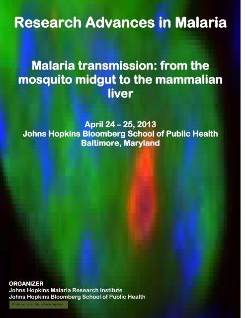
<strong>Research</strong> Advances in <strong>Malaria</strong><br />
<strong>Malaria</strong> transmission: from the<br />
mosquito midgut to the mammalian<br />
liver<br />
April 24 – 25, 2013<br />
<strong>Johns</strong> <strong>Hopkins</strong> Bloomberg School of Public Health<br />
Baltimore, Maryland<br />
ORGANIZER<br />
<strong>Johns</strong> <strong>Hopkins</strong> <strong>Malaria</strong> <strong>Research</strong> <strong>Institute</strong><br />
<strong>Johns</strong> <strong>Hopkins</strong> Bloomberg School of Public Health<br />
Photo courtesy of Dr. Isabelle Coppens
<strong>Research</strong> Advances in <strong>Malaria</strong><br />
<strong>Malaria</strong> Transmission: From the Mosquito Midgut to the Mammalian Liver<br />
DAY 1 – Wednesday, April 24, 2013<br />
8:00a.m. Registration<br />
Coffee<br />
9:00a.m. Dean Michael Klag<br />
Peter Agre<br />
Welcome Address<br />
SESSION I: Moderated by George Dimopoulos and Freddy Frischknecht<br />
9:15 - 10:00a.m. KEYNOTE: Robert Sinden, Imperial College of London and the Jenner<br />
<strong>Institute</strong> Oxford<br />
“Hitting malaria transmission-the sense and the nonsense”<br />
10:10 - 10:30a.m. Marcelo Jacobs-Lorena, <strong>Johns</strong> <strong>Hopkins</strong> Bloomberg School of Public Health<br />
“Fighting malaria with engineered symbiotic bacteria from the mosquito<br />
midgut”<br />
10:40 - 11:05a.m. BREAK<br />
11:10 - 11:30a.m. Carolina Barillas-Mury, National <strong>Institute</strong>s of Health/NIAID<br />
“Plasmodium falciparum evasion of the mosquito immune system”<br />
11:40 - 12:00p.m. George Dimopoulos, <strong>Johns</strong> <strong>Hopkins</strong> Bloomberg School of Public Health<br />
“Targeting malaria in the mosquito gut”<br />
12:10p.m. LUNCH<br />
12:40 - 1:55p.m. LUNCH/POSTERS<br />
SESSION II: Moderated by Rogerio Amino and Marcelo Jacobs-Lorena<br />
2:00 - 2:20p.m. Johannes (Hans) Dessens, London School of Hygiene & Tropical Med.<br />
Dissecting crystalloid biogenesis and function<br />
2:30 - 2:50p.m. Friedrich (Freddy) Frischknecht, Universitats Klinikum<br />
“Molecular and cellular mechanisms of Plasmodium sporozoite migration”<br />
3:00 - 3:20p.m. Rogerio Amino, Institut Pasteur<br />
“In vivo imaging of Plasmodium life and death”<br />
3:30 - 3:55p.m. BREAK<br />
4:00 - 4:20p.m. Jürgen Bosch, <strong>Johns</strong> <strong>Hopkins</strong> Bloomberg School of Public Health<br />
“Inhibitor of the Plasmodium Glideosome designed to stabilize a proteinprotein<br />
interaction”<br />
4:30 - 4:50p.m. Jennifer Stevenson, <strong>Johns</strong> <strong>Hopkins</strong> Bloomberg School of Public Health at<br />
Macha <strong>Research</strong> Trust<br />
“Sampling mosquitoes in the field: where, when, how and what does it all<br />
mean?”<br />
E. Monument St. entrance<br />
Feinstone Hall E2030<br />
Sheldon Hall W1214<br />
Sheldon Hall W1214<br />
Sheldon Hall W1214<br />
Sheldon Hall W1214<br />
Feinstone Hall E2030<br />
Sheldon Hall W1214<br />
Sheldon Hall W1214<br />
Feinstone Hall E2030<br />
Feinstone Hall E2030<br />
Sheldon Hall W1214<br />
Sheldon Hall W1214<br />
Sheldon Hall W1214<br />
Sheldon Hall W1214<br />
Feinstone Hall E2030<br />
Sheldon Hall W1214<br />
Sheldon Hall W1214<br />
5:00 - 6:30p.m. POSTERS & COCKTAIL RECEPTION Feinstone Hall E2030
DAY 2 – Thursday, April 25, 2013<br />
8:00a.m. Registration<br />
Coffee<br />
9:00a.m. Peter Agre<br />
George Dimopoulos<br />
Welcome Address<br />
SESSION III: Moderated by Photini Sinnis and Volker Heussler<br />
9:05 - 9:50a.m. KEYNOTE: Alan Magill, Bill & Melinda Gates Foundation<br />
“Accelerating to zero: an analytic framework for malaria eradication”<br />
10:00 - 10:20a.m. Sangeeta Bhatia, Massachusetts <strong>Institute</strong> of Technology<br />
“Human microlivers for the study of liver stage malaria”<br />
10:30 - 10:55a.m. BREAK<br />
11:00 - 11:20a.m. Laurent Rénia, Singapore Immunology Network (SIgN)<br />
“Human malaria liver stages: from in vitro to humanized mice”<br />
11:30 - 11:50a.m. Photini Sinnis, <strong>Johns</strong> <strong>Hopkins</strong> Bloomberg School of Public Health<br />
“Late liver stages: a role for Falcipain-1 in hepatic merozoite infectivity”<br />
12:00p.m. LUNCH<br />
12:30 - 1:55p.m. LUNCH/POSTERS<br />
SESSION IV: Moderated by Laurent Renia and Fidel Zavala<br />
2:00 - 2:20p.m. Isabelle Coppens, <strong>Johns</strong> <strong>Hopkins</strong> Bloomberg School of Public Health<br />
“The uniqueness of the autophagy process in Plasmodium”<br />
2:30 - 2:50p.m. Volker Heussler, University of Bern<br />
“Plasmodium liver stage parasites induce distinct autophagy-related<br />
events in host hepatocytes”<br />
3:00 - 3:20p.m. Fidel Zavala, <strong>Johns</strong> <strong>Hopkins</strong> Bloomberg School of Public Health<br />
“Effector liver-resident memory CD8+ T cells”<br />
3:30 - 3:55p.m. BREAK<br />
4:00 - 4:20p.m. Jelena Levitskaya, <strong>Johns</strong> <strong>Hopkins</strong> Bloomberg School of Public Health<br />
“<strong>Malaria</strong>-infected hepatocytes as targets for T cell responses”<br />
4:30 - 4:50p.m. Robert Seder, National <strong>Institute</strong>s of Health/NIAID<br />
“Mechanisms of protection following irradiated PfSPZ immunization in<br />
humans”<br />
5:00 - 5:20p.m. Shahid Khan, Leiden University Medical Center<br />
“Preclinical development of genetically attenuated malaria parasites for<br />
vaccine development”<br />
5:30 - 5:45p.m. ANNOUNCEMENTS<br />
E. Monument St. entrance<br />
Feinstone Hall E2030<br />
Sheldon Hall W1214<br />
Sheldon Hall W1214<br />
Sheldon Hall W1214<br />
Sheldon Hall W1214<br />
Feinstone Hall E2030<br />
Sheldon Hall W1214<br />
Sheldon Hall W1214<br />
Feinstone Hall E2030<br />
Feinstone Hall E2030<br />
Sheldon Hall W1214<br />
Sheldon Hall W1214<br />
Sheldon Hall W1214<br />
Feinstone Hall E2030<br />
Sheldon Hall W1214<br />
Sheldon Hall W1214<br />
Sheldon Hall W1214<br />
Sheldon Hall W1214
SPEAKER ABSTRACTS
2013 RESEARCH ADVANCES IN MALARIA<br />
<strong>Malaria</strong> transmission: from the Mosquito Midgut to the Mammalian Liver<br />
SPEAKER ABSTRACTS (name highlighted)<br />
In vivo imaging of Plasmodium life and death<br />
Joana Tavares 1 , Pauline Formaglio 1 , Ian Cockburn 2 , Fidel Zavala 2 , Silvia Boscardin 3 , Robert Ménard 1 and Rogerio<br />
Amino 1 .<br />
1 Institut Pasteur, 2 <strong>Johns</strong> <strong>Hopkins</strong> Bloomberg School of Public Health, 3 University of Sao Paulo.<br />
From the site of inoculation in the skin, Plasmodium sporozoites must accomplish a long journey to the liver, in<br />
order to invade and generate thousands of erythrocyte-infective stages inside hepatocytes. During this perilous<br />
journey, the parasite is confronted with several obstacles, such as the endothelial barriers of cutaneous and hepatic<br />
blood vessels, and humoral and cellular immune effectors. By imaging wild-type and mutant parasites in transgenic,<br />
naïve or immunized animals, we are analyzing the determinants implicated in Plasmodium survival in the skin and<br />
liver of mice. Our goal is to define these determinants at cellular and molecular levels, focused on the requirements<br />
needed to hinder the parasite progression and development in these tissues. I will present how sporozoites evade<br />
innate immunity in the liver sinusoids and how adaptive immunity can efficiently eliminate the parasite in the skin<br />
and in the liver.<br />
Plasmodium falciparum evasion of the mosquito immune system<br />
Alvaro Molina-Cruz, 1 Lindsey S. Garver, 1 Amy Alabaster, 1 Lois Bangiolo, 1 Ashley Haile, 1 Jared Winikor, 1 Corrie<br />
Ortega, 1 Ben C. L. van Schaijk, 2 Robert W. Sauerwein, 2 Emma Taylor-Salmon and Carolina Barillas-Mury 1<br />
1 National <strong>Institute</strong>s of Health, 2 Radboud University Nijmegen Medical Center.<br />
Mosquitoes can mount effective immune responses against Plasmodium parasites. Recent work uncovered a<br />
functional link between epithelial nitration responses to ookinete invasion and activation of the complement-like<br />
system, when parasites emerge from the midgut and come in contact with the mosquito hemolymph. However,<br />
some strains of the human malaria parasite Plasmodium falciparum can infect mosquitoes without activating an<br />
effective immune response. A combination of genetic linkage mapping, linkage group selection and functional<br />
genomics was used to identify a gene from P. falciparum that allows the parasite to infect mosquitoes without<br />
activating the complement-like system. The potential implications of these findings for malaria transmission in the<br />
field will be discussed.<br />
Inhibitor of the Plasmodium Glideosome Designed to Stabilize a Protein-Protein Interaction<br />
Jürgen Bosch<br />
<strong>Johns</strong> <strong>Hopkins</strong> Bloomberg School of Public Health<br />
Plasmodium species, the causative agents of malaria, are obligate intracellular protozoan parasites that rely on an<br />
unusual form of substrate-dependent motility for their migration on and across host-cell membranes and for<br />
invasion and egress. This unusual motility is driven by the "glideosome", an actomyosin intracellular<br />
macromolecular complex anchored to the inner membrane complex of the parasite. A complex of actin, aldolase,<br />
and Thrombospondin-Related Anonymous Protein (TRAP) constitutes the core of the glideosome. Here, we<br />
rationally identified several small molecules that were designed to prevent dis-assembly of the TRAP-aldolase<br />
complex within the glideosome. Two of the compounds markedly disrupted the gliding and invasive abilities<br />
of Plasmodium parasites in vitro, and none of the compounds were toxic to human hepatocytes. Here we present a<br />
high resolution ternary crystal structure composed of aldolase TRAP and compound 24, determined to 2.2 Å<br />
resolution, which now serves as a platform for future ligand optimization. The results validate the glideosome as a<br />
malaria drug target. In addition, we have described and proven the principle of the first rational method to identify<br />
medically promising compounds designed to enhance, rather than inhibit, a protein-protein interface.<br />
The uniqueness of the autophagy process in Plasmodium<br />
Christiane Voss 1# , Bamini Jayabalasingham 1,2# , Julia D. Romano 1 , Maria E. Smith 1 , Karen Ehrenman 1 , David A.<br />
Fidock 2 , Juergen Bosch 1 and Isabelle Coppens 1<br />
1 <strong>Johns</strong> <strong>Hopkins</strong> Bloomberg School of Public Health, 2 Columbia University Medical Center<br />
Autophagy is an adaptive process that ensures the appropriate number and health of organelles. Adaptation of<br />
Plasmodium to various environments is accompanied by changes in their organelle composition and size. During<br />
hepatocyte infection, Plasmodium discards organelles involved in invasion and expands those implicated in
iosyntheses. Plasmodium possesses a rudimentary set of autophagy-related proteins. Plasmodium falciparum<br />
expresses Atg8, the main player in autophagy, in human hepatocytes and erythrocytes, Anopheles midguts and<br />
salivary glands. The members of the Atg8 conjugation system are co-transcribed in all stages of Plasmodium<br />
berghei. PbAtg8 interacts with PbAtg3, but cannot substitute for yeast Atg8 or mammalian LC3, suggesting the<br />
uniqueness of the parasite autophagy machinery. Plasmodium Atg8 mainly localizes to limiting membranes of the<br />
apicoplast. The Atg8 conjugation pathway may serve either to form autophagic structures derived from apicoplast<br />
membranes or to supply lipids required for the enlargement of the apicoplast.<br />
Dissecting Plasmodium crystalloid biogenesis and function<br />
Sadia Saeed and Johannes T. Dessens<br />
London School of Hygiene & Tropical Medicine<br />
During the malaria parasites' transit of vector mosquitoes, the ookinete stage forms distinctive multivesicular<br />
organelles named crystalloids, thought to be involved in molecular trafficking from its upstream gametocyte stage to<br />
its downstream oocyst stage. Thus far, the only parasite molecules linked with this transient organelle are a<br />
gametocyte-expressed family of LCCL-lectin adhesive-like domain proteins (LAPs) that have essential roles in<br />
sporozoite transmission. Using genetically modified parasite lines that express LAP mutants we dissect the process<br />
of crystalloid biogenesis and shed light on the function of the organelle in parasite development. Our data indicate<br />
that Plasmodium crystalloids are essential for sporozoite transmission and could constitute a new chemotherapy<br />
target to control the spread of malaria.<br />
Targeting malaria in the mosquito gut<br />
George Dimopoulos (& Dimopoulos Group)<br />
<strong>Johns</strong> <strong>Hopkins</strong> Bloomberg School of Public Health<br />
Our research program is broadly focused on the innate immune system and gut microbiota of mosquito malaria<br />
vectors and their role in regulating vector competence. The Anopheles immune deficiency (Imd) signaling pathway<br />
has emerged as the most effective pathway in terms of activity against the human malaria parasite. The intestines<br />
of the mosquito vectors harbor a diversity of microbes from bacteria to fungi, some of which can modulate<br />
permissiveness to Plasmodium infection. We are exploring various ways of manipulating the Imd pathway and<br />
modulating the midgut microbiota to achieve resistance to Plasmodium.<br />
Mechanisms of sporozoite migration<br />
Mirko Singer, 1 Misha Kudryashev, 2 Janina Hellmann, 1 Marek Cyrklaff, 1 Ulrich Schwarz, 3 Joachim Spatz 3,4 and<br />
Freddy Frischknecht 1<br />
1 Heidelberg University Medical School, 2 Biocenter Basel, 3 Heidelberg University, 4 Max Planck <strong>Institute</strong> Stuttgart<br />
Plasmodium sporozoites are injected into the dermis during a mosquito bite. In order to establish an infection they<br />
need to migrate through the skin, enter blood vessels and eventually invade hepatocytes. To do so they rely on an<br />
actin-based motility machinery that enables them to migrate at high speed. In addition to actin a number of actinbinding<br />
proteins are essential for movement as are a combination of sporozoite surface proteins, such as TRAP and<br />
related transmembrane-spanning proteins as well as the circumsporozoite protein, CSP. We aim at understanding<br />
the cellular and molecular basis of this motility using a range of microscopy techniques, reverse genetics and new<br />
materials. The talk will present an overview of the different insights the combination of these technologies can<br />
achieve.<br />
Plasmodium liver stage parasites induce distinct autophagy-related events in host hepatocytes<br />
Monica Prado, 1 Nina Eickel, 2 Rebecca Stanway 2 and Volker Heussler 2<br />
1 Bernhard-Nocht-<strong>Institute</strong> for Tropical Medicine, 2 <strong>Institute</strong> of Cell Biology, University of Bern<br />
Autophagy is a process involved in the degradation of internal cell components in order to relocate nutrients during<br />
periods of starvation and stress but autophagy can also control the development of intracellular pathogens. To<br />
investigate autophagy processes in Plasmodium-infected hepatocytes, we infected hepatoma cell lines and<br />
transgenic mice expressing the autophagy marker protein LC3 coupled to GFP with Plasmodium berghei<br />
sporozoites. In vitro and in vivo invasion was accompanied by a strong activation of autophagy and by the attempt<br />
of the host cell to isolate the parasite in an LC3-positive compartment. Interestingly, this membrane is not a newly<br />
generated autophagosome but instead LC3 is incorporated into the parasitophorous vacuole membrane (PVM) and
thus resembles more an LC3-associated phagocytosis (LAP) than a typical autophagy. Parasites successfully<br />
entering late schizogony are able to control LC3 expression in the PVM but host cell autophagy continues. Further<br />
experimental data suggest that host cell autophagy and the observed LAP-like events that result in LC3<br />
accumulation in the PVM are separate events.<br />
Fighting malaria with engineered symbiotic bacteria from vector mosquitoes<br />
Sibao Wang and Marcelo Jacobs-Lorena<br />
<strong>Johns</strong> <strong>Hopkins</strong> School of Public Health<br />
The mosquito microbiota and the most vulnerable stages of the Plasmodium parasite share the midgut<br />
compartment. We are exploring the genetic modification of mosquito symbiotic bacteria for delivering anti-malarial<br />
effector molecules into the mosquito midgut (paratransgenesis). We have shown that the bacterium Pantoae<br />
agglomerans engineered to express and secrete anti-Plasmodium effector molecules strongly inhibits Plasmodium<br />
development in the mosquito by up to 98%.<br />
Recently, we have identified another mosquito symbiotic bacterium of the genus Serratia that is transmitted both<br />
vertically from female to larval progeny and horizontally from male to female during mating. These transmission<br />
properties suggest that it should be possible to introduce recombinant Serratia into field mosquito populations for<br />
the purpose of malaria control. This approach is ‘low-tech’ and does not involve the introduction of a new bacterium<br />
species, since Serratia is a natural component of the African mosquito microbiota.<br />
Preclinical development of genetically attenuated malaria parasites for vaccine development<br />
Takeshi Annoura 1 *, Ben C.L. van Schaijk 2 *, Ivo H.J. Ploemen 2 , Mohammed Sajid 1 , Jing-wen Lin 1 , Martijn W. Vos 2 ,<br />
Cornelus C. Hermsen 2 , Dominique Mazier 3 , Stephen L. Hoffman 4 , Chris J. Janse 1 , Robert W. Sauerwein 2 and<br />
Shahid M. Khan 1<br />
1 Leiden University Medical Center, 2 Radboud University Nijmegen Medical Center, 3 INSERM, U511, 4 Sanaria Inc.<br />
Immunization with Plasmodium irradiated-sporozoites that invade and arrest inside hepatocytes can induce longlasting<br />
sterile protective immunity against malaria in rodent models and in humans. Recently gene deletion mutants,<br />
or genetically attenuated parasites (GAP), have been created in rodent malaria parasites which similarly arrest<br />
during liver-stage development that provoke strong (in some cases even stronger) protective immunity, in mice. By<br />
examining GAPs in rodent-malaria parasites in multiple mice strains, we describe robust and stringent screening<br />
approaches to establish GAP safety (do not produce a blood-stage infection) and GAP potency (immunity with the<br />
fewest parasites/doses). Using information derived from rodent GAPs we have created a multiple gene-deletion P.<br />
falciparum GAP and describe its evaluation during blood-, mosquito- and liver-stage development and have tested<br />
its safety by examining its attenuation in cultured primary human hepatocytes and mice engrafted with human liver<br />
tissue. This P. falciparum GAP is now being prepared for evaluation in Phase I/II human trials.<br />
<strong>Malaria</strong>-infected hepatocytes as targets for T cell responses<br />
Jinxia Ma 1 , Stefanie Trop 1 , Samantha Baer 1 , Elian Rakhmanaliev 1 , Zita Arany 1 , Peter Dumoulin 1 , Hao Zhang 1 , Julia<br />
Romano 1 , Isabelle Coppens 1 , Victor Levitsky 2 and Jelena Levitskaya 1<br />
1 <strong>Johns</strong> <strong>Hopkins</strong> Bloomberg School of Public Health, 2 <strong>Johns</strong> <strong>Hopkins</strong> University School of Medicine<br />
While many intracellular pathogens subvert the MHC class I presentation machinery, its functionality in the course<br />
of malaria replication in hepatocytes has not been investigated. To characterize the MHC class I pathway and its<br />
regulation at different stages of malaria parasite replication, we utilize experimental systems that allow identification<br />
and isolation of relatively large populations of human hepatocytes affected - invaded and/or traversed - by malaria<br />
parasite. Furthermore, our model systems permit detection and separation of malaria parasite developmental<br />
stages distinguished by the cell surface phenotype compatible with the immune escape from T-cell mediated<br />
surveillance. Our data have multiple implications for understanding of the natural T-cell immunity against malaria<br />
and may promote development of novel, efficient anti-malaria vaccines overcoming immune escape of the parasite<br />
in the liver.<br />
Human malaria liver stages: renewed interests for drug development and malaria eradication<br />
Laurent Rénia<br />
Singapore Immunology Network (SIgN), Agency for Science, Technology and <strong>Research</strong> (A*STAR), Singapore
The development of the malaria parasite in the hepatocyte is an obligatory step that dominates the pre-erythrocytic<br />
phase of the malaria infection. Compared with the erythrocytic phase of the infection, our knowledge of the preerythrocytic<br />
stage remains fragmentary. The cost and ethical considerations required to obtain sufficient quantities<br />
of infected cells in order to conduct detailed observations of the natural evolution of the hepatic infection have<br />
restricted detailed investigations to rodent Plasmodium species. In contrast to erythrocytic parasites that cyclically<br />
multiply every 24, 48 or 72 days depending on the species, liver stage development ends after a unique<br />
multiplication step of highly variable duration between Plasmodium species, the shortest for rodent parasites<br />
(around 48 hours) and the longest for human parasites (6 to 15 days). In addition, species such P. vivax, P. ovale<br />
and P. cynomolgi possess dormant liver form called hypnozoites that remains for weeks to months in human or<br />
monkey hepatocytes in vivo and are responsible for recrudescence. Here we review the exciting new approaches<br />
and findings on human liver stage biology and their use for drug development.<br />
Hitting malaria transmission: the sense and the nonsense<br />
Robert E.Sinden<br />
Imperial College London, and the Jenner <strong>Institute</strong>, The University of Oxford.<br />
Publication of the MEG and MalERA analyses of the global malaria research that would be required to underpin<br />
effective programmes for malaria elimination, and potentially eradication, emphasised the essential need to reduce<br />
the number of new infections in endemic populations, i.e. to reduce R0 /Rc to below one.<br />
Experience shows that such campaigns might benefit from careful spatial, temporal and biological targeting, and<br />
highlights the importance of hitting transmission to and from the mosquito as core components of an effective and<br />
sustainable long term strategy.<br />
Developing new tools (vaccines, drugs, and anti-vector measures) to contribute to this effort requires that<br />
appropriate assays for these tools are developed. The talk will focus first on new assays to identify new<br />
transmission blocking drugs targeted to the gametocyte stages of development, highlighting both the potential and<br />
the limitations of these assays to meet the need to identify new drugs that will be useful in endemic settings.<br />
Assays to measure transmission by infection of the mosquito (e.g. the standard membrane feeding assay),<br />
currently describe outputs such as oocyst infection or prevalence. The talk will illustrate how an understanding of<br />
the structure of parasite populations in the mosquito requires a more complete description of the impact to allow<br />
effective inter-lab/experimental comparisons. The talk will also discuss whether enumerating oocyst infections, as<br />
opposed to sporozoites, is the most useful readout.<br />
A new transmission assay, the Population Transmission Assay (Blagborough et al, Nature Communications 2013),<br />
that overcomes many of the theoretical constraints of current assays, and in which important variables can be<br />
individually controlled , will be described. The utility of this assay in providing decision makers with direct evidence<br />
of the impact of any intervention on malaria transmission in populations, and providing critical data required to<br />
undertake effective field trials will be described.<br />
The Cysteine Protease Berghepain-1 Functions During Erythrocyte Invasion by Erythrocytic and Hepatic<br />
Merozoites.<br />
Satish Mishra 1 , Brandy Lee Bennett 2 , Mohammad Sajid 3 , Christine Lehmann 4 , Volker Heussler 4 and Photini<br />
Sinnis 1 .<br />
1 2 3<br />
<strong>Johns</strong> <strong>Hopkins</strong> Bloomberg School of Public Health, New York University School of Medicine, Leiden University<br />
Medical Center, 4 University of Bern<br />
The function of falcipain-1, a papain-family cysteine protease of the human malaria parasite Plasmodium<br />
falciparum, has been the subject of some controversy; while some investigators found a requirement for this<br />
protease during erythrocyte invasion, others used deletion mutants and found no discernible defect in invasion. To<br />
further investigate this, we deleted the P. berghei ortholog, berghepain-1, in the lethal ANKA strain. We found that<br />
berghepain-1 knock-out parasites (BKO) are attenuated in vivo. Blood stage parasites grow more slowly and do not<br />
rapidly kill the host as do wildtype controls. Our data indicate that this is due to a restricted cell tropism, with BKO<br />
merozoites displaying a greater preference for reticulocytes compared to controls. Inducing reticulocytsosis with<br />
phenylhydrazine prior to infection restores both exponential growth and lethality. Interestingly, these parasites also<br />
have a late liver stage phenotype. Sporozoite infectivity is significantly decreased, with prepatent period delays of 4<br />
to 5 days compared to controls. Nonetheless, no differences could be detected in liver stage development, with<br />
normal hepatic merozoite formation and normal merosome budding. The delayed prepatent period observed with<br />
sporozoites, however, could be completely reproduced with the inoculation of BKO merosomes, suggesting that<br />
similar to the blood stage phenotype, hepatic merozoites invaded erythrocytes with lower efficiency.
Sampling mosquitoes in the field: where, when, how and what does it all mean?<br />
Jennifer Stevenson<br />
The <strong>Malaria</strong> <strong>Institute</strong> at Macha, Macha <strong>Research</strong> Trust<br />
Entomological data can be used to estimate malaria risk, to guide and direct interventions and to monitor and<br />
evaluate the effectiveness of malaria control. When designing sampling protocols it is important to consider the<br />
spatial and temporal heterogeneity of malaria transmission. Examples are given of where site selection can result in<br />
estimations of transmission with 10 fold differences and where the time of sampling can result in over or<br />
underestimation of the effectiveness of an intervention. In addition, the methods used to catch mosquitoes have<br />
inherent biases, reliability of vector identification can vary across sites and data may be analysed in multiple ways,<br />
all of which can impact on the conclusions drawn. An example is given of a study in the highlands of western Kenya<br />
, that illustrates where, when and how mosquitoes were sampled in order to compare indoor and outdoor hostseeking<br />
behaviour of mosquitoes.<br />
Effector Liver-Resident Memory CD8 + T cells.<br />
Sze-Wah Tse, Andrea Radtke, Ian A. Cockburn and Fidel Zavala<br />
<strong>Johns</strong> <strong>Hopkins</strong> Bloomberg School of Public Health<br />
Immunization with radiation-attenuated Plasmodium sporozoites elicits effective and long-lived CD8 + T cell<br />
responses that play a crucial role in protection against malaria liver stages. Plasmodium sporozoite-induced<br />
memory CD8 + T exhibits a unique transcriptional profile.<br />
When comparing the of spleen and liver-resident memory CD8 + T cells, we found a large number of differentially<br />
expressed genes most of which are involved in effector immune responses, cell cycle, chemokine binding, sugar<br />
binding, lysosphingolipid and lysophosphatidic acid receptor activity. These changes are likely to influence<br />
significantly the functional properties of these T cells, their tissue migration and the transcriptional activity which<br />
may impact the effector responses of these memory cells.<br />
Liver resident memory cells also express unique surface expression of homing and adhesion molecules, and<br />
chemokine receptors. In fact we show that these memory cell express enhanced levels of CXCR6 and our studies<br />
indicate that the expression of this receptor is critical for the survival of memory CD8 + T cells in the liver.
PARTICIPANT ABSTRACTS
2013 RESEARCH ADVANCES IN MALARIA: <strong>Malaria</strong> transmission:<br />
from the mosquito midgut to the mammalian liver<br />
Participant Abstracts (name highlighted)<br />
#1<br />
Plasmodium berghei Calcium dependent protein kinase 1 is not essential for invasion by sporozoites and<br />
erythrocytic stage parasites<br />
Sylvia Jebiwott, Kavitha Govindaswamy, Amos Mbugua and Purnima Bhanot<br />
University of Medicine and Dentistry of New Jersey – New Jersey Medical School<br />
Apicomplexans express 7 major classes of Calcium dependent protein kinases (CDPKs). CDPK1 is conserved in<br />
different Plasmodium species, Toxoplasma gondii and Cryptosporidium parvum. In Plasmodium, it is expressed in<br />
asexual parasites, sexual stages and sporozoites. In asexual stages, it is implicated in parasite invasion based on<br />
three lines of evidence. First, it phosphorylates components of the motility apparatus in vitro. Second, its inhibitor,<br />
purfalcamine, blocks P. falciparum schizogony. Third, the failure to disrupt CDPK1 in Plasmodium falciparum and P.<br />
berghei is consistent with an essential role in the parasite’s erythrocytic cycle. In sexual stages, CDPK1 controls the<br />
transcription of a subset of translationally-repressed mRNAs, and a knock down of CDPK1 blocks ookinete<br />
development. CDPK1’s role in sporozoites is yet to be determined.<br />
Here we report a comprehensive genetic strategy in Plasmodium berghei to study CDPK1’s function throughout the<br />
parasite lifecycle. We generated both a direct knockout of PbCDPK1 (CDPK1-/-) in which the gene is disrupted in<br />
erythrocytic stages, and a conditional mutant (CDPK1 cKO) in which the gene is disrupted in sporozoites. The<br />
recovery of CDPK1-/- parasites demonstrated that PbCDPK1 is not essential for erythrocytic development. When<br />
CDPK1-/- parasites were passaged into mosquitoes, they formed significantly fewer oocysts in the mosquito midgut.<br />
To better study CDPK1’s role in sporozoites and liver stage parasites, we used CDPK1 cKO parasites in which<br />
oocyst formation and sporozoite development is normal. Equal numbers of sporozoites were recovered from<br />
salivary glands of CDPK1 cKO-infected and control-infected mosquitoes, demonstrating that CDPK1 is not required<br />
for parasite invasion of salivary glands. CDPK1 cKO sporozoites formed normal numbers of liver stages,<br />
demonstrating that CDPK1 is not essential for the parasite’s invasion of hepatocytes or subsequent intrahepatic<br />
development. Equal numbers of merosomes were released from hepatoma cells infected with either CDPK1 cKO or<br />
control parasites, indicating that CDPK1 is not essential for parasite egress from hepatocytes. Mice infected with<br />
CDPK1 cKO sporozoites have a normal pre-patent period of infection. We conclude that despite being expressed<br />
throughout the parasite life-cycle, CDPK1 plays a significant role only during ookinete development.<br />
#2<br />
The Anopheline anti-fungal defense system<br />
Benjamin J. Blumberg, George Barringer III, Andrew Pike, April M. Clayton and George Dimopoulos<br />
<strong>Johns</strong> <strong>Hopkins</strong> Bloomberg School of Public Health<br />
Anopheline mosquitoes rely primarily on their Toll and IMD innate immune systems to defend against pathogen<br />
infection. Although the Toll pathway is implicated in anti-fungal defense, the role of the IMD pathway remains poorly<br />
understood. We used field-collected fungi from Puerto Rico to probe interactions with the mosquito immune system.<br />
Ingestion of two species of filamentous fungi resulted in transient activation of the mosquito’s IMD pathway,<br />
suggesting this pathway could be involved in anti-fungal defense. We observed that Rel2 transgenic A. stephensi<br />
mosquitoes survived significantly longer than wild type controls when infected with the entomopathogenic fungus<br />
Beauveria bassiana, further implicating the IMD pathway in anti-fungal defense. Interestingly, mosquitoes that<br />
ingested filamentous fungi became more susceptible to Plasmodium infection. These results have implications for<br />
our understanding of Anopheline innate immunity. We are further investigating if exposure to fungi could account<br />
for disparities in Plasmodium susceptibility in nature.<br />
#3<br />
Secretion of malaria transmission-blocking proteins from Asaia bogorensis<br />
Nicholas Bongio and David Lampe<br />
Duquesne University<br />
Many effector proteins have been identified which are capable of blocking transmission of Plasmodium falciparum<br />
by mosquitoes, thereby preventing the spread of malaria. These proteins include both antibodies targeting the<br />
parasite surface proteins and antimicrobial peptides that disrupt the membrane of the parasite. Delivery of these
proteins and peptides in the mosquito midgut by bacteria can disrupt Plasmodium development in a technique<br />
called paratransgenesis. Asaia bogorensis is a bacterial species that naturally inhabits the mosquito midgut and<br />
is vertically and horizontally transmitted through the host population, making it a good potential paratransgenic<br />
delivery mechanism for effector molecules. In order to utilize this species, native secretion signals from the<br />
genome have been identified using an alkaline phosphatase (PhoA) reporter. Random Asaia genomic DNA<br />
fragments have been cloned into a vector in frame with PhoA, to create a protein fusion library. Phosphatase<br />
activity has been selected for on media containing BCIP which turns blue in the presence of secreted, active PhoA.<br />
This allowed for the selection of PhoA fusions with secreted proteins only. Clones have been sequenced to identify<br />
the unique secretion signals used by Asaia. Thirteen unique secreted proteins were identified in the genetic screen,<br />
with two in particular showing strong secretion. These two were demonstrated to secrete PhoA into the<br />
supernatant in an active form. These secreted proteins were then further modified to secrete anti-malarial fusion<br />
proteins into the environment. Two native secretion signals have been identified from Asaia bogorensis. These<br />
signals have the potential to be used as a delivery system for anti-malarial effector proteins within the mosquito<br />
midgut. The anti-malarial constructs that have been produced will be tested in vivo against both mouse and human<br />
malaria strains for transmission-blocking activity. This strategy has the potential to block transmission of the<br />
malaria parasite through the mosquito host, thereby preventing the mosquito from being a vector for the disease.<br />
#4<br />
Purification strategies and assay development for an essential Plasmodial protease<br />
Lauren E. Boucher, Mike Martin and Jürgen Bosch<br />
<strong>Johns</strong> <strong>Hopkins</strong> Bloomberg School of Public Health<br />
Plasmodium falciparum is the parasite responsible for the most deadly cases of malaria. Emerging drug resistance<br />
to current therapies requires research to develop new antimalarials. A potential target of new drugs is the<br />
proteolytic egress cascade, which has been identified as essential to the blood stage parasite. A key protein in the<br />
cascade is subtilisin-like protease 1 (SUB1). This protease undergoes two self-cleavage events to separate the Nterminal<br />
prodomain, necessary for correct folding, from the C-terminal, catalytic domain. To obtain protein suitable<br />
for crystallization, we have expressed SUB1 both in bacterial and insect cell systems. However, recovery is difficult<br />
and yield is often low. To improve the yield and purity of the protein, we employ a selective affinity purification<br />
scheme developed in the lab. Our SPR validation assays indicate specific binding of a protein originating from<br />
either insect cell or bacterial expression of recombinant SUB1. The possibility of producing active SUB1 in bacterial<br />
systems after undergoing limited proteolysis is also being explored and validated via SPR analysis. Determining<br />
dissociation conditions and residues important to the interaction via mutational studies will provide a more<br />
comprehensive understanding of this protease and aid in future structure based drug design.<br />
#5<br />
Targeting the Plasmodium subtilisin-like protease 2 for antimalarial drug design<br />
Daisy D. Colón-López, Eric Simko and Jürgen Bosch<br />
<strong>Johns</strong> <strong>Hopkins</strong> Bloomberg School of Public Health<br />
<strong>Malaria</strong> is a vector-borne infectious disease caused by obligate intracellular protozoan parasites of the Plasmodium<br />
genus. Severe and untreated clinical cases can be fatal. It is estimated that more than 1 million fatal clinical cases<br />
of malaria occur every year. Drug treatment has been available for centuries however, the continuous emergence<br />
of drug-resistant strains calls for a new multifunctional drug treatment. The life cycle of malaria parasites is complex<br />
involving highly regulated host cell invasion steps, thereby allowing the parasites development in mosquito and<br />
mammalian hosts. Proteases are required to process and shed Plasmodium surface antigens. Parasites treated<br />
with a serine protease inhibitor fail to invade RBCs in vivo. SUB2 is an essential membrane anchored serine<br />
protease that cleaves Plasmodium surface antigens and is required for RBC invasion. The overall goal of this<br />
project is to design an inhibitory probe targeting Plasmodium SUB2 shown to be required for RBC invasion.<br />
Like other subtilisins, SUB2 contains a prodomain that acts as a SUB2-specific inhibitor and keeps the protein<br />
stable upon its activation. Thus, SUB2 prodomain can be used as an inhibitor in biochemical assays. In the present<br />
study we characterized and crystallized the P. berghei SUB2 prodomain. The P. berghei SUB2 prodomain was<br />
expressed soluble in E. coli, purified near homogeneity and crystallized. Also, the thermal stability of this construct<br />
was assessed via a fluorescence-based thermal stability assay. SUB2 prodomain crystals will be used for<br />
crystallization experiments for seeding a SUB2 construct that contains the prodomain and catalytic domain of SUB2.<br />
This study will provide insights of the role of SUB2 during RBC invasion. Moreover, an inhibitor targeting PfSUB2<br />
will constitute a promising future drug candidate for malaria treatment and prevention. The same inhibitor could be<br />
used to control the spread of the parasite since PfSUB2 is also involved in invasion of the mosquito’s gut during<br />
sexual stages.
#6<br />
Seasonality of malaria vectors in Nchelenge District, Zambia<br />
Smita Das, Laura C. Norris and Douglas E. Norris<br />
<strong>Johns</strong> <strong>Hopkins</strong> Bloomberg School of Public Health<br />
As part of the International Centers of Excellence in <strong>Malaria</strong> <strong>Research</strong> (ICEMR) project, mosquito collections were<br />
conducted in April 2012 and September 2012 in Nchelenge District in Luapula Province, Zambia. Nchelenge<br />
experiences hyperendemic malaria despite continued implementation of indoor residual spraying (IRS) and longlasting<br />
insecticide nets (LLINs) as control measures. Center for Disease Control light trap (CDC LT) and pyrethroid<br />
spray catch (PSC) collections performed during the wet season in April 2012 revealed the presence of<br />
both Anopheles gambiae s.s. and An. funestus s.s. Both species were highly anthropophilic and the Plasmodium<br />
falciparum sporozoite infection rate in An. funestus was higher compared to An. gambiae. In the dry season<br />
collections, An. funestus continued to be the dominant species with even fewer An. gambiae caught compared to<br />
the wet season. Due to the abundance of An. funestus and high human malaria infection rates in Nchelenge, it is<br />
predicted that the human blood index and entomological inoculation rate for An. funestus is higher than that of An.<br />
gambiae in both seasons. The multiple blood feeding behavior and insecticide resistance status of both malaria<br />
vectors will also be explored. The vector data in Nchelenge present unique opportunities to further our<br />
understanding of malaria transmission and the implications for malaria control in high-risk areas.<br />
#7<br />
Antibody-based vectored immunoprophylaxis provides high-titer Plasmodium falciparum circumsporozoite<br />
antibodies<br />
Cailin Deal 1 , Alejandro B. Balazs 2 , Fidel Zavala 1 , David Baltimore 2 and Gary Ketner 1<br />
1 <strong>Johns</strong> <strong>Hopkins</strong> Bloomberg School of Public Health, 2 California <strong>Institute</strong> of Technology<br />
Pre-erythrocytic immunity, which attacks a bottle-neck phase of the Plasmodium parasite life cycle, would eliminate<br />
both pathology and transmission of the parasite. Sporozoites, the infectious form of the parasite injected into<br />
humans by a mosquito bite, can be neutralized in vitro by monoclonal antibodies against the major surface protein,<br />
circumsporozoite protein (CSP). Passively transferred monoclonal antibodies against P. falciparum CSP can block<br />
transgenic rodent parasite P. berghei sporozoites expressing the P. falciparum CSP from invading the liver, thus<br />
preventing infection in mice. Previous attempts at targeting CSP for a vaccine have fallen short of expectations, in<br />
part due to inability to induce durable high-titer antibodies. A single un-neutralized sporozoite can initiate infection,<br />
necessitating sustained high-titer neutralizing antibodies for lasting protection.<br />
Recently, Dr. David Baltimore’s lab has developed an adeno-associated virus serotype 8 (AAV8) platform to deliver<br />
pre-formed monoclonal antibodies in vivo. Mice injected with a single intramuscular dose of AAV8 expressing a<br />
neutralizing HIV monoclonal antibody produced high-titer antibody expression for at least 52 weeks. Humanized<br />
mice were protected against an HIV challenge as monitored by CD4 + T cell loss. In collaboration with Dr.<br />
Baltimore’s lab, we have developed and administered to mice an AAV8 vector expressing either 2A10 or 2C11<br />
humanized monoclonal antibodies that recognize the CSP protein of P. falciparum. Mice developed high titer<br />
human IgG antibodies as early as 1 week post inoculation, persisting for at least 8 weeks. Antibody levels exceed<br />
those previously shown to protect mice from a malaria challenge. Thus, this vectored immunoprophylaxis (VIP) has<br />
the potential to be an effective form of malaria control.<br />
#8<br />
Antibodies against the C-terminal region of Plasmodium falciparum circumsporozoite protein significantly<br />
protect against in vivo sporozoite invasion<br />
Diego A. Espinosa 1 , Kazutoyo Miura 2 , Emily Locke 3 , Sanjay Singh 4 , Carole A. Long 2 and Fidel Zavala 1<br />
1 <strong>Johns</strong> <strong>Hopkins</strong> Bloomberg School of Public Health, 2 National <strong>Institute</strong> of Allergy and Infectious Disease, National<br />
<strong>Institute</strong>s of Health, 3 PATH <strong>Malaria</strong> Vaccine Initiative, 4 Gennova Biopharmaceuticals Ltd.<br />
Early studies in animals have shown that antibodies against the circumsporozoite protein (CSP) can neutralize<br />
sporozoite infectivity and confer sterile immunity. More recently, studies in humans vaccinated with the RTS,S<br />
malaria vaccine candidate indicate that protection among vaccinees correlates with titers of antibodies against the<br />
repeat domain of CSP. However, while epitopes at the repeat domain of CSP are capable of inducing strong<br />
antibody responses, the protective effect of antibodies targeting epitopes outside the repeat domain has not been<br />
evaluated. Using novel chimeric P. berghei sporozoites expressing the C-terminal region of the P. falciparum CSP<br />
(Pb-Pf CT), we have characterized and assessed the in vivo protective capacity of antibodies specific against the<br />
C-terminal region of P. falciparum CSP. We demonstrate that the tested anti-CSP C-terminus monoclonal<br />
antibodies react only to epitopes at the P. falciparum CSP C-terminus and that passive transfer of these antibodies
into naïve mice significantly inhibits sporozoite invasion of hepatocytes. Overall, our results demonstrate that<br />
antibodies targeting CSP outside the repeat region are highly protective and provide a framework for studies to<br />
evaluate a possible synergistic effect between antibodies of different epitope specificities to inhibit in vivo<br />
sporozoite invasion.<br />
#9<br />
The JNK pathway as a key regulator of antiplasmodial immunity in the midgut and hemocytes of Anopheles<br />
gambiae<br />
Lindsey S. Garver, Giselle de Almeida Oliveira 1 , Alvaro Molina-Cruz and Carolina Barillas-Mury<br />
National <strong>Institute</strong> of Allergy and Infectious Diseases, National <strong>Institute</strong>s of Health, 1 Current affiliation: Laboratório de<br />
Entomologia Médica, Instituto René Rachou, Fiocruz,<br />
The mosquito innate immune response of has a dramatic effect on the ability of Anopheles to transmit Plasmodium.<br />
Experiments revealing that suppression of the JNK pathway greatly enhanced Plasmodium berghei infection; while<br />
overactivation of this cascade had the opposite effect suggest that this pathway is part of that immune response.<br />
We have found that the JNK pathway limits infection via two coordinated responses. It induces the expression of<br />
two enzymes (HPx2 and NOX5) that potentiate midgut epithelial nitration in response to Plasmodium invasion and<br />
regulates expression of two key hemocyte-derived immune effectors (TEP1 and FBN9). This suggests that the<br />
nitration and complement-like effectors work in concert to kill parasites under the direction of the JNK pathway.<br />
Furthermore, we show that components of the JNK pathway and downstream effector genes are overe-expressed<br />
in the refractory An. gambiae L3-5 strain and that this cascade mediates, to a large extent, the drastic parasite<br />
elimination exhibited by L3-5 mosquitoes. Interestingly, the human malaria parasite P. falciparum (NF54 wt)<br />
appears to be refractory to this JNK-coordinated response; mosquitoes do not up-regulate JNK pathway members,<br />
HPx2 and NOX5 expression as well as midgut nitration is suppressed and parasite survival is not influenced by<br />
silencing JNK pathway members or effectors. Parallel experiments performed with pfs47KO (NF54 background) P.<br />
falciparum reveal that lack of pfs47 restores the mosquito’s JNK-directed response, suggesting the JNK pathway is<br />
such a potent antiplasmodial immune signaling pathway that it is a target of pfs47-dependent immune evasion<br />
during midgut invasion.<br />
#10<br />
Disrupting the Regulation of Fatty Acid Synthesis Blocks Parasite Transmission in P. berghei<br />
Christopher D. Goodman, Vanessa Mollard, Angelika Sturm and Geoffrey I. McFadden<br />
Plant Cell Biology <strong>Research</strong> Centre, School of Botany, University of Melbourne<br />
The Plasmodium de novo fatty acid synthesis (FAS) pathway is essential for the liver stage of the parasite life cycle<br />
but is dispensable in the blood stages. Several rodent malaria parasites lines lacking FAS pathway components<br />
can complete the insect stage, so it appears that FAS is not essential for the parasite development in the mosquito.<br />
However, deleting the first committed step in FAS, acetyl-CoA carboxylase (ACC), interferes with oocyst<br />
development in the midgut of P. berghei. Without functional ACC, infective sporozoites do not develop. Oocysts<br />
are present at normal levels but fail to completely mature and no sporozoites are found in the salivary glands.<br />
Transmission from mosquito to mouse is completely blocked. A “fluorescent genetic” approach was used to create<br />
double knockout P. berghei strains lacking both ACC and pyruvate dehydrogenase, an upstream components of<br />
the FAS pathway. Analysis of these double mutant parasites indicates that ACC functions as an essential regulator<br />
of substrate flux through the FAS pathway; a role played by ACC enzymes in other organisms. It also suggests<br />
that the mosquito stage in P. berghei is extremely sensitive to disruptions of metabolic regulation in the apicoplast.<br />
#11<br />
Inhibitors of the IL-2 signaling pathway prevent the development of murine cerebral malaria without<br />
reducing systemic inflammation<br />
Emile B. Gordon 1 , Michael Waisberg 1 , Severin Zinöcker 1 , Elisaveta Voynova 1 , Mirna Pena 1 , Martin Lizak 2 , Silvia<br />
Bolland 1 and Susan K. Pierce 1<br />
1 National <strong>Institute</strong> of Allergy and Infectious Disease, 2 National <strong>Institute</strong> of Neurological Disorders and Stroke<br />
Severe malaria, including cerebral malaria (CM), results in the deaths of nearly a million children each year in Africa<br />
alone and at present there are no effective adjunctive therapies to treat CM. Clearly the development of such<br />
therapies would benefit from a better understanding of the cellular and molecular mechanisms underlying CM. CM<br />
is consistently associated with a hyper-inflammatory immune response including excessive production of<br />
proinflammatory cytokines by T cells. Sirolimus (rapamycin) and tacrolimus (FK-506) are potent<br />
immunosuppressant drugs that function to block T cell responses by inhibiting IL-2 signaling via the mTOR<br />
pathway. We investigated the effect on sirolimus and tacrolimus on CM using an experimental mouse model of
cerebral malaria (ECM). Mice were infected with Plasmodium berghei ANKA and either left untreated or treated one<br />
or four days post infection. Whereas all untreated mice died 6 to 7 days after infection, mice treated with sirolimus<br />
(p
#14<br />
Interaction of the Plasmodium sporozoite with the host innate immune response at the inoculation site<br />
Christine S. Hopp and Photini Sinnis<br />
<strong>Johns</strong> <strong>Hopkins</strong> Bloomberg School of Public Health<br />
The Plasmodium sporozoite (spz) is injected into the dermis of a vertebrate host by an anopheline mosquito and<br />
takes up to three hours to leave the skin: the spz moves within the dermis, penetrates the vascular endothelium to<br />
enter the blood vessel and travel to the liver. Within minutes of inoculation, the spz has to face the forces of the<br />
host innate immune response. Mast cells (MCs) and neutrophils (NPs) are pivotal innate effector cells with<br />
functions in surveillance and the first line of defense against many pathogens. MCs are often placed close to blood<br />
and lymphatic vessels and within seconds of exposure to PAMPs or DAMPs, they can respond through release of<br />
pre-synthesized mediators stored in cytoplasmic granules. Activation of MCs or macrophages stimulates the<br />
vascular endothelium, which allows NPs to bind and gain entry into tissues, where they contribute through<br />
phagocytosis of pathogens and the release of reactive oxygen species and proteases. Although numerous studies<br />
have focused on the biology of MCs and NPs, their role in the earliest stage of infection by the Plasmodium spz is<br />
still elusive. Work by Demeure et al. found that in response to the bite of an uninfected mosquito, MCs degranulate<br />
and thereby induce a NP influx, which has recently been supported by Choumet et al. describing polynuclear and<br />
MCs associated with mosquito saliva deposits in the dermis. Similarly, Peters et al. found NP recruitment to the bite<br />
site of a Leishmania major-infected sand fly. Dual mutations in the stem-cell factor receptor (c-Kit w/w-v ) result in MCdeficiency<br />
in mice and using this tool, as well as Ly6G-specific antibody-dependent depletion of NPs, we are<br />
exploring how lack of this early innate response affects the number of Plasmodium spz that successfully reaches<br />
the liver. With intravital imaging of P. berghei spz injected during a mosquito bite we are investigating whether MCdeficiency<br />
or NP-depletion influences spz motility.<br />
#15<br />
Testing a molecular marker of age in African malaria mosquitoes<br />
Diana L. Huestis 1 , Monica L. Artis 1 , Adama Dao 2 , Alpha S. Yaro 2 and Tovi Lehmann 1<br />
1 National <strong>Institute</strong>s of Health, 2 University of Bamako<br />
<strong>Malaria</strong> transmission depends on mosquito age, since only mosquitoes older than 10 days might have completed<br />
the extrinsic incubation period required for plasmodium development. Therefore, estimating the age of wild<br />
mosquitoes allows for better assessment of the risk of malaria transmission. Additionally, the age structure of<br />
mosquitoes is central in the assessment of vector-control efforts and in studies of mosquito life history and<br />
aestivation. Several methods for mosquito age-grading have been proposed, but they were seldom tested using<br />
wild mosquitoes. Variation in cuticular hydrocarbons (CHCs) have also been used to age-grade mosquitoes;<br />
however, these studies were typically restricted to unmated, unfed, laboratory-reared females, and thus are<br />
probably underestimating the variance due to genetic and environmental sources as well as to mating status and<br />
gonotrophic state. We conducted a laboratory study to examine the effects of age on CHCs of male and female<br />
Anopheles gambiae, while accommodating the effects of mating status and gonotrophic status. We identified CHCs<br />
that had significant relationships with age and used these to generate predictive models for age alone and age in<br />
the presence of mating and/or female feeding status. These models were tested using an independent,<br />
confirmatory dataset from a different cohort of lab mosquitoes. Finally, the age-grading models were tested using<br />
wild-collected mosquitoes of known age, generated by a mark-release-recapture experiment in Mali, West Africa. A<br />
preliminary analysis revealed a substantial deterioration in the reliability of age-grading wild mosquitoes,<br />
questioning its utility. This approach provides the most realistic assessment of the application of CHC data for agegrading<br />
wild Anopheles gambiae.<br />
#16<br />
Potential of Plasmodium knowlesi, the fifth malarial pathogen, in Bangladesh<br />
Nazrul Islam 1,2 , Stefanos Bonovas 3 and Georgios K. Nikolopoulos 2,3,4<br />
1 School of Population and Public Health, University of British Columbia, 2 Cyprus International <strong>Institute</strong> for<br />
Environmental and Public Health, 3 Hellenic Centre for Disease Control and Prevention, 4 National Development and<br />
<strong>Research</strong> <strong>Institute</strong>s, Inc.<br />
Having approximately 34% of its population at risk of malaria, Bangladesh is one of the four major malaria-endemic<br />
countries in South-East Asia. Most of the malarial cases come from 13 endemic districts of the country.<br />
Plasmodium falciparum is the predominant malarial parasite in Bangladesh; however, P. vivax, P. malariae, and P.<br />
ovale have also been reportedly found in the country. Based on tests of human cases and archived blood,<br />
Plasmodium knowlesi has recently been suggested as the fifth malarial pathogen in humans, which can cause
severe and fatal malaria. This is of great public health concern owing to its fatal nature, especially in resource-poor<br />
countries (e.g., Bangladesh). Though P. knowlesi has not been found in Bangladesh yet, which could possibly be<br />
due to difficulties in identifying it by microscopy, and in distinguishing it from P. malariae, the country is at potential<br />
risk of this parasite due to its geographic position. Myanmar, a smaller part of India, and a significant part of<br />
Bangladesh have been mapped within the geographic distribution of Anopheles leucosphyrus, the main vector for P.<br />
knowlesi. Moreover, several Macaque species including the usual hosts of P. knowlesi have their habitats in<br />
Bangladesh. Macaca fascicularis, one of the critically endangered Macaque species, is known to be naturally<br />
available in extreme southeastern areas of the country.<br />
#17<br />
Naturally acquired transmission blocking activity: An early look at field isolates from Mali<br />
Sujith K. Joseph 1 , Olga Muratova 1 , Mahamadoun Hamady Assadou 2 , Agnes Guindo 2 , Joan Aebig 1 , Issaka<br />
Sagara 2 , Patrick E. Duffy 1 and Yimin Wu 1<br />
1 2<br />
National <strong>Institute</strong> of Allergy and Infectious Diseases, National <strong>Institute</strong>s of Health, <strong>Malaria</strong> <strong>Research</strong> and Training<br />
Center, Bamako, Mali<br />
A longitudinal study was undertaken between June 2011 and June 2012 at Bancoumana, Mali to measure naturally<br />
acquired transmission blocking activity against Plasmodium falciparum malaria. Standard membrane feeding assay<br />
(SMFA) was performed using the human plasma and purified IgG samples to assess antibody mediated TBA in this<br />
malaria endemic cohort. Among the 93 samples tested, 8 were observed to have more than 50% transmission<br />
blocking activity. Interestingly, one sample demonstrated 100% transmission blocking activity, when tested in<br />
SMFA using plasma as well as purified IgG with or without human complement. Purified IgG from the samples<br />
recognized gametocyte antigens in indirect ELISA as well as immunofluorescence assay. ELISA performed against<br />
leading recombinant transmission blocking vaccine candidates yielded mild reactivity to Pfs48/45 and no reactivity<br />
to either Pfs230 or Pfs25. We postulate that naturally acquired transmission blocking activity may be targeted<br />
against other parasite antigens not described as vaccine candidates and hence their identification will yield novel<br />
targets for transmission blocking vaccine development for malaria.<br />
#18<br />
Evaluation of an adenovirus-vectored malaria vaccine in monkeys<br />
Kasey A. Karen, Cailin Deal, Carolyn M. Nielsen, Cameron Ward and Gary Ketner<br />
<strong>Johns</strong> <strong>Hopkins</strong> Bloomberg School of Public Health<br />
Live adenovirus (Ad) delivered orally to the gastrointestinal tract has been used as a vaccine against adenovirusinduced<br />
respiratory illness for decades with great success. These Ad vaccines are safe, effective, and economical.<br />
In an effort to extend the advantages of live Ad to immunization against malaria, a viable recombinant Ad vector<br />
has been constructed that displays a protective peptide B- and T-cell epitope (NANPNVDP(NANP)3) from the<br />
Plasmodium falciparum circumsporozoite protein (CSP) on the virus capsid by inserting the peptide sequence into<br />
a hypervariable region of the major Ad capsid protein, hexon. This recombinant virus was previously shown to<br />
induce high titers of sporozoite-neutralizing antibodies in a murine model, but mice cannot support replication of the<br />
recombinant and these experiments will not accurately reflect events following human immunization. An evaluation<br />
of the immunogenicity of this recombinant virus is therefore underway in the non-human primate species, Macaca<br />
mulatta and Aotus nancymaae, where replication is expected to occur. Indeed, preliminary results from Western<br />
blots for antibodies against late non-structural viral proteins suggest replication has taken place post-immunization.<br />
To assess the humoral response, ELISAs were completed to determine antibody titers against Ad and CSP<br />
following a series of two or three immunizations. All the Aotus monkeys responded well to both antigens. The<br />
macaques responded to Ad antigens at a lower level to the Aotus and only two animals has minimal antibody titers<br />
for CSP, while the other two had none. Immunoblot and immunofluorescence assays show that in Aotus,<br />
antibodies recognize CSP from whole sporozoites and in its native form. Neutralization assays were performed<br />
using tissue culture hepatocytes and revealed that one of the four Aotus monkeys showed significant levels of<br />
neutralization in vitro. Passive immunization assays where monkey plasma is injected into mice along with<br />
sporozoites are currently being performed to verify whether the induced anti-CSP antibodies detected in the<br />
ELISAs are capable of inhibiting liver cell infection in vivo. Future work will include an assessment of the cellmediated<br />
immune response by intracellular cytokine staining and flow cytometry.<br />
#19<br />
Fundamental insights towards the in vitro analyses of hepatocyte invasion and exo-erythrocytic<br />
development of human Plasmodium<br />
Jonas King 1* , Dingyin Tao 1* , Ubaida Mohien Ceereena 2 , Philipp Jost 3 , Justin Boddey 4 and Rhoel R. Dinglasan 1
1 <strong>Johns</strong> <strong>Hopkins</strong> Bloomberg School of Public Health, 2 <strong>Johns</strong> <strong>Hopkins</strong> University School of Medicine; 3 III.<br />
Medizinische Klinik, Klinikum rechts der Isar, 4 The Walter and Eliza Hall <strong>Institute</strong><br />
*Authors contributed equally to the work.<br />
Our overarching aim is to reveal the protein repertoire deployed by the exo-erythrocytic (EEF) liver-stages of<br />
Plasmodium and to understand how these proteins manipulate host cells. We are especially interested in factors<br />
involved in cellular invasion and modulation of host cell death, as the ability to prevent cell death is likely a primary<br />
selective pressure on the parasite. Here, we address basic questions of cellular biology regarding a human cell line,<br />
HC-04, which is capable of supporting Plasmodium falciparum and P. vivax EEF development at low intensities.<br />
Using various cell culture techniques combined with focused proteomics, transcriptomics and confocal microscopy,<br />
we have begun answering fundamental questions regarding (i) the optimization of in vitro P. falciparum invasion<br />
and development, (ii) HC-04 transcriptomic and proteomic profiles in the context of apoptosis vs. proliferation, and<br />
(iii) the comparative cell surface, and (iv) cytosolic glycoproteomic analysis of HC-04 relative to human hepatocyte<br />
lines that do not support the development of human malaria EEF stages. Upon completion, these results will form a<br />
framework for more directed experiments aimed at understanding Plasmodium invasion and manipulation of host<br />
cells.<br />
#20<br />
Uncovering a mystery: Why culicine mosquitoes do not transmit human malarias.<br />
Julia Knöckel 1 , Alvaro Molina-Cruz 1 , Elizabeth Fischer 2 , Olga Muratova 3 , Ashley Haile 1 , Carolina Barillas-Mury 1<br />
and Louis H. Miller 1<br />
1 National <strong>Institute</strong> of Allergy and Infectious Diseases, National <strong>Institute</strong>s of Health, 2 Rocky Mountain Laboratories,<br />
National <strong>Institute</strong>s of Allergy and Infectious Diseases, National <strong>Institute</strong>s of Health, 3 National <strong>Institute</strong> of Allergy and<br />
Infectious Diseases, National <strong>Institute</strong>s of Health<br />
Anopheles mosquitoes are the vectors for human Plasmodium spp.. However, there are also other mosquito<br />
species—among them culicines (Culex spp., Aedes spp.)—present in malaria-endemic areas. Culicine mosquitoes<br />
transmit arboviruses and filarial worms to humans and are vectors for avian Plasmodium spp., but have never been<br />
observed to transmit human Plasmodium spp.. Why are parasites not transmitted when these mosquitoes take up<br />
gametocyte-containing blood from an infected human? When ingested by a culicine mosquito, parasites could<br />
either face an environment that does not allow development due to biologic incompatibility, or they could be actively<br />
killed by the mosquito’s immune system. In the latter case, the molecular mechanism of killing must be sufficiently<br />
powerful that Plasmodium is not able to overcome it. We infected Culex quinquefasciatus with Plasmodium<br />
falciparum NF54 and monitored development of parasites in the blood bolus and midgut epithelium at different time<br />
points after blood feeding of the mosquito. Our results reveal that ookinetes develop in the midgut lumen of<br />
C. quinquefasciatus in slightly lower numbers compared to Anopheles gambiae G3. After 30 hours, parasites have<br />
invaded the midgut and can be observed on the basal side of the midgut epithelium by confocal and transmission<br />
electron microscopy. However, very few of the parasites in C. quinquefasciatus are alive, most of them are lysing.<br />
Eight days after the mosquito’s blood meal, no oocysts can be found in C. quinquefasciatus. Our results suggest<br />
that the mosquito immune system could be involved in parasite killing early in development after ookinetes have<br />
crossed the midgut epithelium and come in contact with the mosquito hemolymph. Further experiments are<br />
underway to identify the C. quinquefasciatus immune genes that prevent P. falciparum development in the<br />
mosquito.<br />
#21<br />
The Anopheles gambiae cE5, a tight- and fast-binding thrombin inhibitor with post-transcriptionally<br />
regulated salivary-restricted expression<br />
Raffaele Ronca 1* , Michalis Kotsyfakis 2* , Fabrizio Lombardo 3* , Cinzia Rizzo 3 , Chiara Currà 4 , Marta Ponzi 4 ,<br />
Gabriella Fiorentino 1 , Josè M.C. Ribeiro 5 and Bruno Arcà 1,3<br />
1 2<br />
“Federico II” University <strong>Institute</strong> of Parasitology, Biology Centre of the Academy of Sciences of Czech Republic,<br />
3 4 5<br />
Sapienza University, Istituto Superiore di Sanità, National <strong>Institute</strong> of Allergy and Infectious Diseases, National<br />
<strong>Institute</strong> of Health<br />
*equal contribution<br />
Mosquito saliva carries a large number of factors with anti-hemostatic, anti-inflammatory and immuno-modulatory<br />
activities. The cE5 protein was initially identified during an Anopheles gambiae salivary gland transcriptome study<br />
and later shown to share sequence similarity with anophelin, a thrombin inhibitor from the saliva of the New World<br />
mosquito Anopheles albimanus. The cE5 gene was found to encode different mRNA isoforms coexisting in several<br />
tissues of both male and female mosquitoes, a highly unusual profile for a gene potentially encoding an antithrombin<br />
and involved in blood feeding. Expression of the cE5 protein and assessment of its activity and inhibitory<br />
properties showed that it is a highly specific and tight-binding thrombin inhibitor, which differs from the A. albimanus<br />
orthologue for the fast-binding kinetics. Despite the widespread occurrence of cE5 transcripts in different mosquito
tissues the corresponding protein was only found in female salivary glands, where it undergoes post-translational<br />
modification. Therefore, tissue-specific restriction of the A. gambiae cE5 is not achieved by transcriptional control,<br />
as common for mosquito salivary genes involved in hematophagy, but by post-trascriptional gene regulatory<br />
mechanisms. Our observations provide a paradigm of post-transcriptional regulation as key determinant of tissue<br />
specificity for a protein from an important disease vector and point out that transcriptomic data should be<br />
interpreted with caution in the absence of concomitant proteomic support.<br />
#22<br />
Multigravid Anti-DBL2χ, DBL3χ and DBL5ε IgGs Mediate the Phagocytosis of VAR2CSA-Expressing<br />
Plasmodium falciparum-infected Erythrocytes<br />
Lester H. Lambert 1 , Kazutoyo Miura 1 , Jeanee L. Bullock 2 , Sharma T. Cook 2 , David N. Garboczi 2 , Mahamadou<br />
Diakite 3 , Rick M. Fairhurst 1 , Kavita Singh 2 and Carole A. Long 1<br />
1 National <strong>Institute</strong> of Allergy and Infectious Diseases, National <strong>Institute</strong>s of Health, 2 National <strong>Institute</strong> of Allergy and<br />
Infectious Diseases, National <strong>Institute</strong>s of Health, 3 University of Bamako<br />
Pregnancy-associated malaria (PAM) is a significant health risk to mothers and their infants in endemic areas. The<br />
resistance to PAM observed in multigravid women has been primarily attributed to the development of antibodies<br />
recognizing a variant of the Plasmodium falciparum erythrocyte membrane protein-1 (PfEMP-1), designated<br />
VAR2CSA, present on the surface of infected erythrocytes. These antibodies are thought to block the adhesion of<br />
infected erythrocytes to chondroitin sulfate A (CSA) in the placenta. Despite previous studies associating the<br />
resistance of multigravid women with increasing antibody recognition of the Duffy binding-like (DBL) domains of<br />
VAR2CSA, little consensus exists as to which domains of this large and complex protein are essential for immunity.<br />
An important yet understudied aspect of acquired immunity to PAM is antibody mediated phagocytosis of infected<br />
erythrocytes by mononuclear phagocytes. We investigated what DBL domains are targeted by the opsonizing<br />
antibodies of multigravid women living in a P. falciparum endemic area of Mali for phagocytic clearance by human<br />
monocytes. When tested by ELISA, antisera from multigravid women recognized five DBL domains of VAR2CSA<br />
(DBL2χ, DBL3χ, DBL4ε, DBL5ε and DBL6ε), and we showed that IgG1 subclass antibodies from these women bind<br />
to the surface of VAR2CSA-expressing infected erythrocytes. We also developed a flow cytometry assay to<br />
measure the proportion of human monocytes that phagocytosed infected erythrocytes. IgGs from multigravid<br />
Malian women mediated significantly higher phagocytosis of VAR2CSA-expressing infected erythrocytes than IgGs<br />
from men tested with both THP-1 monocytic cells and primary monocytes. To identify the targets of these<br />
opsonizing IgGs, we introduced the novel approach of antigen reversal of opsonization—preincubating IgGs with P.<br />
falciparum antigens before incubation with infected erythrocytes for opsonization by monocytes. These studies<br />
indicate that DBL3χ, DBL5ε and to a lesser degree DBL2χ are the primary targets of multigravid opsonizing<br />
antibodies directed to VAR2CSA.<br />
#23<br />
Roles of the first Aquaglyceroporin in Anopheles gambiae<br />
Kun Liu 1 *, Hitoshi Tsujimoto 2 , Jason L. Rasgon 2 and Peter Agre 1<br />
1 <strong>Johns</strong> <strong>Hopkins</strong> Bloomberg School of Public Health, 2 The Pennsylvania State University<br />
Anopheles gambiae is a major vector of human malaria and it can survive hot environment in Africa. Aquaporins<br />
(AQPs) are a family of channel proteins facilitating water, glycerol, and/or urea movement across cell membrane.<br />
Although studied for over twenty years in many other species, AQPs in malaria vector mosquitoes have not been<br />
reported until our recent paper on the first water-selective AQP in A. gambiae (AgAQP1). We have cloned six AQPs<br />
(AgAQPs) from female mosquito cDNA and here AgAP010326 is the major focus because it is the only AQP that<br />
carries conserved sequences similar to known aquaglyceroporins which transport glycerol and/or urea. Glyerol is a<br />
key player in membrane synthesis and stress responses such as heat and freezing. Urea is a metabolic waste to<br />
be removed for normal physiology. Therefore aquaglyceroporin may be important for mosquito physiology and<br />
related to parasite growth in vector. In this study we have characterized the in vitro selectivities, the expression<br />
profiles, and the in vivo functions of AgAP010326. When expressed in Xenopus oocytes, AgAP010326 transports<br />
water, glycerol and urea. Its expression is variable between sexes, among developmental stages, and in different<br />
tissues. It is highly expressed in the malpighian tubules (MTs) and gut of female adults. Using RNA interference<br />
(RNAi), we have successfully reduced mRNA level of AgAP010326 to as low as 2% of the normal expression on<br />
day-2 post injection. Mosquitoes with reduced AgAP010326 survived significantly shorter than control mosquitoes<br />
in a hot environment (39°C vs. normally 25°C). After an infectious bloodmeal, mosquitoes with silenced<br />
AgAP010326 had significantly fewer P. falciparum oocysts compared to oocysts in mock-injected mosquitoes. Our<br />
results suggest that AgAP010326 plays an important role in heat adaptation of A. gambiae and also limits malaria<br />
parasite growth during the oocyst stage in vector.
#24<br />
A malaria DNA vaccine construct with markedly enhanced immunogenicity and protective capabilities<br />
Kun Luo 1 , Hong Zhang 1 , Diego A.Espinosa 1 , Fidel Zavala 1 , Arya Biragyn 2 , Robert Adams 3 and Richard Markham 1<br />
1 2 3<br />
<strong>Johns</strong> <strong>Hopkins</strong> Bloomberg School of Public Health, The National <strong>Institute</strong> on Aging, <strong>Johns</strong> <strong>Hopkins</strong> University<br />
School of Medicine<br />
DNA vaccines offer the advantage of inexpensive preparation costs, stability under adverse storage conditions,<br />
inability to cause disease in potentially immunocompromised populations and the ability to induce cell-mediated, as<br />
well as humoral, immune responses. However, these vaccines have been found not to be highly immunogenic in<br />
non-human primates and humans. The current studies are designed to enhance the response to DNA vaccines by<br />
treating the immunization process as requiring two independent steps: 1) attracting antigen-presenting cells to the<br />
site of immunization and 2) specifically directing vaccine antigen to the immature dendritic cells that initiate the<br />
immune response. To attract cells to the site of immunization we have used the adjuvant Vaxfectin, which has<br />
been shown to be effective for DNA vaccines. We have further enhanced the response by including DNA encoding<br />
the chemokine MIP3α, the ligand for the CCR6 receptor on immature dendritic cells, Our results demonstrate that<br />
this strategy results in profound enhancement the protective antibody response against malaria sporozoite<br />
challenge. Use in a murine challenge model of the adjuvant Vaxfectin® with a DNA construct encoding either the<br />
human or murine chemokine MIP-3αfused to CSP from either P. yoelii or P. falciparum resulted in a greater than<br />
one log reduction in liver parasite burden compared to use of the construct without the chemokine and a reduction<br />
of greater than five orders of magnitude compared to that observed in CSP naïve mice. Where protection was<br />
compared to that achieved by immunization with irradiated sporozoites, the level of protection was equivalent.<br />
Further, the chemokine-adjuvant-CSP combination provided significant 80-100% protection against the<br />
development of blood stage malaria, a result not obtained with the regimen lacking the chemokine component (0-<br />
40% protection).These data suggest that a DNA construct expressing full-length CSP fused to MIP3α formulated with<br />
Vaxfectin® offers a simple vaccine formulation to be used for preventing malaria infection. Further studies to evaluate<br />
this platform in non-human primates are planned, given initial success in pilot studies evaluating immunogenicity in Aotus<br />
monkeys.<br />
#25<br />
Characterization of the MHC class I processing and presentation pathway in the course of malaria parasite<br />
replication in human hepatocytes<br />
Jinxia Ma 1 , Stefanie Trop 1 , Samantha Baer 1 , Elian Rakhmanaliev 1 , Zita Arany 1 , Peter Dumoulin 1 , Hao Zhang 1 ,<br />
Isabelle Coppens 1 , Victor Levitsky 2 and Jelena Levitskaya 1 *<br />
1 <strong>Johns</strong> <strong>Hopkins</strong> Bloomberg School of Public Health, 2 <strong>Johns</strong> <strong>Hopkins</strong> University School of Medicine<br />
While many intracellular pathogens subvert the MHC class I presentation machinery, its functionality in the course<br />
of malaria replication in hepatocytes has not been characterized. Using novel experimental systems we found that<br />
malaria-infected hepatocytes retain largely unaltered expression of MHC class I pathway molecules until very late<br />
stages of parasite development. Furthermore, infected cells exhibited no obvious defects in the capacity to<br />
upregulate expression of different molecular components of the MHC class I machinery in response to proinflammatory<br />
lymphokines or trigger direct activation of allo-specific as well as peptide-specific human CD8+ T-cells.<br />
In addition, we identified supra-cellular structures, which arose at late stages of parasite replication, possessed the<br />
characteristic morphology of merosomes and exhibited nearly complete loss of surface MHC class I expression.<br />
These data have multiple implications for our understanding of natural T-cell immunity against malaria and may<br />
promote development of malaria vaccines overcoming immune escape of the parasite in the liver.<br />
#26<br />
Positive/negative selection combined with flow cytometry for rapid generation of gene knockout parasites<br />
in Plasmodium berghei and Plasmodium yoelii<br />
Giulia Manzoni, Sylvie Briquet, Selma Topçu, Veronica Risco-Castillo and Olivier Silvie<br />
INSERM / UPMC UMRS 945<br />
Experimental genetics have been widely used to explore the biology of the malaria parasites, especially in the<br />
rodent malaria species P. berghei and P. yoelii. Targeted gene deletion can provide insights into the function of<br />
Plasmodium genes. Parasites can be engineered to express heterologous transgenes, such as fluorescent or<br />
luminescent reporter probes to analyse parasite-host interactions in vitro and in vivo. Currently, only two drug<br />
resistance genes are available for selection of recombinant parasites: T. gondii DHFR/TS and human DHFR, which<br />
confer resistance to pyrimethamine or both pyrimethamine and WR99210, respectively. These two markers can be<br />
used in successive transfections but multiple modifications of the parasite genome are difficult to achieve and
equire multiple steps and large numbers of mice. Recently, a positive/negative selection procedure, using yeast<br />
bifunctional enzyme FCU fused to hDHFR, has been developed to recycle the selectable marker and thus allow<br />
successive modifications of the parasite genome. A major bottleneck in the generation of pure recombinant parasite<br />
populations is the mandatory in vivo cloning step, performed in mice. Recently, FACS sorting of GFP-expressing<br />
transgenic parasites has been used to isolate isogenic populations of P. berghei mutants without the need for in<br />
vivo cloning. We combined these recent advances into a single procedure for the production and selection of<br />
transgenic rodent malaria parasites. This strategy, which combines positive/negative selection and flow cytometryassisted<br />
sorting of fluorescent parasites, allows the rapid generation of marker-free knockout parasites expressing<br />
a GFP or GFP-luciferase cassette. This strategy will facilitate genetic screens in P. berghei and P. yoelii, allowing<br />
combined genetic modifications and phenotypic analysis of mutants by fluorescence or bioluminescence imaging.<br />
#27<br />
Mechanisms of a Mosquito-based <strong>Malaria</strong> Transmission Blocking Vaccine<br />
Jennifer S. Armistead 1 , Isabelle Morlais 2 , Derrick K. Mathias 1 , Juliette G. Jardim 1 , Arthur Fridman 3 , Adam C.<br />
Finnefrock 3 , Natalie A. Borg 4 , Jetsumon Sattabongkot 5 , and Rhoel R. Dinglasan 1<br />
1 <strong>Johns</strong> <strong>Hopkins</strong> Bloomberg School of Public Health, 2 Laboratoire de Recherche sur le Paludisme, Institut de<br />
Recherche pour le Développement IRD-OCEAC, 3 Merck <strong>Research</strong> Laboratories, 4 School of Biomedical Sciences,<br />
Monash University, 5 Mahidol Vivax <strong>Research</strong> Center, Mahidol University<br />
<strong>Malaria</strong> transmission-blocking vaccines (TBVs) represent a promising approach for the elimination and eradication<br />
of this deadly disease. AnAPN1 is a lead TBV candidate that targets a surface antigen on the midgut of the<br />
obligate vector of human malaria parasites, the Anopheles mosquito. Antibodies targeting AnAPN1 block<br />
transmission of Plasmodium falciparum and Plasmodium vivax across distantly related anopheline species in<br />
malaria endemic countries. Given the patent biological differences between P. falciparum and P. vivax, AnAPN1<br />
has the potential to be cross-protective against all five of the Plasmodium species responsible for malaria in<br />
humans. Using a biochemical and immunological approach we determined that the mechanism of action for the<br />
transmission-blocking phenomenon stems from antibody recognition of a single protective epitope on AnAPN1,<br />
which we found to be immunogenic in murine and non-human primate models, eliciting high titer antibodies for 150<br />
days, and to be highly conserved among Old World and New World anopheline vectors. These data indicate that<br />
AnAPN1 meets the established target product profile for TBVs and suggests a potential key role for an AnAPN1based<br />
pan-malaria TBV in the effort to eliminate and eradicate malaria.<br />
#28<br />
A relic of the TOR pathway is required for Plasmodium falciparum survival upon amino acid starvation<br />
Kyle Jarrod McLean and Marcelo Jacobs-Lorena<br />
<strong>Johns</strong> <strong>Hopkins</strong> Bloomberg School of Public Health<br />
Recent work has demonstrated that Plasmodium falciparum tolerates deprivation of the essential amino acid<br />
isoleucine for upwards of 72hr by slowing its cell cycle and entering a hibernatory state. The molecular basis for this<br />
stress response remains unknown. We have identified a transposon insertion mutant of Plasmodium falciparum<br />
that displays a ~90% reduction in survival relative to wild type upon 72hr Ile-deprivation, suggesting the affected<br />
gene may play a role in the hibernatory response. Sequence analysis and modeling revealed the disrupted gene to<br />
be the Plasmodium homolog of Maf1, an effector of the TOR pathway that has been demonstrated in yeast and<br />
mammalian cells to inhibit Pol III transcription upon TOR inhibition. TOR is a highly conserved nutrient sensing<br />
pathway that is integral to the nutrient stress response in most studied eukaryotes, including the Apicomplexan<br />
parasite Toxoplasma gondii. The Plasmodium genera, however, has lost TOR kinase and other members of the<br />
pathway, leaving Maf1 as the only clearly identifiable remaining component. Complementation studies in yeast<br />
revealed that PfMaf1 rescues yeast null mutants only when the regulatory regions of the protein are replaced with<br />
the native yeast sequences. In line with this data, preliminary evidence suggests that PfMaf1 is phosphorylated in<br />
vivo, suggesting it undergoes phosphoregulation in an analogous, though divergent manner to its mammalian and<br />
fungal homologs. Ongoing efforts aim to understand the mechanisms of phosphoregulation in greater detail, and to<br />
decipher the role Maf1 inhibition of Pol III plays in the response to nutrient deprivation in Plasmodium falciparum.<br />
#29<br />
Selection for chloroquine-sensitive Plasmodium falciparum by Anopheles arabiensis in southern Zambia<br />
Sungano Mharakurwa 1,2 , Mavis Sialumano 1 , Philip E. Thuma 1, 2 , Khun Liu 2 , and Peter Agre 2<br />
1 Macha <strong>Research</strong> Trust, 2 <strong>Johns</strong> <strong>Hopkins</strong> Bloomberg School of Public Health<br />
The emergence of Plasmodium falciparum drug resistance poses a major obstacle for malaria control and<br />
elimination. Public health strategies are needed to delay or minimize escalation. Field observations point to a link
etween mosquito control and the prevalence of P. falciparum drug resistance, the origin of which has remained<br />
unclear. Here we show field evidence for natural selection of wild type chloroquine-sensitive malaria parasites by<br />
An. arabiensis in southern Zambia. We screened 753 An. arabiensis by PCR, of which 8% and 10% were positive<br />
for salivary gland and mid-gut P. falciparum infections, respectively. We typed P. falciparum in humans and<br />
mosquitoes at the chloroquine resistance conferring amino acid codon 76 of the PfCRT gene. Our data showed that<br />
despite being acquired from humans within a few weeks, P. falciparum infections in mosquitoes were up to 10X<br />
more likely to bear wild type PfCRT K76 than in humans (OR [95%CI]: 10 [4.3 – 25.3], p < 0.001, n = 370). We<br />
concluded that a sporogonic selection occurs against mutated PfCRT 76T-bearing P. falciparum in mosquitoes,<br />
presumably owing to altered biological fitness. This strong selection would seem to explain the association seen in<br />
the field between mosquito control and prevalence of drug resistance. We hypothesize that through this sporogonic<br />
selection, mosquitoes contribute to restoration of chloroquine-sensitive K76 parasites after suspension of drug use<br />
in humans. Understanding the nature and direction of the sporogonic selection could be instrumental in rational<br />
curtailment of drug resistance in integrated malaria control or elimination programmes.<br />
#30<br />
Plasmodium falciparum histidine-rich protein 2 is a cerebrospinal fluid biomarker for cerebral malaria<br />
Kei Mikita 1 , Kiran Thakur 2 , Kim A. Piera 3 , Carlos Pardo-Villamizar 2 , J. Brice Weinberg 4 , Nicholas M. Anstey 3 ,<br />
Jackson Mukemba 5,6 , Salvatore Florence 5,6 , Esther D. Mwaikambo 5 , Donald L. Granger 5 and David Sullivan 1<br />
1 <strong>Johns</strong> <strong>Hopkins</strong> Bloomberg School of Public Health, 2 <strong>Johns</strong> <strong>Hopkins</strong> University School of Medicine, 3 Menzies<br />
School of Health <strong>Research</strong>, Charles Darwin University 4 V.A. & Duke University Medical Centers, 5 Hubert Kairuki<br />
Memorial University, 6 University of Utah School of Medicine and Veterans Affairs Medical Center<br />
Background: Presently cerebral malaria is diagnosed on the basis of P. falciparum parasitemia, altered mental<br />
status and retinal changes. The examination of the cerebrospinal fluid in cerebral malaria cases shows absent<br />
pleocytosis, normal protein and glucose.<br />
Methods: The immuno-PCR technique was adapted for malaria use by coupling a 60 base pair oligonucleotide to a<br />
monoclonal histidine-rich protein 2(HRP2) antibody. A primary HRP2 antibody was coated onto wells, followed by<br />
blocking, then sample incubation. The conjugated HRP2 antibody-oligonucleotide was added and after incubation<br />
nonbinding antigen-antibody complexes were washed. The antibody-antigen complexes were released into hot<br />
water and then input into real-time PCR. The limit of detection was demonstrated to be 1 picogram per sample with<br />
excellent specificity over 90%.<br />
Results: We demonstrate in a group of Tanzanian children that HRP2 is present by a novel sensitive immune-PCR<br />
HRP2 detection in 98% of (82/84) samples. A RDT using 10 microL of CSF was positive in 81% (68/84) of the<br />
samples. The geometric mean HRP2 concentration in CSF was 20 picograms / microL with an average and median<br />
of 10 picograms/microL. In a small set of 11 matched plasma and CSF samples the ratio of plasma to CSF HRP 2<br />
was 168. The Plasma levels of HRP2 averaged 1550 picograms / microL.<br />
Conclusions: HRP2 is present in the CSF of patients with cerebral malaria in the range of 10 picograms / microL for<br />
which some RDT could be adapted to verify the diagnosis of cerebral malaria. Exclusion of HRP2 in the CSF of<br />
patients with severe malaria anemia and elevated peripheral HRP2 needs to be determined.<br />
#31<br />
High guandinium permeability reveals unusual plasmodial surface anion channel selectivity and allows<br />
rapid synchronization of cultures<br />
Neida K. Mita-Mendoza, Abdullah A.B. Bokhari, and Sanjay Desai<br />
National <strong>Institute</strong>s of Health, National <strong>Institute</strong> of Allergy and Infectious Diseases<br />
Erythrocytes infected with malaria parasites have increased permeability to various solutes. These changes are<br />
mediated by an unusual small conductance channel known as the plasmodial surface anion channel (PSAC). We<br />
found that the organic cation guanidinium (Gdm + ) has remarkably high permeability into infected erythrocytes as<br />
determined by osmotic fragility studies. While uninfected cells were osmotically stable in Gdm-Cl, trophozoitestage<br />
infected cells underwent lysis with a half-time of 0.89 ± 0.08 min. This rapid lysis indicates a Gdm +<br />
permeability 8-fold greater than that of sorbitol (half-time of 6.7 ± 0.5 min, P < 10 -8 ). Studies with specific PSAC<br />
inhibitors indicated that uptake was mediated by this parasite channel. Comparison to the permeabilities of group<br />
1A cations revealed an inverse-size relationship with Gdm + having the greatest uptake; this rank order suggests<br />
that permeation of cations is limited by the energy required for dehydration in bulk solution. The high Gdm +<br />
permeability may be useful for synchronization of in vitro parasite cultures through channel-mediated selective lysis.<br />
When compared to sorbitol, Gdm-Cl yielded rapid and stringent synchronizations that should benefit cellular and<br />
molecular studies of this parasite. These findings have implications for our understanding of solute selectivity and<br />
permeation though PSAC.
#32<br />
Pfs47 Mediates Evasion of the Mosquito Immune System<br />
Alvaro Molina-Cruz 1 , Lindsey S. Garver 1 , Amy Alabaster 1 , Lois Bangiolo 1 , Ashley Haile 1 , Jared Winikor 1 ,<br />
Corrie Ortega 1 , Ben C. L. van Schaijk 2 , Robert W. Sauerwein2, Emma Taylor-Salmon 1 and Carolina Barillas-Mury 1, *<br />
1 2<br />
National <strong>Institute</strong> of Allergy and Infectious Diseases, National <strong>Institute</strong>s of Health. Radboud University Nijmegen<br />
Medical Center<br />
Some Plasmodium falciparum lines are able to infect the refractory Anopheles gambiae L3-5 strain by evading its<br />
immune system. A Quantitative Trait Loci (QTL) mapping was carried out to identify the P. falciparum gene(s) that<br />
allow evasion of the A. gambiae immune system. The gene mapping was done in a cross between P. falciparum<br />
GB4 (successfully infects the A.g. L3-5 strain) and 7G8 line ( is eliminated in A.g. L3-5 by melanotic encapsulation).<br />
QTL analysis identified one main significant locus associated with the phenotype, this locus was confirmed<br />
independently by linkage group selection. Based on the expression differences and sequence polymorphisms,<br />
candidate genes were selected for detailed genetic analysis. Pfs47, a member of the 6-cys protein family, was<br />
identified as the main gene mediating evasion of the A. gambiae immune system. Pfs47 is expressed in the female<br />
gametocyte and was found to remain in the in the surface of the ookinete. Pfs47 may be important to understand<br />
adaptation of the parasite to different Anopheline mosquitos around the world, and could be a target for<br />
transmission blocking strategies.<br />
#33<br />
Synthesis and Antimalarial Efficacy of Two-Carbon Linked, Artemisinin-Derived, Trioxane Dimers in<br />
Combination with Known Antimalarial Drugs<br />
Bryan T. Mott 1 , Abhai Tripathi 2 , Maxime A. Siegler 1 , Cathy D. Moore 1 , David J. Sullivan 2 1, 2<br />
, and Gary H. Posner<br />
1 2<br />
The <strong>Johns</strong> <strong>Hopkins</strong> University, <strong>Johns</strong> <strong>Hopkins</strong> Bloomberg School of Public Health, <strong>Johns</strong> <strong>Hopkins</strong> <strong>Malaria</strong><br />
<strong>Research</strong> <strong>Institute</strong><br />
We report, for the first time, synthesis of a series of two-carbon linked artemisinin-derived dimers, their unique<br />
structural features, and demonstration of their antimalarial efficacy via single oral dose administration in two 60-day<br />
survival studies of Plasmodium berghei-infected mice. Several of the new endoperoxide chemical entities<br />
consistently demonstrated excellent antimalarial efficacy, and combinations with two non-peroxide antimalarial<br />
drugs have been studied.<br />
#34<br />
Mapping loci associated with parasite clearance and virulence using murine models of malaria.<br />
Sethu C.Nair 1 , Sittiporn Pattaradilokrat 2 , Martine Zilversmit 1 , Jian Wu 1 , Richard Eastman 1 , Vijayaraj Nagarajan 3 ,<br />
Wenming Xiao 3 , John Tan 4 and Xin-zhuan Su 1<br />
1 National <strong>Institute</strong> of Allergy and Infectious Disease, National <strong>Institute</strong>s of Health, 2 Faculty of Science,<br />
Chulalongkorn University, 3 National <strong>Institute</strong> of Allergy and Infectious Diseases, National <strong>Institute</strong>s of Health. 4 The<br />
Eck <strong>Institute</strong> of Global Health, University of Notre Dame<br />
<strong>Malaria</strong> is the most deadly parasitic disease of mankind, killing more than half a million children each year. A<br />
better understanding of the molecular basis governing disease pathology will help reduce mortality and morbidity.<br />
Murine malaria parasites have served as important models for studying host-parasite interaction and disease<br />
pathogenesis. Here we have genetically crossed two Plasmodium yoelii parasites that have different in growth<br />
characteristics and cause different host mortality to identify novel parasite factors that can influence host response<br />
and disease severity. Forty-six recombinant progeny were cloned and identified from the cross after typing the<br />
progeny with microsatellite markers. Using a new assembly of the P. yoelii genome and Next Generation<br />
Sequencing data of multiple isolates we generated, we designed a genotyping microarray containing approximately<br />
11,000 single nucleotide polymorphisms between the two parental strains for quick and accurate assessment of<br />
progeny genotypes. Quantitative trait loci analysis (QTL) using disease phenotypes and genotypes from<br />
recombinant progeny identified two chromosomal loci on Chromosome 7 and 13 that are linked to parasite growth<br />
and disease severity. Further analysis of the loci revealed significant additive interactions between the loci<br />
potentially involved in controlling parasite growth. We are in the process of validating candidate genes in the loci<br />
that could potentially be explored for vaccine development.
#35<br />
A small molecule glycosaminoglycan mimetic blocks Plasmodium invasion of the mosquito midgut<br />
Derrick K. Mathias 1,6 , Rebecca Pastrana-Mena 1,6 , Elisabetta Ranucci 2 , Paolo Ferruti 2 , Corrie Ortega 1 , Gregory O.<br />
Staples 3 , Joseph Zaia 3 , Eizo Takashima 4 , Takafumi Tsuboi 4 , Natalie A. Borg 5 , Luisella Verotta 2 and Rhoel R.<br />
Dinglasan 1 .<br />
1 2 3<br />
<strong>Johns</strong> <strong>Hopkins</strong> Bloomberg School of Public Health, University of Milan, Boston University School of Medicine,<br />
4 5<br />
Ehime University, School of Biomedical Sciences, Monash University<br />
6<br />
Authors contributed equally.<br />
<strong>Malaria</strong> transmission-blocking (T-B) interventions are essential for malaria elimination. Small molecules that inhibit<br />
Plasmodium ookinete-to-oocyst transition in the midgut of Anopheles mosquitoes, thereby blocking sporogony,<br />
represent one approach to achieving this goal. To this end, we have synthesized a polysulfonated polymer called<br />
VS1, a molecular mimetic of chondroitin sulfate glycosaminoglycans present on the apical surface of the Anopheles<br />
gambiae midgut epithelium and thought to serve as ligands for Plasmodium falciparum ookinete binding. As<br />
hypothesized, VS1 repeatedly reduced midgut oocyst development by as much as 99% (P < 0.0001) in mosquitoes<br />
fed with P. falciparum and Plasmodium berghei. Through direct-binding assays, we observed that VS1 bound to<br />
two critical ookinete micronemal proteins, each containing at least one von Willebrand factor A (vWA) domain: (i)<br />
circumsporozoite protein and thrombospondin-related anonymous protein-related protein (CTRP) and (ii) vWA<br />
domain-related protein (WARP). Immunofluorescence microscopy showed that VS1 stains permeabilized P.<br />
falciparum and P. berghei ookinetes but does not stain P. berghei CTRP knockouts or transgenic parasites lacking<br />
the vWA domains of CTRP while retaining the thrombospondin repeat region. We produced structural homology<br />
models of the first vWA domain of CTRP and identified putative GAG-binding sites on CTRP that align closely with<br />
those predicted for the human vWA A1 domain and the Toxoplasma gondii MIC2 adhesin. Importantly, the models<br />
also identified patches of electropositive residues that may extend CTRP’s GAG-binding motif and thus potentiate<br />
VS1 binding. Our study lays the framework for a high-throughput screen of existing libraries of safe compounds to<br />
identify those with potent T-B activity. We envision that such compounds when used as partner drugs with current<br />
antimalarial regimens and with RTS,S vaccine delivery could prevent the transmission of drug-resistant and<br />
vaccine-breakthrough strains.<br />
#36<br />
The Rel2 regulated transcriptome and proteome of Rel2 overexpressing Anopheles stephensi<br />
Andrew Pike, Alekhya Vadlamani, Simone Sandiford and George Dimopoulos<br />
<strong>Johns</strong> <strong>Hopkins</strong> University Bloomberg School of Public Health<br />
Mosquitoes possess an innate immune system capable of fighting off infection by many pathogens that they carry,<br />
including the Plasmodium parasites responsible for malaria. Our group has previously developed multiple mosquito<br />
lines overexpressing the IMD pathway regulated transcription factor Rel2 and shown that these mosquitoes are<br />
resistant to P. falciparum. While some IMD pathway regulated anti-Plasmodium effectors are known, many<br />
components of this defense system remain unknown. To better characterize this immune response, we quantified<br />
changes in transcript and protein abundance following overexpression of Rel2 in transgenic mosquitoes and chose<br />
a number of genes to test for anti-Plasmodium activity.<br />
Midguts and fat bodies were collected from transgenic and wild type mosquitoes before and after a blood meal.<br />
mRNA levels were measured using whole genome microarrays and total protein levels were analyzed by iTRAQ.<br />
Sequences were assigned putative functions and gene ontology (GO) terms based on homology to previously<br />
annotated A. gambiae genes. Selected up-regulated genes from multiple GO categories were tested for anti-<br />
Plasmodium action using RNA interference (RNAi).<br />
Many genes were differentially expressed following Rel2 overexpression. While multiple immune genes were<br />
upregulated, the majority of differentially regulated genes have no known immune functions. A greater number of<br />
both transcripts and proteins had altered expression in the midgut following Rel2 overexpression than in the fat<br />
body, possibly indicating a greater role for the IMD pathway in the mosquito midgut. Increased expression of an<br />
immune pathway transcription factor affects the expression of many diverse genes, both within and out of the<br />
canonical innate immune pathways, and multiple novel anti-Plasmodium effectors were identified by RNAi.<br />
#37<br />
Migration of Plasmodium berghei sporozoites to the skin-draining lymph nodes is required for CD8 + T cell<br />
priming
Andrea Radtke 1 , Wolfgang Kastenmüller 2,3 , Diego Espinosa 1 , Sze-Wah Tse 1 , Victor Popoola 1 , Photini Sinnis 1 ,<br />
Fidel Zavala 1 and Ian Cockburn 1,4<br />
1 <strong>Johns</strong> <strong>Hopkins</strong> Bloomberg School of Public Health, 2 National <strong>Institute</strong> of Allergy and Infectious Diseases, National<br />
<strong>Institute</strong>s of Health, 3 <strong>Institute</strong>s of Molecular Medicine and Experimental Immunology (IMMEI), University of Bonn,<br />
4 ANU College of Medicine, Australian National University<br />
The priming and differentiation of CD8 + T cells depends on the first interactions between naïve T cells and<br />
professional antigen presenting cells. Using a murine model of malaria infection, we have demonstrated that<br />
antigen-specific CD8 + T cells are primed by dendritic cells in the lymph nodes draining the site of inoculation. To<br />
determine whether sporozoite migration to the draining lymph nodes and/or lymph node migration of antigen<br />
bearing skin-derived dendritic cells are required for CD8 + T cell priming, we created a mutant transgenic<br />
Plasmodium berghei parasite that is unable to exit the dermis and therefore does not reach the draining lymph<br />
nodes or liver (P. berghei CS 5M∆N ). Intradermal inoculation of P. berghei CS 5M∆N sporozoites failed to induce robust<br />
antigen specific CD8 + T cell responses in wild type mice as compared to control sporozoites. Furthermore,<br />
inhibition of sporozoite migration to the draining lymph nodes by antibody and Fab monomer treatment resulted in<br />
reduced CD8 + T cell priming following intradermal immunization. These studies strongly suggest that sporozoite<br />
migration to the skin-draining lymph nodes is a critical factor for CD8 + T cell priming following sporozoite<br />
immunization.<br />
Zavala is supported by NIH grant # AI44375. Radtke is supported by a pre-doctoral fellowship from the <strong>Johns</strong><br />
<strong>Hopkins</strong> <strong>Malaria</strong> <strong>Research</strong> <strong>Institute</strong>. The authors are grateful for the support of the Bloomberg Family Foundation.<br />
#38<br />
Hemocyte differentiation in Innate Immune Memory under the regulation of Immune signaling pathways<br />
Jose L. Ramirez 1 , Lindsey S. Garver 1 , Giselle de Almeida Oliveira 2 and Carolina Barillas-Mury 1<br />
1 2<br />
National <strong>Institute</strong> of Allergy and Infectious Disease, National <strong>Institute</strong>s of Health, Centro de Pesquisas Rene<br />
Rachou – Fiocruz<br />
The innate immune memory triggered by gut microbiota during Plasmodium infection confers the mosquito the<br />
ability to indirectly control subsequent Plasmodium infections. Phenotypically, the acquired innate immune memory<br />
is characterized by hemocyte differentiation, in particular an increase in the granulocyte population of challenged<br />
mosquitoes. Transferring cell-free hemolymph from challenged to naïve mosquitoes leads to hemocyte<br />
differentiation in the recipient mosquitoes and renders them resistant to Plasmodium infection. A hemocyte<br />
differentiation factor inherent to the hemolymph of challenged mosquitoes appears to drive the change in hemocyte<br />
populations of the recipient naïve mosquitoes.<br />
Immune signaling pathways, such as the Toll, Imd, Jak-STAT and JNK pathways, are critical to the establishment<br />
of a robust immune response to invading pathogens. Their importance in hematopoiesis has been observed in<br />
Drosophila, but little is known about the role they play in hemocyte differentiation in the adult mosquito. In this study,<br />
RNAi-based gene silencing of the transcription factors, Rel-1, and STAT-A and JNK abolished the cell’s ability to<br />
respond to the hemocyte differentiation factor, while silencing of Rel2 did not alter the hemocyte populations of<br />
recipient mosquitoes. This indicates that the Toll, JNK and Jak-STAT pathway are important components of the<br />
innate immune memory; and they might be involved in hemocyte activation or in direct changes in cellular<br />
morphology and differentiation.<br />
#39<br />
Developing a conditional localization tool to control trafficking of apicoplast targeted proteins in<br />
Plasmodium falciparum<br />
Aleah Roberts and Sean T. Prigge<br />
<strong>Johns</strong> <strong>Hopkins</strong> Bloomberg School of Public Health<br />
The apicoplast is an essential organelle of P. falciparum that is the target of antibiotic treatments such as<br />
azithromycin and ciprofloxacin. These treatments disrupt apicoplast maintenance so that the organelle is not<br />
passed on to daughter cells when the parasite divides during schizogeny. Currently, there are no molecular tools to<br />
identify and validate specific essential apicoplast proteins. We have therefore been working to develop a new<br />
molecular tool to probe the function of specific apicoplast targeted proteins to determine their requirement for<br />
apicoplast maintenance. Our Conditional Localization system will allow us to control the localization of apicoplasttargeted<br />
proteins.<br />
Nuclear encoded proteins that are targeted to the apicoplast encode an N-terminal bipartite signaling motif that<br />
includes a signal sequence and a transit peptide domain. The signal sequence directs proteins into the<br />
endoplasmic reticulum while the transit peptide assists in sorting apicoplast proteins into vesicles that fuse with the<br />
apicoplast outer membrane. Previous studies done in our lab (Gallagher et al) showed that transit peptides have a
unique requirement that they be unstructured during apicoplast import. These studies also found that, formation of<br />
structure in the transit peptide region actually blocks apicoplast import. It is this feature of the transit peptide regions<br />
that we alter in our Conditional Localization system to control the localization of apicoplast-targeted proteins. We<br />
have tested a conditionally destabilized version of the DHFR protein (borrowed from a conditional degradation<br />
system that has previously been used in P. falciparum) that binds Trimethoprim to stabilize structure as a potential<br />
conditional localization domain. Thus far, this protein does not appear to efficiently traffic proteins to the apicoplast<br />
as determined by fluorescence microscopy experiments used to determine protein localization. We plan to test<br />
multiple proteins as potential conditional localization domains, the most successful of which will be used to tag<br />
endogenous apicoplast targeted proteins to control their localization.<br />
#40<br />
A One-step process for cross-linking recombinant Plasmodium falciparum surface antigen 25 (Pfs25) and<br />
ExoProtein A (EPA) from Pseudomonas aeruginosa as a strategy for manufacturing a transmissionblocking<br />
vaccine against malaria<br />
Christopher G. Rowe, Beth B. Chen, David S. Jones and Patrick E. Duffy<br />
National <strong>Institute</strong>s of Allergy and Infectious Diseases, National <strong>Institute</strong>s of Health<br />
Pfs25 is a sexual-stage surface antigen of Plasmodium falciparum and a viable target for a transmission-blocking<br />
vaccine against malaria. Although recombinant Pfs25 is a poor immunogen in animal models when formulated on<br />
alum, we have previously shown that immunogenicity is significantly increased upon conjugation of Pfs25 to<br />
recombinant ExoProtein A (Pfs25-EPA) via thioether bonds formed between linkers attached to primary amines. A<br />
robust, four-step process for producing cGMP-grade Pfs25-EPA was developed and used to manufacture vaccine<br />
material for an ongoing Phase I clinical trial. In order to improve upon the efficiency of manufacturing Pfs25-EPA, a<br />
one-step method was developed and optimized using EDC and NHS to form zero-length amide bonds between<br />
carboxyls and primary amines. The Pfs25-EPA conjugates made via thioether (“TE”) and zero-length amide<br />
(“ZLA”) cross-linking were compared biochemically and biophysically and determined to be similar in composition<br />
and structure. Following their formulation on alum and injection into mice, both conjugates induced significantly<br />
better anti-Pfs25 antibody titers by ELISA than Pfs25 alone. The one-step ZLA cross-linking method has been<br />
applied to other antigen-carrier combinations and may one day be used to manufacture a cGMP-grade conjugate<br />
vaccine against malaria.<br />
#41<br />
P. falciparum Hemolysin III: A unique pore-forming protein demonstrates hemolytic activity and may play a<br />
role in severe malaria anemia<br />
Natalie Sanders 1 , Shannon Moonah 2 and David J. Sullivan 1<br />
1 <strong>Johns</strong> <strong>Hopkins</strong> Bloomberg School of Public Health, 2 University of Virginia<br />
Plasmodium falciparum malaria contributes to global morbidity and mortality due to severe disease manifestations<br />
such as cerebral malaria and severe malaria anemia (SMA). While SMA is the result of both dyserythropoeisis and<br />
destruction of infected erythrocytes, several studies suggest clearance of uninfected erythrocytes is the main<br />
contributor to anemia and that clearance occurs by an antibody-independent mechanism. Our lab has identified a<br />
putative hemolysin III in the genome of P. falciparum (PfHlyIII, PF14_0528), with homologues in P. vivax, P. yoelii,<br />
P. berghei, P. chabaudi, as well as other apicomplexans such as Toxoplasma gondii. For initial characterization a<br />
codon-optimized, HIS-tagged recombinant PfHlyIII was expressed in E coli and purified, resulting in an active<br />
hemolysin protein capable of lysing 80% of 1% hematocrit uninfected red blood cells. Recombinant PfHlyIII driven<br />
hemolysis was temperature dependent, inhibited by high molecular weight polyethylene glycol (PEG1500-6000),<br />
and partially inhibited by a potassium channel blocker Glibenclamide. In addition, transfection of GFP-tagged<br />
PfHlyIII into the P. falciparum Dd2attB strain followed by IFA showed localization of the over-expressed protein to<br />
the digestive vacuole of the parasite, with Western blot confirming a soluble form of the protein. A swollen vacuole<br />
phenotype was also noted. Here we demonstrate hemolytic activity of P. falciparum parasite lysates that is dosedependent<br />
and also inhibited by high molecular weight PEG. Further confirmation of PfHlyIII pore formation was<br />
demonstrated by expression of PfHlyIII in Xenopus laevis oocytes which ruptured in hypotonic buffer unless<br />
inhibited by PEG, similar to oocytes expressing human aquaporin one. We hypothesize soluble PfHlyIII is an active,<br />
pore-forming hemolysin expressed in the digestive vacuole of the asexual and sexual stages of the intraerythrocytic<br />
parasite and may play a role in anemia by lysing uninfected erythrocytes upon schizont rupture and residual body<br />
release.<br />
#42<br />
Characterizing the Anopheles gambiae late-phase immune responses that limit Plasmodium oocyst<br />
survival
Ryan C. Smith 1 , Carolina Barillas-Mury 2 and Marcelo Jacobs-Lorena 1<br />
1 <strong>Johns</strong> <strong>Hopkins</strong> Bloomberg School of Public Health, 2 National <strong>Institute</strong> of Allergy and Infectious Diseases, National<br />
<strong>Institute</strong>s of Health<br />
Mosquitoes of the genus Anopheles serve as the obligate vectors of the malaria parasite Plasmodium. During its<br />
development within the mosquito host, several factors and developmental bottlenecks limit parasite success,<br />
including two distinct phases of the mosquito innate immune response. Emerging evidence suggests that parasite<br />
numbers are largely influenced by an “early-phase” that targets ookinetes as they reach the basal lamina of the<br />
mosquito midgut through the exposure to complement-like components of the hemolymph and a “late-phase”<br />
response that limits oocyst survival through the production of nitric oxide mediated by the STAT pathway. Recently,<br />
we have identified a novel LITAF-like transcription factor (LL3) in Anopheles gambiae that is an important<br />
component of the mosquito response to Plasmodium. Following LL3-silencing, oocyst numbers are significantly<br />
increased and preliminary evidence suggests that LL3 is involved in the “late-phase” response by limiting oocyst<br />
survival. Experiments to determine the relationship between the “late-phase” response for LL3 and those previously<br />
described for the STAT pathway imply that the LL3 and STAT phenotypes occur through independent, yet closely<br />
related mechanisms involving hemocyte function. Current experiments aim to further dissect LL3 function to<br />
address a critical gap in our knowledge of the mosquito late-phase immune response and the mechanisms that limit<br />
oocyst survival in its mosquito host.<br />
#43<br />
Sporozoite antibody titer profiling in longitudinal cohort studies of the development of natural immunity to<br />
malaria in Kilifi District, Kenya<br />
Vittoria Offeddu 1 , Faith Osier 2 , Ally I. Olutu 2 , Kai Matuschewski 1 , Kevin Marsh 2 and Vandana Thathy 2<br />
1 2<br />
Max Planck <strong>Institute</strong> for Infection Biology, Kenya Medical <strong>Research</strong> <strong>Institute</strong> (KEMRI)-Wellcome Trust <strong>Research</strong><br />
<strong>Program</strong>me<br />
RTS,S/AS01 is a pediatric malaria vaccine candidate in an advanced phase III trial and has consistently shown<br />
partial protection against clinical malaria episodes. Immunizations elicit sustained high antibody titres and CD4+ T<br />
cells that recognize Plasmodium falciparum (Pf) circumsporozoite protein (CSP). The resulting inactivation of<br />
sporozoites and subsequent liver stages is central for protective efficacy.<br />
Previous immune-epidemiological studies typically revealed slow, age-dependent acquisition of natural anti-<br />
Pfsporozoite and -PfCSP responses in malaria-endemic regions. However, a causal relationship between<br />
signatures of pre-erythrocytic immunity and protection against clinical malaria and re-infections remains to be<br />
demonstrated. Understanding acquisition of pre-erythrocytic immunity in malaria-endemic countries remains a<br />
research priority. We analyzed serum levels of naturally acquired antibodies to whole Pf sporozoites and assessed<br />
the association between anti-sporozoite antibody levels and risk of clinical malaria episodes in an extensively<br />
characterized Kenyan epidemiological cohort actively monitored for episodes of uncomplicated malaria for 6<br />
months between October 2000 and March 2001. Serum samples from 522 healthy individuals (aged 3 months - 82<br />
years) obtained during a cross-sectional bleed at the start of the malaria transmission season were screened for<br />
antibodies against air-dried Pf sporozoites by a classical immunofluorescence assay. A clear age distribution of<br />
anti-sporozoite antibody responses in the population was revealed. Intriguingly, the prevalence of anti-Pfsporozoite<br />
reactivity ranged between 47% and 95% in children 1-2 years old and adults over 40 years, respectively. Although<br />
serum end-titers displayed an overall correlation with age, we identified a few high responders among younger<br />
individuals. The prevalence of anti-sporozoite responses was substantially higher among individuals that harbored<br />
asymptomatic parasitemias (slide-positive) at the time of blood sampling. Anti-Pfsporozoite antibodies appear to<br />
serve as correlates of exposure to Pf-infected Anopheles mosquitoes. However, we also report a strong interaction<br />
between the effect of antibody levels to Pfsporozoites and asymptomatic parasitaemia on the risk of clinical malaria,<br />
warranting further investigation.<br />
#44<br />
Immune biology of hepatocytes traversed by Plasmodium falciparum sporozoites<br />
Stefanie Trop, Jinxia Ma, Peter Dumoulin, Samantha Baer and Jelena Levitskaya<br />
<strong>Johns</strong> <strong>Hopkins</strong> Bloomberg School of Public Health<br />
Following injection into the skin, Plasmodium sporozoites traverse through the dermis and other non-target cells en<br />
route to the liver, where they ultimately invade hepatocytes. These traversed cells undergo a unique process of<br />
membrane damage and healing, contain sporozoite antigens, and are proximal to infected cells. Altogether,<br />
traversed cells play an important yet previously unexplored role in local immune responses to Plasmodium liver<br />
stage parasites.<br />
During In vitro P. falciparum infection, traversing sporozoites breach the plasma membranes of hepatocytes and<br />
can deposit the immunogen circumsporozoite protein. Though many traversed cells are distinct from infected cells
containing developing EEFs, they retain deposited CSP for at least five days, providing a long-lived source of<br />
antigen for the induction of CSP-specific adaptive immune responses. Through the use of chemical inhibitors, we<br />
have shown that multiple proteolytic systems contribute to the processing of CSP, providing evidence for the<br />
availability of CSP-derived peptides for presentation to CD8+ T cells by MHC class I on the surface of traversed<br />
hepatocytes. In the context of vaccination or natural infection, this suggests that traversed cells could potentially<br />
recruit CSP-specific T cells to neighboring infected cells for elimination. Additionally, gene expression studies<br />
indicate that traversed cells transcribe a set of genes involved in inflammation, expanding local immune responses.<br />
Together, our findings indicate that sporozoite traversal through hepatocytes may produce a local<br />
microenvironment surrounding infected cells with altered immune responses and the capacity to recruit and<br />
stimulate CSP-specific CD8+ T cells.<br />
#45<br />
Autophagy in Plasmodium Liver Stage<br />
Christiane Voss and Isabelle Coppens<br />
<strong>Johns</strong> <strong>Hopkins</strong> Bloomberg School of Public Health<br />
Plasmodium parasites undergo diverse conditions as they cycle between their vertebrate host and mosquito vector.<br />
Adaptation to these distinct environments requires the malaria parasite to drastically change its morphology and<br />
metabolism. Our main focus is to study the molecular mechanisms involved in the metamorphosis and the<br />
development of Plasmodium within liver cells. We are particularly interested in the autophagic process, which helps<br />
cells to survive extraordinary conditions, e.g. differentiation or nutrient starvation, by organelle and protein<br />
degradation. We observed in liver forms that micronemes and rhoptries are localized within multi-layered<br />
membrane vesicles. Their uptake induces an autophagy-related process to selectively eliminate these organelles.<br />
The expression of the autophagic marker protein Atg8p starts right after induction of autophagy to initiate the<br />
formation of the autophagosomes. Mitochondria, endoplasmic reticulum, plasma membrane, and the Golgi<br />
apparatus are known membrane sources for autophagosomes. Our recent data show the Golgi as a possible<br />
membrane source for autohagosomes in early Plasmodium liver stages. Interestingly, we observed that Atg8containing<br />
structures and their content are distributed with Golgi protein containing structures. Since no acidic<br />
compartments are present in Plasmodium liver stage, and microneme-containing vesicles were found close to the<br />
plasma membrane we propose that an autophagosome-like secretion may exist in Plasmodium liver stage.<br />
#46<br />
Uncovering conservation of Drosophila salivary gland development signaling in Anopheles mosquitoes<br />
Michael Wells and Deborah Andrew<br />
<strong>Johns</strong> <strong>Hopkins</strong> University School of Medicine<br />
Mosquitoes are the primary vector for transmission of multiple devastating human diseases, which include yellow<br />
fever, dengue fever, and malaria. In these cases, the salivary glands (SGs) are critical for both the parasite life<br />
cycle and for infection. Very little is known regarding the genetic and molecular mechanisms governing mosquito<br />
salivary gland development and homeostasis. However, the core regulators and much of the mechanisms<br />
regulating development and maintenance of the salivary glands are known for another member of the order Diptera,<br />
Drosophila melanogaster. The Drosophila SGs are two polarized epithelia consisting of about 100 cells each that<br />
migrate as collectives from an invaginating placode, normally without further replication or cell death, during midembryogenesis.<br />
In this work, we ask whether homologs of Drosophila salivary gland and additional developmental<br />
regulators are present, and to what degree mosquito SG development mirrors fly SG development, in the malarial<br />
vector mosquito species Anopheles gambiae and Anopheles stephensi. By homology search, we were able to<br />
identify mosquito genes that show a high degree of homology to Drosophila SG regulators (Scr, Fkh, Sage, CrebA,<br />
Hkb, Rib, and Sens) as well as several other fly developmental regulators and proteins of interest (α-spectrin, En,<br />
and Crb). We are currently optimizing our tissue staining methodologies and working to provide descriptions of<br />
Anopheles embryogenesis by visualizing DAPI-stained nuclei. Using antibodies that recognize the fly homologs of<br />
SG regulators, we are also beginning to assay these proteins by immunohistochemistry in mosquito salivary glands<br />
at both embryonic and adult stages. Our results suggest that the majority of SG regulators are conserved at the<br />
protein level between flies and mosquitoes. Given the potential for functional conservation that this implies, we will<br />
determine the extent of similarity between fly and mosquito SG regulator functions, SG development, and SG<br />
maintenance. Studies of Anopheles SG development and maintenance could provide meaningful insights into the<br />
role of mosquitoes as vectors for malaria parasites and indicate SG-directed strategies for disease control.<br />
#47<br />
<strong>Malaria</strong> parasite CLAG3, a protein linked to nutrient channels, forms high molecular weight complexes at<br />
the host cell membrane
Kayvan Zainabadi and Sanjay Desai<br />
National <strong>Institute</strong>s of Health, National <strong>Institute</strong>s of Allergy and Infectious Diseases<br />
Erythrocytes infected with malaria parasites have increased permeability to many solutes required for intracellular<br />
pathogen growth, as mediated by the plasmodial surface anion channel (PSAC). The P. falciparum clag3 genes<br />
have recently been identified as determinants of PSAC, but how their product contributes to channel formation and<br />
whether additional proteins are required is unknown. Here, we performed co-immunoprecipitations using a epitopetagged<br />
CLAG3 to search for associated proteins. Using non-denaturing Blue Native PAGE with conditions that<br />
preferentially recover membrane-associated CLAG3, we identified two high molecular weight complexes (480 and<br />
720 kDa) that were not detected in untransfected parasites, indicating specific recovery. Western blotting revealed<br />
that both complexes contain CLAG3. Cross-linking with NHS esters prevented dissociation of these complexes<br />
under harsh conditions and identified an additional 800 kDa band. These high molecular weight complexes<br />
suggest a multi-subunit ion channel containing CLAG3 as a key component. Mass spectrometry is being used to<br />
identify the putative subunits, which may contribute to pore formation, modulate channel activity, and/or deliver the<br />
subunits to the erythrocyte membrane.<br />
#48<br />
Increased Apoptosis of Intrahepatic Central Memory CD8 T cells in the Absence of IL-15 is Attributed to the<br />
Loss of Long-Lasting Protection against <strong>Malaria</strong> Infection<br />
Stasya Zarling and Urszula Krzych<br />
Walter Reed Army <strong>Institute</strong> of <strong>Research</strong><br />
Ag-specific memory T cell responses elicited by infections or vaccinations are inextricably linked to long-lasting<br />
protective immunity. Studies of protective immunity amongst residents of malaria endemic areas indicate that<br />
memory responses to Plasmodia antigens are not adequately developed or maintained. In contrast, multiple<br />
exposures to radiation-attenuated Plasmodia sporozoites (γ-spz) induce long-lasting protective immunity to<br />
experimental sporozoite challenge. In the P. berghei (Pb) γ-spz mouse model, lasting protection is associated with<br />
the presence of intrahepatic CD8 T cells that comprise two phenotypically and functionally discrete sets:<br />
effector/effector memory (TE/EM) cells that produce IFN-γ, and central memory (TCM) cells which express elevated<br />
levels of IL-15Rβ, suggesting that CD8 TCM cells depend upon IL-15 for maintenance and/or function. We asked<br />
whether immunization with Pb γ-spz could protect IL-15 KO mice against sporozoite challenge. The results<br />
demonstrate that although protective immunity is inducible in IL-15 KO mice, protection is short-lived, owing to<br />
reduced expression of Bcl-2 and increased apoptosis of proliferating intrahepatic CD8 TCM in the absence of IL-15.<br />
Therefore, we hypothesize that the maintenance of long-lasting protection induced by Pb γ-spz depends on a<br />
process whereby intrahepatic CD8 TCM cells, maintained by IL-15-mediated survival and basal proliferation, are<br />
conscripted into CD8 TE/EM cell pool during subsequent infections.<br />
This work was supported in part by a grant NIH AI 46438(UK) and by US Army Materiel Command.<br />
#49<br />
Dephosphorylation of the eukaryotic initiation factor 2α (eIF2α) by UIS2 is essential for exo-erythrocytic<br />
stage development of Plasmodium.<br />
Min Zhang 1 , Satish Mishra 2 and Victor Nussenzweig 1<br />
1 New York University School of Medicine, 2 <strong>Johns</strong> <strong>Hopkins</strong> Bloomberg School of Public Health.<br />
Protein translation is repressed in Plasmodium sporozoites by the phosphorylation of eukaryotic initiation factor 2α<br />
(eIF2α). The eIF2α is dephosphorylated and translational repression is alleviated when sporozoites are injected into<br />
the mammalian host and transform into exo-erythrocytic stages. The transition between sporozoites and exoerythrocytic<br />
stages is controlled by an eIF2α kinase eIK2 (UIS1) and a phosphotase. Here we identified UIS2 as the<br />
eIF2α phosphatase, but not PP1 which dephosphorylates eIF2α-P in human and yeast. UIS2 was<br />
immunoprecipitated with eIF2α-P, and the UIS2 phosphatase domain dephosphorylated eIF2α-P in vitro. The UIS2<br />
phosphatase is Mn2+ dependent. The eIF2α phosphorylation level was enhanced in the uis2 conditional knockout<br />
(cKO) sporozoites. The uis2 cKO sporozoites invaded hepatocytes, while their liver stage development was<br />
abolished. Together, the results suggest that the dephosphorylation of eIF2α-P by the phosphatase UIS2 is<br />
essential for exo-erythrocytic stage development of Plasmodium.<br />
#50<br />
ComPar: A fast and flexible pipeline for analysis of malaria genome variation using next-generation<br />
sequencing data<br />
Martine M. Zilversmit, Sethu Nair, Mariam Quinones, Andrew Oler, Sittiporn Pattaradilokrat, Maria J López-<br />
Barragánand and Xin-zhuan Su
National <strong>Institute</strong> of Allergy and Infectious Disease, National <strong>Institute</strong>s of Health<br />
In malaria biology, genetic variants such as single nucleotide polymorphisms (SNPs) have been used to identify<br />
genomic loci associated with virulence, drug resistance, and immune evasion in multiple Plasmodium species. By<br />
applying standard methods for variant analysis plus Sanger sequencing validation, we show that the commonly<br />
used systems for next-generation sequence mapping output a notable number of both false positive and false<br />
negative SNPs when applied to the malaria parasite P. yoelii. To examine this problem with accuracy, we<br />
conducted a comparative analysis of standard, custom (non-statistical), and modified standard methods. From<br />
these experiments, we find that we can produce results with 98.4% accuracy using a specialized application of<br />
standard methods, over 83% using just standard or non-statistical methods. This system (specialized for use with<br />
AT-rich, haploid, eukaryotic genomes) is implemented using the ComPar pipeline, a UNIX-based bash shell script.<br />
This program quickly and accurately maps raw reads on to an annotated genome reference to produce organized<br />
outputs of annotated variants.<br />
#51<br />
Deciphering the expressed antibody V gene repertoire in malaria<br />
Severin Zinöcker 1 , Jean-Nicolas Schickel 2 , Eric Meffre 2 and Susan K. Pierce 1<br />
1 National <strong>Institute</strong>s of Health, 2 Yale University<br />
Individuals living in malaria endemic areas gradually acquire conventional parasite-specific memory B cells (MBC)<br />
as well as a large population of atypical MBCs that are associated with chronic infectious diseases, including AIDS.<br />
At present, we know little about the molecular mechanisms underlying the generation of conventional and atypical<br />
MBCs in response to malaria. To gain insight into these processes we are sequencing the variable (V) segments of<br />
the heavy (H) and light (L) chain genes from hundreds of individual conventional and atypical MBCs from the<br />
peripheral blood of children and adults living in a malaria endemic area of Mali. The analyses of these paired VH<br />
and VL sequences will allow us to determine the germline VH and VL gene usage in the conventional and atypical<br />
MBC population and the relationship between the two. In addition, the number and nature of somatic<br />
hypermutations in the VH and VL genes will provide insights to the role of antigen-driven selection in the<br />
development of conventional and atypical MBCs. By this analysis we aim to gain a better understanding of the<br />
generation of both conventional and atypical MBC during the acquisition of antibody-dependent immunity in malaria.<br />
#52<br />
Transient expression of CSP in Human Hepatocytes reveals distinct immunogenicity of two natural<br />
variants of CSP antigen<br />
Peter Dumoulin, Jinxia Ma, Samantha Baer, Jelena Levitskaya<br />
<strong>Johns</strong> <strong>Hopkins</strong> Bloomberg School of Public Health<br />
During the natural course of P. falciparum infection in human hepatocytes the circumsporozoite protein (CSP) can<br />
be found in two forms: the native full length 380aa protein and a “mature” cleavage product giving rise to 299aa<br />
long polypeptide. Immune responses to the exoerythrocytic antigens of the Plasmodium lifecycle is at least in part,<br />
mediated by CD8 T-cells targeting CSP. The contribution of the two form of CSP to the CD8 T-cells response<br />
against infected hepatocytes is unknown. We have developed a method for ectopic, transient expression of P.<br />
falciparum 3D7 CSP in human hepatocytes. To evaluate antigen processing and presentation of CSP by the host<br />
hepatocytes, we constructed DNA plasmids coding for CSP full length or mature CSP, both expressing<br />
immunodominant HLA-A2 restricted T-cell epitopes from either influenza virus M1 protein (GILGFVFTL) or BZLF-1<br />
protein encoded by Epstein Barr Virus (GLCTLVAML). Ectopic expression of CSPfull and CSPshort in human<br />
hepatocytes generates comparable amounts of chimeric proteins with the similar expression kinetics. However, Tcell<br />
mediated killing of hepatocytes expressing CSPshort was reduced as compared to targets expressing CSPfull.<br />
The latter could not be explained by differences in MHC class I and ICAM-1 expression of targets. Using a synthetic<br />
peptide titration curve, we quantified the amount of processed peptide to be five times greater at the surface of cells<br />
expressing full length CSP as compared to short. Inhibition of major proteolytic machineries in targets expressing<br />
CSP chimeras resulted in different patterns and kinetics of CSPfull and CSPshort expression, indicating that distinct<br />
proteolytic machineries may be involved into the processing and generation of immunogenic determinants from two<br />
CSP forms.
PARTICIPANT DIRECTORY
Adjei, Baron Ablorh<br />
Bolinga Nation<br />
P.O. Box 8165<br />
Tema GT, Accra Ghana 00233<br />
Phone:<br />
Email: nanaboro120@yahoo.com<br />
Alaro, James<br />
National <strong>Institute</strong>s of Health/NIAID<br />
12735 Twinbrook Pkwy<br />
Rockville, MD USA 20852<br />
Phone: 301-451-7330<br />
Email: alarojr@niaid.nih.gov<br />
Andrew, Deborah<br />
<strong>Johns</strong> <strong>Hopkins</strong> University School of Medicine<br />
725 N. Wolfe Street<br />
Baltimore, MD USA 21205<br />
Phone: 410-614-2722<br />
Email: dandrew@jhmi.edu<br />
Appiah, Benard Kofi<br />
Bolinga Nation<br />
P.O. Box 8165<br />
Tema GT, Accra Ghana 00233<br />
Phone:<br />
Email: nanaboro120@yahoo.com<br />
Bakshi, Rahul<br />
<strong>Johns</strong> <strong>Hopkins</strong> University School of Medicine<br />
600 N. Wolfe Street<br />
Osler 525<br />
Baltimore, MD USA 21287<br />
Phone: 410-502-3252<br />
Email: rbakshi@jhmi.edu<br />
Barillas-Mury, Carolina<br />
National <strong>Institute</strong>s of Health/NIAID<br />
12735 Twinbrook Parkway<br />
Twinbrook III, Room 2E20<br />
Bethesda, MD USA 20892<br />
Phone: 301-496-3066<br />
Email: cbarillas@niaid.nih.gov<br />
Bennett, Jason<br />
Walter Reed Army <strong>Institute</strong> of <strong>Research</strong><br />
503 Robert Grant Avenue<br />
Silver Spring, MD USA 20910<br />
Phone: 301-319-9577<br />
Email: evelina.angov@us.army.mil<br />
Agre, Peter<br />
<strong>Johns</strong> <strong>Hopkins</strong> Bloomberg School of Public Health<br />
615 N. Wolfe Street<br />
Room E5132<br />
Baltimore, MD USA 21205<br />
Phone: 443-287-8745<br />
Email: tward@jhsph.edu<br />
Amino, Rogerio<br />
Institut Pasteur<br />
25, 28 rue du Docteur Roux<br />
Paris, Cedex 15 France 75724<br />
Phone: 33-1443-89459<br />
Email: rogamin@gmail.com<br />
Angov, Evelina<br />
Walter Reed Army <strong>Institute</strong> of <strong>Research</strong><br />
503 Robert Grant Avenue<br />
Division of <strong>Malaria</strong> Vaccine Development<br />
Silver Spring, MD USA 20910<br />
Phone: 301-319-9614<br />
Email: evelina.angov@us.army.mil<br />
Baah, Prince<br />
Bolinga Nation<br />
P.O. Box 8165<br />
Tema GT, Accra Ghana 00233<br />
Phone:<br />
Email: nanaboro120@yahoo.com<br />
Balaban, Amanda<br />
<strong>Johns</strong> <strong>Hopkins</strong> Bloomberg School of Public Health<br />
929 N. Wolfe Street<br />
Apt. 1302<br />
Baltimore, MD USA 21205<br />
Phone: 301-466-9196<br />
Email: abalaban@jhsph.edu<br />
Becker, Katja<br />
National <strong>Institute</strong>s of Health/NIAID<br />
12735 Twinbrook Parkway<br />
Rockville, MD USA 20852<br />
Phone: 301-435-2969<br />
Email: katja.becker@nih.gov<br />
Bergmann-Leitner, Elke<br />
Walter Reed Army <strong>Institute</strong> of <strong>Research</strong><br />
503 Robert Grant Avenue<br />
Silver Spring, MD USA 20910<br />
Phone: 301-319-9278<br />
Email: elke.bergmannleitner@us.army.mil
Bhanot, Purnima<br />
UMDNJ - New Jersey Medical School<br />
225 Warren Street<br />
Room E450D<br />
Newark, NJ USA 7103<br />
Phone: 973-972-3273<br />
Email: bhanotpu@umdnj.edu<br />
Blankson, Emma<br />
Bolinga Nation<br />
P.O. Box 8165<br />
Tema GT, Accra Ghana 00233<br />
Phone:<br />
Email: nanaboro120@yahoo.com<br />
Bongio, Nicholas<br />
Duquesne University<br />
915 Bluff St<br />
Pittsburgh, PA USA 15282<br />
Phone: 412-439-7481<br />
Email: bongio881@duq.edu<br />
Bosch, Jürgen<br />
<strong>Johns</strong> <strong>Hopkins</strong> Bloomberg School of Public Health<br />
615 N. Wolfe Street<br />
Room W8710<br />
Baltimore, MD USA 21205<br />
Phone: 410-614-4742<br />
Email: jubosch@jhsph.edu<br />
Breman, Joel<br />
National <strong>Institute</strong>s of Health/FIC<br />
16 Center Drive<br />
Bethesda, MD USA 20892<br />
Phone: 301-496-4162<br />
Email: bremanj@nih.gov<br />
Canepa, Gaspar<br />
National <strong>Institute</strong>s of Health/NIAID<br />
12735 Twinbrook Pkwy<br />
Rockville, MD USA 20852<br />
Phone: 301-451-0142<br />
Email: gaspar.canepa@nih.gov<br />
Chakravarty, Sumana<br />
Sanaria Inc.<br />
9800 Medical Center Drive<br />
Rockville, MD USA 20850<br />
Phone: 301-512-5792<br />
Email: schakravarty@sanaria.com<br />
Bhatia, Sangeeta<br />
Massachusetts <strong>Institute</strong> of Technology<br />
77 Massachusetts Ave<br />
Cambridge, MA USA 2139<br />
Phone: 617-480-1743<br />
Email: sbhatia@mit.edu<br />
Blumberg, Benjamin<br />
<strong>Johns</strong> <strong>Hopkins</strong> Bloomberg School of Public Health<br />
615 N. Wolfe Street<br />
Room E5132<br />
Baltimore, MD USA 21205<br />
Phone: 410-955-3223<br />
Email: bblumber@jhsph.edu<br />
Boroo, Nana<br />
Bolinga Nation<br />
P.O. Box 8165<br />
Tema GT, Accra Ghana 00233<br />
Phone:<br />
Email: nanaboro120@yahoo.com<br />
Boucher, Lauren<br />
<strong>Johns</strong> <strong>Hopkins</strong> Bloomberg School of Public Health<br />
615 N. Wolfe Street<br />
Room W8716<br />
Baltimore, MD USA 21205<br />
Phone: 410-614-4894<br />
Email: lboucher@jhu.edu<br />
Campos-Chagas, Andrezz<br />
National <strong>Institute</strong>s of Health/NIAID<br />
12735 Twinbrook Pkwy<br />
Rockville, MD USA 20851<br />
Phone: 301-256-2233<br />
Email: andrezza.campos-chagas@nih.gov<br />
Cha, Sung-Jae<br />
<strong>Johns</strong> <strong>Hopkins</strong> Bloomberg School of Public Health<br />
615 N. Wolfe Street<br />
Room W4008<br />
Baltimore, MD USA 21205<br />
Phone: 443-287-0789<br />
Email: scha@jhsph.edu<br />
Chen, Yang<br />
<strong>Johns</strong> <strong>Hopkins</strong> Bloomberg School of Public Health<br />
615 N. Wolfe Street<br />
Baltimore, MD USA 21205<br />
Phone: 410-955-3223<br />
Email: yachen@jhsph.edu
Clayton, April<br />
<strong>Johns</strong> <strong>Hopkins</strong> Bloomberg School of Public Health<br />
615 N. Wolfe Street<br />
Baltimore, MD USA 21205<br />
Phone: 410-955-3223<br />
Email: aclayton@jhmi.edu<br />
Coppens, Isabelle<br />
<strong>Johns</strong> <strong>Hopkins</strong> Bloomberg School of Public Health<br />
615 N. Wolfe Street<br />
Baltimore, MD USA 21205<br />
Phone: 443-287-1589<br />
Email: icoppens@jhsph.edu<br />
Davidson, Silas<br />
Walter Reed Army <strong>Institute</strong> of <strong>Research</strong><br />
503 Robert Grant Ave<br />
Silver Spring, MD USA 20910<br />
Phone: 301-319-3083<br />
Email: SILAS.DAVIDSON@US.ARMY.MIL<br />
Degelo, Giovana<br />
<strong>Johns</strong> <strong>Hopkins</strong> Bloomberg School of Public Health<br />
615 N. Wolfe Street<br />
Baltimore, MD USA 21205<br />
Phone: 443-287-0789<br />
Email: gdegelo@jhsph.edu<br />
Dennison, Nathan<br />
<strong>Johns</strong> <strong>Hopkins</strong> Bloomberg School of Public Health<br />
615 N. Wolfe Street<br />
Baltimore, MD USA 21205<br />
Phone: 443-454-5357<br />
Email: ndenniso@jhsph.edu<br />
Dessens, Johannes<br />
London School of Hygiene & Tropical Medicine<br />
Keppel Street<br />
Room 384<br />
London, England UK WC1E 7HT<br />
Phone: 44-20-761-27865<br />
Email: johannes.dessens@lshtm.ac.uk<br />
Diouf, Ababacar<br />
National <strong>Institute</strong>s of Health/NIAID<br />
12735 Twinbrook Pkwy<br />
Rockville, MD USA 20852<br />
Phone: 301-443-5762<br />
Email: dioufa@niaid.nih.gov<br />
Colon-Lopez, Daisy<br />
<strong>Johns</strong> <strong>Hopkins</strong> Bloomberg School of Public Health<br />
615 N. Wolfe Street<br />
Room W8716<br />
Baltimore, MD USA 21205<br />
Phone: 787-516-3409<br />
Email: ddcolonl@Jhsph.edu<br />
Crompton, Peter<br />
National <strong>Institute</strong>s of Health/NIAID<br />
12441 Parklawn Dr<br />
Twinbrook 2, Rm 125<br />
Rockville, MD USA 20852<br />
Phone: 301-496-2959<br />
Email: pcrompton@niaid.nih.gov<br />
Deal, Cailin<br />
<strong>Johns</strong> <strong>Hopkins</strong> Bloomberg School of Public Health<br />
615 N. Wolfe Street<br />
Baltimore, MD USA 21205<br />
Phone: 410-502-2521<br />
Email: cdeal@jhsph.edu<br />
Deng, Bingbing<br />
National <strong>Institute</strong>s of Health/NIAID<br />
12735 Twinbrook Pkwy<br />
Rockville, MD USA 20850<br />
Phone: 301-435-2178<br />
Email: dengb@niaid.nih.gov<br />
Desai, Sanjay<br />
National <strong>Institute</strong>s of Health/NIAID<br />
12735 Twinbrook Pkwy<br />
Rockville, MD USA 20852<br />
Phone: 301-435-7552<br />
Email: sdesai@niaid.nih.gov<br />
Dimopoulos, George<br />
<strong>Johns</strong> <strong>Hopkins</strong> Bloomberg School of Public Health<br />
615 N. Wolfe Street<br />
RoomE3630<br />
Baltimore, MD USA 21205<br />
Phone: 443-287-0128<br />
Email: gdimopou@jhsph.edu<br />
Dong, Yuemei<br />
<strong>Johns</strong> <strong>Hopkins</strong> Bloomberg School of Public Health<br />
615 N. Wolfe Street<br />
Room E3209<br />
Baltimore, MD USA 21205<br />
Phone: 410-955-3223<br />
Email: ydong@jhsph.edu
Dumoulin, Peter<br />
<strong>Johns</strong> <strong>Hopkins</strong> Bloomberg School of Public Health<br />
615 N. Wolfe Street<br />
Baltimore, MD USA 21205<br />
Phone: 518-527-9285<br />
Email: peter.dumoulin@gmail.com<br />
Dutta, Sheetij<br />
Walter Reed Army <strong>Institute</strong> of <strong>Research</strong><br />
503 Robert Grant Avenue<br />
Silver Spring, MD USA 20910<br />
Phone: 301-319-9154<br />
Email: sheetij.dutta@us.army.mil<br />
Ferrer, Patricia<br />
<strong>Johns</strong> <strong>Hopkins</strong> Bloomberg School of Public Health<br />
615 N. Wolfe Street<br />
Baltimore, MD USA 21205<br />
Phone: 410-614-1562<br />
Email: ferrerp@gmail.com<br />
Garver, Lindsey<br />
National <strong>Institute</strong>s of Health/NIAID<br />
12735 Twinbrook Pkwy<br />
Rockville, MD USA 20852<br />
Phone: 301-451-0142<br />
Email: garverls@niaid.nih.gov<br />
Gisselberg, Jolyn<br />
<strong>Johns</strong> <strong>Hopkins</strong> Bloomberg School of Public Health<br />
615 N.Wolfe Street<br />
Room E4410<br />
Baltimore, MD USA 21210<br />
Phone: 443-287-6695<br />
Email: jgisselb@jhsph.edu<br />
Goodman, Christopher Dean<br />
University of Melbourne<br />
School of Botany<br />
Parkville, Victoria Australia 3010<br />
Phone: 61-3-8344-5053<br />
Email: deang@unimelb.edu.au<br />
Haile, Ashley<br />
University of Nebraska<br />
1700 Rockville Pike<br />
Suite 400<br />
Rockville, MD USA 20852<br />
Phone: 301-881-3939<br />
Email: aaron@mscweb.com<br />
Duncan Gooden, Elizabeth<br />
Walter Reed Army <strong>Institute</strong> of <strong>Research</strong><br />
503 Robert Grant Avenue<br />
Room 3W13<br />
Silver Spring, MD USA 20910<br />
Phone: 301-319-9075<br />
Email: elizabeth.duncan@us.army.mil<br />
Espinosa, Diego<br />
<strong>Johns</strong> <strong>Hopkins</strong> Bloomberg School of Public Health<br />
615 N.Wolfe Street<br />
Room E4402<br />
Baltimore, MD USA 21205<br />
Phone: 443-287-1768<br />
Email: despinos@jhsph.edu<br />
Frischknecht, Friedrich<br />
Univ ersityof Heidelberg Medical School<br />
Dept of Infect Diseases<br />
IM Neuenheimer Feld 672<br />
Heidelberg, Germany Germany 69120<br />
Phone: 49-62-215-66537<br />
Email: freddy.frischknecht@med.uni-heidelberg.de<br />
Genest, Paul-Andre<br />
Elsevier<br />
360 Park Ave South<br />
New York, NY USA 10010<br />
Phone: 212-633-3781<br />
Email: P.Genest@elsevier.com<br />
Goldberg, Daniel<br />
Washington University<br />
660 S. Euclid Avenue<br />
Campus Box 8230<br />
St. Louis, MO 63110<br />
Phone: 314-362-1514<br />
Email: Goldberg@borcim.wustl.edu<br />
Gordon, Emile<br />
National <strong>Institute</strong>s of Allergy and Infectious Disease<br />
12441 Parklawn Drive<br />
Rockville, MD USA 20852<br />
Phone: 480-516-6514<br />
Email: emile.gordon@nih.gov<br />
Hain, Adelaide Ulricke<br />
<strong>Johns</strong> <strong>Hopkins</strong> Bloomberg School of Public Health<br />
615 N. Wolfe Street<br />
Room W8716<br />
Baltimore, MD USA 21205<br />
Phone: 410-614-4894<br />
Email: ahain@jhsph.edu
Hanson, Kirsten<br />
Instituto de Medicina Molecular<br />
Av. Prof. Egas Moniz<br />
Lisbon, Lisbon Portugal 1649-028<br />
Phone: 351217999514<br />
Email: khanson@fm.ul.pt<br />
Hayibor, Rodney Yaw<br />
Bolinga Nation<br />
P.O. Box 8165<br />
Tema GT, Accra Ghana 00233<br />
Phone:<br />
Email: nanaboro120@yahoo.com<br />
Holman, Jane<br />
215 W. 116th Street<br />
Apt. 2E<br />
New York, NY USA 10026<br />
Phone:<br />
Email: mary.jane.holman@gmail.com<br />
Huestis, Diana<br />
National Institues of Health/NIAID<br />
12735 Twinbrook Pkwy<br />
Room 2W-13A<br />
Rockville, MD USA 20852<br />
Phone: 301-496-4880<br />
Email: diana.huestis@nih.gov<br />
Ikwuobe, John<br />
Liverpool School of Tropical Diseases<br />
Pembroke place<br />
Liverpool, Merseyside United Kingdom L3 5QA<br />
Phone: 44-151-705-3179<br />
Email: ojgamma@hotmail.com<br />
Jacobs-Lorena, Marcelo<br />
<strong>Johns</strong> <strong>Hopkins</strong> Bloomberg School of Public Health<br />
615 N. Wolfe Street<br />
Baltimore, MD USA 21205<br />
Phone: 443-287-0839<br />
Email: mlorena@jhsph.edu<br />
Joy, Diedre<br />
National <strong>Institute</strong>s of Health/NIAID<br />
6610 Rockledge Drive<br />
Room 3123<br />
Bethesda, MD USA 20892<br />
Phone: 301-451-3312<br />
Email: djoy@mail.nih.gov<br />
Hauns, Kevin<br />
Walter Reed Army <strong>Institute</strong> of <strong>Research</strong><br />
503 Robert Grant Avenue<br />
Silver Spring, MD USA 20910<br />
Phone: 301-319-9577<br />
Email: evelina.angov@us.army.mil<br />
Heussler, Volker<br />
University of Bern<br />
Dept of Parasitology & Cell Biology<br />
Baltzerstrasse4<br />
Bern, Switzerland Switzerland CH-3012<br />
Phone:<br />
Email: volker.heussler@izb.unibe.ch<br />
Hopp, Christine<br />
<strong>Johns</strong> <strong>Hopkins</strong> Bloomberg School of Public Health<br />
615 N. Wolfe Street<br />
Baltimore, MD USA 21205<br />
Phone: 443-890-1156<br />
Email: chopp@jhsph.edu<br />
Hurley, Frank<br />
RRD International, Inc<br />
7361 Calhoun Place<br />
Suite 150<br />
Rockville, MD USA 20855<br />
Phone: 301-762-6100<br />
Email: fhurley@hdconsulting.com<br />
Islam, Nazrul<br />
University of British Columbia<br />
B03-5568 Osler Street<br />
Vancouver, BC Canada V6M 2W9<br />
Phone: 778-879-0671<br />
Email: nazrul.islam@ubc.ca<br />
Joseph, Sujith<br />
National <strong>Institute</strong>s of Health/NIAID<br />
5640 Fishers Lane<br />
Rockville, MD USA 20852<br />
Phone: 301-435-3058<br />
Email: josephsk@niaid.nih.gov<br />
Jupatanakul, Natapong<br />
<strong>Johns</strong> <strong>Hopkins</strong> Bloomberg School of Public Health<br />
615 N.Wolfe Street<br />
Room E3209<br />
Baltimore, MD USA 21210<br />
Phone: 410-955-3223<br />
Email: njupatan@jhsph.edu
Kang, Seokyoung<br />
<strong>Johns</strong> <strong>Hopkins</strong> Bloomberg School of Public Health<br />
615 N.Wolfe Street<br />
Baltimore, MD USA 21205<br />
Phone: 410-955-3223<br />
Email: skang@jhsph.edu<br />
Karen, Kasey<br />
<strong>Johns</strong> <strong>Hopkins</strong> Bloomberg School of Public Health<br />
615 N.Wolfe Street<br />
Room E-5643<br />
Baltimore, MD USA 21205<br />
Phone: 410-502-2521<br />
Email: kkaren@jhsph.edu<br />
Kelly, Tom<br />
Sloan-Kettering <strong>Institute</strong><br />
1000 North Village Avenue<br />
Rockville Centre, NY USA 11570<br />
Phone: 212-639-8614<br />
Email: tkelly@mskcc.org<br />
Khund Sayeed, Syed<br />
National <strong>Institute</strong>s of Health/NIAID<br />
12735 Twinbrook Pkwy<br />
Twinbrook III, Room 3W-05<br />
Rockville, MD USA 20852<br />
Phone: 301-435-2969<br />
Email: sayeed.syed@nih.gov<br />
Kizito, Christopher<br />
<strong>Johns</strong> <strong>Hopkins</strong> Bloomberg School of Public Health<br />
615 N.Wolfe Street<br />
Baltimore, MD USA 21205<br />
Phone: 443-287-0789<br />
Email: ckizito@jhsph.edu<br />
Knoeckel, Julia<br />
National <strong>Institute</strong>s of Health/NIAID<br />
12735 Twinbrook Pkwy<br />
Rockville, MD USA 20852<br />
Phone: 301-402-6869<br />
Email: julia.knoeckel@nih.gov<br />
Kotsyfakis, Michail<br />
Biology Center ASCR v.v.i.<br />
Branisovska 1160/31<br />
Ceske Budejovice, Czech Republic Czech Republic 37005<br />
Phone: 420-387-775-491<br />
Email: mich_kotsyfakis@yahoo.com<br />
Kansler, Emily<br />
<strong>Johns</strong> <strong>Hopkins</strong> Bloomberg School of Public Health<br />
615 N.Wolfe Street<br />
Baltimore, MD USA 21205<br />
Phone: 410-502-9134<br />
Email: emily.kansler@gmail.com<br />
Ke, Hangjun<br />
Drexel University<br />
2900 Queen Lane<br />
Philadelphia, PA USA 19129<br />
Phone: 215-991-8502<br />
Email: hk84@drexel.edu<br />
Khan, Shahid<br />
Leiden University Medical Center<br />
P.O. Box 9600 Postzone L4-Q<br />
Bldg 1, Room E4-60<br />
Leiden, The Netherlands The Netherlands 2300 RC<br />
Phone: 31-715-261405<br />
Email: s.m.khan@lumc.nl<br />
King, Jonas<br />
<strong>Johns</strong> <strong>Hopkins</strong> Bloomberg School of Public Health<br />
615 N.Wolfe Street<br />
Baltimore, MD USA 21231<br />
Phone: 615-440-3706<br />
Email: joking@jhsph.edu<br />
Klag, Dean<br />
<strong>Johns</strong> <strong>Hopkins</strong> Bloomberg School of Public Health<br />
615 N.Wolfe Street<br />
Baltimore, MD USA 21205<br />
Phone:<br />
Email: dsims@jhsph.edu<br />
Kobayashi, Tamaki<br />
<strong>Johns</strong> <strong>Hopkins</strong> Bloomberg School of Public Health<br />
615 N.Wolfe Street<br />
Baltimore, MD USA 21205<br />
Phone:<br />
Email: tkobayas@jhsph.edu<br />
Krzych, Urszula<br />
Walter Reed Army <strong>Institute</strong> of <strong>Research</strong><br />
503 Robert Grant Avenue<br />
Silver Spring, MD USA 20910<br />
Phone: 301-379-9558<br />
Email: urszula.krzych@amedd.army.mil
Lacerda-Queiroz, Norinne<br />
National <strong>Institute</strong>s of Health/NIAID<br />
12735 Twinbrook Pkwy<br />
Bethesda, MD USA 20852-8132<br />
Phone: 301-575-7927<br />
Email: norinne.lacerdaqueiroz@nih.gov<br />
Lanar, David<br />
Walter Reed Army <strong>Institute</strong> of <strong>Research</strong><br />
503 Robert Grant Avenue<br />
Silver Spring, MD USA 20910<br />
Phone: 301-319-9577<br />
Email: David.Lanar@amedd.army.mil<br />
Lim, Pharath<br />
National <strong>Institute</strong>s of Health/NIAID<br />
12735 Twinbrook Pkwy<br />
Rockville, MD USA 20852<br />
Phone: 301-402-7131<br />
Email: limp@niaid.nih.gov<br />
Locke, Emily<br />
PATH <strong>Malaria</strong> Vaccine Initiative<br />
455 Massachussetts Ave. NW<br />
Ste. 1000<br />
Washington, DC USA 20001<br />
Phone: 20-28-220-033<br />
Email: elocke@path.org<br />
Luo, Kun<br />
<strong>Johns</strong> <strong>Hopkins</strong> Bloomberg School of Public Health<br />
615 N.Wolfe Street<br />
Baltimore, MD USA 21205<br />
Phone: 410-955-9280<br />
Email: kluo@jhsph.edu<br />
Ma, Jinxia<br />
<strong>Johns</strong> <strong>Hopkins</strong> Bloomberg School of Public Health<br />
615 N.Wolfe Street<br />
Baltimore, MD USA 21205<br />
Phone: 410-502-9134<br />
Email: jima@jhsph.edu<br />
Magill, Alan<br />
Gates Foundation<br />
<strong>Malaria</strong> Global Health <strong>Program</strong><br />
500 Fifth Ave, North<br />
Seattle, WA USA 98109<br />
Phone: 206-770-2475<br />
Email: Alan.magill@gatesfoundation.org<br />
Lambert, Lester<br />
National <strong>Institute</strong>s of Health<br />
12735 Twinbrook Pkwy<br />
Rockville, MD USA 20852<br />
Phone: 347-247-9223<br />
Email: ll284@cornell.edu<br />
Levitskaya, Jelena<br />
<strong>Johns</strong> <strong>Hopkins</strong> Bloomberg School of Public Health<br />
615 N.Wolfe Street<br />
Room E2622<br />
Baltimore, MD USA 21205<br />
Phone: 410-502-9131<br />
Email: jelevits@jhsph.edu<br />
Liu, Kun<br />
<strong>Johns</strong> <strong>Hopkins</strong> Bloomberg School of Public Health<br />
615 N.Wolfe Street<br />
Room E5132<br />
Baltimore, MD USA 21205<br />
Phone: 410-614-4737<br />
Email: kuliu@Jhsph.edu<br />
Long, Carole<br />
National <strong>Institute</strong>s of Health/NIAID<br />
12735 Twinbrook Pkwy<br />
Rockville, MD USA 20852<br />
Phone: 301-496-3655<br />
Email: clong@niaid.nih.gov<br />
Ma, Dongying<br />
National <strong>Institute</strong>s of Health/NIAID<br />
12735 Twinbrook Pkwy<br />
Rockville, MD USA 20852<br />
Phone: 301-402-5872<br />
Email: dongying.ma@nih.gov<br />
Madan, Damian<br />
Intellectual Ventures Labs<br />
1555 132nd Ave NE<br />
Bellevue, WA USA 98005<br />
Phone: 425-777-9619<br />
Email: dmadan@intven.com<br />
Manzoni, Giulia<br />
Inserm UMRS945<br />
80 Bd diderot<br />
Paris, France France 75012<br />
Phone: 33-66-296-6397<br />
Email: giuliamanzoni88@yahoo.it
Markham, Richard<br />
<strong>Johns</strong> <strong>Hopkins</strong> Bloomberg School of Public Health<br />
615 N.Wolfe Street<br />
Room E5150<br />
Baltimore, MD USA 21205<br />
Phone: 410-955-9601<br />
Email: rmarkham@jhsph.edu<br />
Matthews, Krista<br />
<strong>Johns</strong> <strong>Hopkins</strong> Bloomberg School of Public Health<br />
615 N.Wolfe Street<br />
Room E4410<br />
Baltimore, MD USA 21205<br />
Phone: 443-287-6695<br />
Email: kamatthe@jhsph.edu<br />
McAllaster, Michael<br />
<strong>Johns</strong> <strong>Hopkins</strong> Bloomberg School of Public Health<br />
615 N.Wolfe Street<br />
Baltimore, MD USA 21231<br />
Phone: 214-733-1616<br />
Email: mmcallas@jhsph.edu<br />
Mensah, Ishmael Akyea<br />
Bolinga Nation<br />
P.O. Box 8165<br />
Tema GT, Accra Ghana 00233<br />
Phone:<br />
Email: nanaboro120@yahoo.com<br />
Mikita, Kei<br />
<strong>Johns</strong> <strong>Hopkins</strong> Bloomberg School of Public Health<br />
615 N.Wolfe Street<br />
Room W4612<br />
Baltimore, MD USA 21205<br />
Phone: 410-502-2522<br />
Email: kmikita@jhsph.edu<br />
Mishra, Satish<br />
<strong>Johns</strong> <strong>Hopkins</strong> Bloomberg School of Public Health<br />
615 N.Wolfe Street<br />
Room E4209<br />
Baltimore, MD USA 21205<br />
Phone: 443-287-0129<br />
Email: samishra@jhsph.edu<br />
Miura, Kazutoyo<br />
National <strong>Institute</strong>s of Health/NIAID<br />
12735 Twinbrook Pkwy<br />
Rockville, MD USA 20852<br />
Phone: 301-435-2180<br />
Email: kmiura@niaid.nih.gov<br />
Mathias, Derrick<br />
<strong>Johns</strong> <strong>Hopkins</strong> Bloomberg School of Public Health<br />
615 N.Wolfe Street<br />
Room W4104<br />
Baltimore, MD USA 21205<br />
Phone: 410-614-4760<br />
Email: dmathias@jhsph.edu<br />
Mazzone, Jennifer<br />
<strong>Johns</strong> <strong>Hopkins</strong> University<br />
3400 N. Charles Street<br />
Remsen Hall<br />
Baltimore, MD USA 21218<br />
Phone: 410-516-6491<br />
Email: jmazzon2@jhu.edu<br />
McLean, Kyle<br />
<strong>Johns</strong> <strong>Hopkins</strong> Bloomberg School of Public Health<br />
615 N.Wolfe Street<br />
Room E5132<br />
Baltimore, MD USA 21205<br />
Phone: 443-287-0789<br />
Email: kjmclean@jhsph.edu<br />
Mharakurwa, Sungano<br />
Macha <strong>Research</strong> Trust<br />
P.O. Box 630166<br />
Choma, Southern Province Zambia 630166<br />
Phone: 26-09-793-12796<br />
Email: smharaku@jhsph.edu<br />
Miraftab-Zadeh, Haleh<br />
National <strong>Institute</strong>s of Health/NIAID<br />
6610 Rockledge Drive<br />
Room 3104B<br />
Bethesda, MD USA 20892<br />
Phone:<br />
Email: haleh.miraftabzadeh@nih.gov<br />
Mita-Mendoza, Neida<br />
National <strong>Institute</strong>s of Health/NIAID<br />
12735 Twinbrook Pkwy<br />
Rockville, MD USA 20852<br />
Phone: 301-435-2969<br />
Email: mitamendozan@niaid.nih.gov<br />
Mlambo, Godfree<br />
<strong>Johns</strong> <strong>Hopkins</strong> Bloomberg School of Public Health<br />
615 N.Wolfe Street<br />
Room E5132<br />
Baltimore, MD USA 21205<br />
Phone: 410-502-7744<br />
Email: gmlambo@jhsph.edu
Molina-Cruz, Alvaro<br />
National <strong>Institute</strong>s of Health/NIAID<br />
12735 Twinbrook Pkwy<br />
Rockville, MD USA 20852<br />
Phone: 301-435-2706<br />
Email: amolina-cruz@niaid.nih.gov<br />
Morin, Merribeth<br />
PATH <strong>Malaria</strong> Vaccine Initiative<br />
455 Massachussetts Ave., NW<br />
Ste. 1000<br />
Washington, DC USA 20001<br />
Phone: 202-822-0033<br />
Email: mmorin@path.org<br />
Mott, Bryan<br />
<strong>Johns</strong> <strong>Hopkins</strong> University School of Medicine<br />
Department of Chemistry<br />
3400 N. Charles St<br />
Baltimore, MD USA 21218<br />
Phone: 410-516-6491<br />
Email: bmott2@live.johnshopkins.edu<br />
Nguyen, Megan<br />
American Red Cross<br />
15601 Crabbs Branch Way<br />
Rockville, MD USA 20855<br />
Phone: 240-314-3537<br />
Email: Megan.Nguyen@redcross.org<br />
Norris, Douglas<br />
<strong>Johns</strong> <strong>Hopkins</strong> Bloomberg School of Public Health<br />
615 N.Wolfe Street<br />
Room E3628<br />
Baltimore, MD USA 21205<br />
Phone: 410-614-2710<br />
Email: dnorris@jhsph.edu<br />
Nussenzweig, Ruth<br />
New York University School of Medicine<br />
110 Bleecker<br />
Apt 14d<br />
New York, NY USA 10012<br />
Phone: 212-982-8808<br />
Email: victor.nussenzweig@nyumc.org<br />
Ockenhouse, Chris<br />
Walter Reed Army <strong>Institute</strong> of <strong>Research</strong><br />
503 Robert Grant Avenue<br />
Silver Spring, MD USA 20910<br />
Phone: 301-319-9473<br />
Email: chris.ockenhouse@amedd.army.mil<br />
Moretz, Samuel<br />
National <strong>Institute</strong>s of Health/NIAID<br />
12735 Twinbrook Pkwy<br />
Rockville, MD USA 20852<br />
Phone: 301-443-5760<br />
Email: moretzs@mail.nih.gov<br />
Morrisey, Joanne<br />
Drexel University College of Medicine<br />
2900 Queen Lane<br />
Philadelphia, PA USA 19129<br />
Phone: 215-991-8502<br />
Email: Joanne.Morrisey@drexelmed.edu<br />
Nair, Sethu<br />
National <strong>Institute</strong>s of Health<br />
12735 Twinbrook Pkwy<br />
Room 3E-20B<br />
Rockville, MD USA 20851<br />
Phone: 301-402-7660<br />
Email: sethu.nair@nih.gov<br />
Nixon, Genevieve<br />
<strong>Johns</strong> <strong>Hopkins</strong> Bloomberg School of Public Health<br />
615 N.Wolfe Street<br />
Room E5132<br />
Baltimore, MD USA 21205<br />
Phone: 410-614-4883<br />
Email: gnixon@jhsph.edu<br />
Nortey, Joyce Abrokwah<br />
Bolinga Nation<br />
P.O. Box 8165<br />
Tema GT, Accra Ghana 00233<br />
Phone:<br />
Email: nanaboro120@yahoo.com<br />
Nussenzweig, Victor<br />
New York University School of Medicine<br />
110 Bleecker<br />
Apt 14d<br />
New York, NY USA 10012<br />
Phone: 212-982-8808<br />
Email: victor.nussenzweig@nyumc.org<br />
Padron, Alejandro<br />
National <strong>Institute</strong>s of Health<br />
12735 Twinbrook Pkwy<br />
Rockville, MD USA 20852<br />
Phone: 786-797-3247<br />
Email: alejandro.padron@nih.gov
Pastrana-mena, Rebecca<br />
<strong>Johns</strong> <strong>Hopkins</strong> Bloomberg School of Public Health<br />
615 N.Wolfe Street<br />
Baltimore, MD USA 21205<br />
Phone:<br />
Email: rpastran@jhsph.edu<br />
Pershing, April<br />
Drexel University<br />
2900 W. Queen Lane<br />
Philadelphia, PA USA 19129<br />
Phone: 215-991-8504<br />
Email: amp361@gmail.com<br />
Pike, Andrew<br />
<strong>Johns</strong> <strong>Hopkins</strong> Bloomberg School of Public Health<br />
615 N.Wolfe Street<br />
Room E3209<br />
Baltimore, MD USA 21224<br />
Phone: 443-287-0128<br />
Email: apike@jhsph.edu<br />
Posner, Gary<br />
<strong>Johns</strong> <strong>Hopkins</strong> University School of Medicine<br />
3400 N. Charles St<br />
Baltimore, MD USA 21218<br />
Phone: 410-516-4670<br />
Email: ghp@jhu.edu<br />
Promeneur, Dominique<br />
<strong>Johns</strong> <strong>Hopkins</strong> Bloomberg School of Public Health<br />
615 N.Wolfe Street<br />
Room E5132<br />
Baltimore, MD USA 21205<br />
Phone: 410-614-4737<br />
Email: dpromene@jhsph.edu<br />
Ragheb, Daniel<br />
<strong>Johns</strong> <strong>Hopkins</strong> Bloomberg School of Public Health<br />
615 N.Wolfe Street<br />
Room E4209<br />
Baltimore, MD USA 21205<br />
Phone: 443-287-0129<br />
Email: dragheb@jhsph.edu<br />
Ramphul, Urvashi<br />
National <strong>Institute</strong>s of Health/NIAID<br />
12735 Twinbrook Pkwy<br />
Room 2E-20B<br />
Rockville, MD USA 20852<br />
Phone: 301-435-3182<br />
Email: urvashi.ramphul@nih.gov<br />
Pavlovic Djuranovic, Slavica<br />
Washington University School of Medicine<br />
660 S. Euclid Ave<br />
McDonnell Sci Bld Rm 514<br />
St Louis, MO USA 63110<br />
Phone: 314-362-9706<br />
Email: spavlov@wustl.edu<br />
Pichugin, Alexander<br />
Walter Reed Army <strong>Institute</strong> of <strong>Research</strong><br />
503 Robert Grant Avenue<br />
Room 3W110<br />
Silver Spring, MD USA 20910<br />
Phone: 301-319-9039<br />
Email: alexander.pichugin@amedd.army.mil<br />
Pomykala, Lindsay<br />
Drexel University College of Medicine<br />
2900 W. Queen Lane<br />
Philadelphia, PA USA 19129<br />
Phone: 215-991-8506<br />
Email: LEP48@drexel.edu<br />
Prigge, Sean<br />
<strong>Johns</strong> <strong>Hopkins</strong> Bloomberg School of Public Health<br />
615 N.Wolfe Street<br />
Baltimore, MD USA 21205<br />
Phone: 443-287-4822<br />
Email: sprigge@jhsph.edu<br />
Radtke, Andrea<br />
<strong>Johns</strong> <strong>Hopkins</strong> Bloomberg School of Public Health<br />
615 N.Wolfe Street<br />
Suite E4402<br />
Baltimore, MD USA 21205<br />
Phone: 443-287-1768<br />
Email: aradtke@jhsph.edu<br />
Ramirez, Jose Luis<br />
National <strong>Institute</strong>s of Health/NIAID<br />
12735 Twinbrook Pkwy<br />
Twinbrook III Rm # 2E-20 12735 Twinbrook Pkwy<br />
Rockville, MD USA 20852<br />
Phone: 443-825-6727<br />
Email: ramirezjl@niaid.nih.gov<br />
Rao, Malla<br />
National <strong>Institute</strong>s of Health/NIAID<br />
6610 Rockledge Drive<br />
Bethesda, MD USA 20892<br />
Phone: 301-451-3749<br />
Email: mrao@niaid.nih.gov
Rayavara, Kempaiah<br />
National <strong>Institute</strong>s of Health/NIAID<br />
12735 Twinbrook Pkwy<br />
Room 3W-05<br />
Rockville, MD USA 20852<br />
Phone: 301-402-9256<br />
Email: krayavara@niaid.nih.gov<br />
Renia, Laurent<br />
Singapore Immunology Network<br />
Agency for Science Technology & Res. (A*STAR)<br />
8 A Biomedical Grove, Immunos Bldg, Level 4<br />
Singapore, Singapore Singapore 138648<br />
Phone: 65-64-070-005<br />
Email: renia_laurent@immunol.a-star.edu.sg<br />
Roepe, Paul<br />
Georgetown University Medical Center<br />
Department Biochemistry<br />
3900 Reservoir Rd NW<br />
Washington, DC USA 20057<br />
Phone: 202-687-7300<br />
Email: roepep@georgetown.edu<br />
Rowe, Christopher<br />
National <strong>Institute</strong>s of Health/NIAID<br />
5640 Fishers Lane<br />
TWBK I, Room 1205<br />
Rockville, MD USA 20852<br />
Phone: 410-978-2114<br />
Email: rowechris@niaid.nih.gov<br />
Sacci, John<br />
University of Maryland School of Medicine<br />
685 W. Baltimore St.<br />
HSF I, Rm 380<br />
Baltimore, MD USA 21201<br />
Phone: 410-706-4071<br />
Email: jsacc001@umaryland.edu<br />
Sandiford, Simone<br />
<strong>Johns</strong> <strong>Hopkins</strong> Bloomberg School of Public Health<br />
615 N.Wolfe Street<br />
Baltimore, MD USA 21205<br />
Phone: 410-955-3223<br />
Email: ssandifo@jhsph.edu<br />
Schoomaker, Eric<br />
Uniformed Services University of the Health Sciences<br />
4301 Jones Bridge Rd<br />
Room E2034<br />
Bethesda, MD USA 20814<br />
Phone: 301-295-1656<br />
Email: eric.schoomaker@usuhs.edu<br />
Regules, Jason<br />
Walter Reed Army <strong>Institute</strong> of <strong>Research</strong><br />
503 Robert Grant Avenue<br />
Silver Spring, MD USA 20910<br />
Phone: 301-319-9577<br />
Email: evelina.angov@us.army.mil<br />
Roberts, Aleah<br />
<strong>Johns</strong> <strong>Hopkins</strong> Bloomberg School of Public Health<br />
615 N.Wolfe Street<br />
Room E5132<br />
Baltimore, MD USA 21205<br />
Phone: 443-287-6695<br />
Email: adrobert@jhsph.edu<br />
Roizman, Bernard<br />
University of Chicago<br />
910 East 58th Street<br />
MKL 107<br />
Chicago, IL USA 60637<br />
Phone: 773-702-1898<br />
Email: Bernard@bds.uchicago.edu<br />
Ruben, Adam<br />
Sanaria Inc<br />
9800 Medical Center Dive<br />
Suite 209a<br />
Rockville, MD USA 20850<br />
Phone: 240-403-2754<br />
Email: aruben@sanaria.com<br />
Sanders, Natalie<br />
<strong>Johns</strong> <strong>Hopkins</strong> Bloomberg School of Public Health<br />
615 N.Wolfe Street<br />
Room W4612<br />
Baltimore, MD USA 21205<br />
Phone: 512-508-1854<br />
Email: sandersn11@gmail.com<br />
Santos, Andre<br />
<strong>Johns</strong> <strong>Hopkins</strong> Bloomberg School of Public Health<br />
615 N.Wolfe Street<br />
Baltimore, MD USA 21205<br />
Phone:<br />
Email: alasantos2007@gmail.com<br />
Scott, Alan<br />
<strong>Johns</strong> <strong>Hopkins</strong> Bloomberg School of Public Health<br />
615 N.Wolfe Street<br />
Baltimore, MD USA 21205<br />
Phone: 410-955-3430<br />
Email: ascott@jhsph.edu
Seder, Robert<br />
National <strong>Institute</strong>s of Health/NIAID<br />
40 Convent Drive<br />
Bldg. 40, Room 3512<br />
Bethesda, MD USA 20814<br />
Phone: 301-594-8483<br />
Email: rseder@mail.nih.gov<br />
Shah, Keerti<br />
<strong>Johns</strong> <strong>Hopkins</strong> Bloomberg School of Public Health<br />
615 N.Wolfe Street<br />
Baltimore, MD USA 21205<br />
Phone: 410-955-3189<br />
Email: kvshah@jhsph.edu<br />
Shiff, Clive<br />
<strong>Johns</strong> <strong>Hopkins</strong> Bloomberg School of Public Health<br />
615 N.Wolfe Street<br />
Room E5132<br />
Baltimore, MD USA 21205<br />
Phone: 410-955-1263<br />
Email: cshiff@jhsph.edu<br />
Sina, Barbara<br />
National Institues of Health/FIC<br />
Building 31 B2C39<br />
Bethesda, MD USA 29892<br />
Phone: 301-402-9467<br />
Email: sinab@mail.nih.gov<br />
Sinnis, Photini<br />
<strong>Johns</strong> <strong>Hopkins</strong> Bloomberg School of Public Health<br />
615 N.Wolfe Street<br />
Baltimore, MD USA 21205<br />
Phone: 410-502-6918<br />
Email: psinnis@jhsph.edu<br />
Smith, Maryann<br />
<strong>Johns</strong> <strong>Hopkins</strong> Bloomberg School of Public Health<br />
615 N.Wolfe Street<br />
Baltimore, MD USA 21205<br />
Phone: 443-287-4853<br />
Email: mksmith@jhsph.edu<br />
Sommer, Alfred<br />
<strong>Johns</strong> <strong>Hopkins</strong> Bloomberg School of Public Health<br />
615 N.Wolfe Street<br />
Baltimore, MD USA 21205<br />
Phone:<br />
Email: rpickard@jhsph.edu<br />
Serrano, Adelfa E<br />
University of Puerto Rico School of Medicine<br />
P.O.Box 365067<br />
San Juan, PR Puerto Rico 00936-5067<br />
Phone:<br />
Email: adelfa.serrano@upr.edu<br />
Shields, Alicia<br />
<strong>Johns</strong> <strong>Hopkins</strong> Bloomberg School of Public Health<br />
615 N.Wolfe Street<br />
Room E3209<br />
Baltimore, MD USA 21205<br />
Phone: 410-955-3223<br />
Email: ashields@jhsph.edu<br />
Short, Sarah<br />
<strong>Johns</strong> <strong>Hopkins</strong> Bloomberg School of Public Health<br />
615 N.Wolfe Street<br />
Room E5132<br />
Baltimore, MD USA 21205<br />
Phone: 410-955-3223<br />
Email: sshort@jhsph.edu<br />
Sinden, Robert<br />
Imperial College of London<br />
Sir Alexander Fleming Bldg<br />
Imperial College Road<br />
London, England UK Sw7 2AZ<br />
Phone: 44-207-594-5425<br />
Email: r.sinden@imperial.ac.uk<br />
Slack, Rachel<br />
<strong>Johns</strong> <strong>Hopkins</strong> University<br />
3400 N. Charles Street<br />
Remsen Hall<br />
Baltimore, MD USA 21218<br />
Phone: 410-516-6491<br />
Email: rslack2@jhu.edu<br />
Smith, Ryan<br />
<strong>Johns</strong> <strong>Hopkins</strong> Bloomberg School of Public Health<br />
615 N.Wolfe Street<br />
Department of Molecular Microbiology and Immunology<br />
Baltimore, MD USA 21205<br />
Phone: 443-287-0789<br />
Email: rcsmith@jhsph.edu<br />
Srinivasan, Prakash<br />
National <strong>Institute</strong>s of Health/NIAID<br />
12735 Twinbrook Pkwy<br />
Rockville, MD USA 20852<br />
Phone: 301-402-6828<br />
Email: srinivasanp@niaid.nih.gov
St. Laurent, Brandy<br />
National <strong>Institute</strong>s of Health<br />
12735 Twinbrook Pkwy<br />
Room 2W-13<br />
Rockville, MD USA 20852<br />
Phone: 301-594-5561<br />
Email: brandyce.stlaurent@nih.gov<br />
Stewart, V. Ann<br />
Uniformed Services University of the Health Sciences<br />
4301 Jones Bridge Road<br />
Bethesda, MD USA 20814<br />
Phone: 301-295-3139<br />
Email: astewart@usuhs.mil<br />
Sullivan, David<br />
<strong>Johns</strong> <strong>Hopkins</strong> Bloomberg School of Public Health<br />
615 N. Wolfe Street<br />
Baltimore, MD USA 21205<br />
Phone: 410-502-2522<br />
Email: dsulliva@jhsph.edu<br />
Taylor-Salmon, Emma<br />
National <strong>Institute</strong>s of Health<br />
12735 Twinbrook Pkwy<br />
Rockville, MD USA 20852<br />
Phone: 301-435-3356<br />
Email: emma.taylor-salmon@nih.gov<br />
Thiruvengadam, Girija<br />
National <strong>Institute</strong>s of Health/NIAID<br />
12735 Twinbrook Pkwy<br />
Rockville, MD USA 20852<br />
Phone: 301-435-2969<br />
Email: girija_6@yahoo.com<br />
Tripathi, Abhai<br />
<strong>Johns</strong> <strong>Hopkins</strong> Bloomberg School of Public Health<br />
615 N.Wolfe Street<br />
Room W5315<br />
Baltimore, MD USA 21043<br />
Phone: 410-502-7744<br />
Email: atripath@jhsph.edu<br />
Vadlamani, Alekhya<br />
<strong>Johns</strong> <strong>Hopkins</strong> Bloomberg School of Public Health<br />
1415 Duckens Street<br />
Unit #108<br />
Odenton, MD USA 21113<br />
Phone: 443-220-1710<br />
Email: alvadlam@jhsph.edu<br />
Stevenson, Jennifer<br />
Macha <strong>Research</strong> Trust<br />
Choma Namwala Road<br />
Box 630166<br />
Choma, Zambia<br />
Phone:<br />
Email: jennycstevenson80@gmail.com<br />
Storme, Casey<br />
Walter Reed Army <strong>Institute</strong> of <strong>Research</strong><br />
503 Robert Grant Avenue<br />
Silver Spring, MD USA 20910<br />
Phone: 530-410-4948<br />
Email: casey.k.storme.ctr@us.army.mil<br />
Sun, Yang<br />
<strong>Johns</strong> <strong>Hopkins</strong> Bloomberg School of Public Health<br />
615 N.Wolfe Street<br />
Room E5132<br />
Baltimore, MD USA 21210<br />
Phone: 443-287-0789<br />
Email: yasun@jhsph.edu<br />
Thathy, Vandana<br />
KEMRI-Wellcome Trust <strong>Research</strong> <strong>Program</strong>me<br />
P.O. BOX 230<br />
Kilifi, N/A Kenya 80108<br />
Phone: 254-721-788-227<br />
Email: vthathy@kemri-wellcome.org<br />
Thuma, Phil<br />
Macha <strong>Research</strong> Trust<br />
Choma-Namwala Rd<br />
Box 630166<br />
Choma, Zambia 0<br />
Phone: 717-432-1775<br />
Email: phil.thuma@macharesearch.org<br />
Trop, Stefanie<br />
<strong>Johns</strong> <strong>Hopkins</strong> Bloomberg School of Public Health<br />
615 N.Wolfe Street<br />
Room E5132<br />
Baltimore, MD USA 21231<br />
Phone: 410-502-9134<br />
Email: strop@jhsph.edu<br />
Vega-Rodriguez, Joel<br />
<strong>Johns</strong> <strong>Hopkins</strong> Bloomberg School of Public Health<br />
615 N.Wolfe Street<br />
Room E5132<br />
Baltimore, MD USA 21205<br />
Phone: 443-287-0789<br />
Email: jvegarod@jhsph.edu
Voss, Christiane<br />
<strong>Johns</strong> <strong>Hopkins</strong> Bloomberg School of Public Health<br />
615 N.Wolfe Street<br />
Room E2209<br />
Baltimore, MD USA 21205<br />
Phone: 443-287-1588<br />
Email: cvoss@jhsph.edu<br />
Wells, Michael<br />
<strong>Johns</strong> <strong>Hopkins</strong> University School of Medicine<br />
725 N. Wolfe St.<br />
Baltimore, MD USA 21205-2196<br />
Phone: 410-614-2645<br />
Email: mwells24@jhmi.edu<br />
Wohlgemuth, Nicholas<br />
<strong>Johns</strong> <strong>Hopkins</strong> Bloomberg School of Public Health<br />
615 N.Wolfe Street<br />
Baltimore, MD USA 21205<br />
Phone:<br />
Email: nwohlgem@jhsph.edu<br />
Yadava, Anjali<br />
Walter Reed Army <strong>Institute</strong> of <strong>Research</strong><br />
503 Robert Grant Avenue<br />
Room3W63<br />
Silver Spring, MD USA 20910<br />
Phone: 301-319-9577<br />
Email: Anjali.Yadava@amedd.army.mil<br />
Yee, Alyson<br />
National <strong>Institute</strong>s of Health/NIAID<br />
12735 Twinbrook Pkwy<br />
Rockville, MD USA 20852<br />
Phone: 301-435-2969<br />
Email: alyson.yee@nih.gov<br />
Zarling, Stasya<br />
Walter Reed Army <strong>Institute</strong> of <strong>Research</strong><br />
503 Robert Grant Ave<br />
Silver Spring, MD USA 20910<br />
Phone: 301-319-7143<br />
Email: stasya.zarling.ctr@us.army.mil<br />
Zeituni, Amir<br />
National <strong>Institute</strong>s of Health<br />
12735 Twinbrook Pkwy<br />
Building Twinbrook 3, Room 3W-13<br />
Rockville, MD USA 20852<br />
Phone: 619-742-0786<br />
Email: amir.zeituni@nih.gov<br />
Waisberg, Michael<br />
National <strong>Institute</strong>s of Health/NIAID<br />
1620 E Jefferson Street<br />
Apt 434<br />
Rockville, MD USA 20852<br />
Phone: 240-281-9297<br />
Email: waisberg@gmail.com<br />
White, Regina<br />
National <strong>Institute</strong>s of Health/NIAID<br />
5640 Fishers Lane<br />
TWB1, Room 1121<br />
Rockville, MD USA 20852<br />
Phone: 301-443-2064<br />
Email: whiteregi@niaid.nih.gov<br />
Wu, Jian<br />
National <strong>Institute</strong>s of Health/NIAID<br />
12735 Twinbrook Pkwy<br />
Room 3E20B<br />
Rockville, MD USA 20852<br />
Phone: 301-402-0252<br />
Email: jian.wu2@nih.gov<br />
Yeboah, Isaac Opare<br />
Bolinga Nation<br />
P.O. Box 8165<br />
Tema GT, Accra Ghana 00233<br />
Phone:<br />
Email: nanaboro120@yahoo.com<br />
Zainabadi, Kayvan<br />
National <strong>Institute</strong>s of Health/NIAID<br />
12735 Twinbrook Pkwy<br />
Rockville, MD USA 20852<br />
Phone: 301-435-2969<br />
Email: kayvan.zainabadi@nih.gov<br />
Zavala, Fidel<br />
<strong>Johns</strong> <strong>Hopkins</strong> Bloomberg School of Public Health<br />
615 N.Wolfe Street<br />
Baltimore, MD USA 21205<br />
Phone: 443-287-1769<br />
Email: fzavala@jhsph.edu<br />
Zhang, Min<br />
New York University School of Medicine<br />
550 First Avenue<br />
MSB131<br />
New York, NY USA 10016<br />
Phone: 212-263-5346<br />
Email: min.zhang@nyumc.org
Zhou, Luwen<br />
National <strong>Institute</strong>s of Health/NIAID<br />
12735 Twinbrook Pkwy<br />
Rockville, MD USA 20582<br />
Phone: 301-451-4947<br />
Email: luwen.zhou@nih.gov<br />
Zinocker, Severin<br />
National <strong>Institute</strong>s of Health/NIAID<br />
12441 Parklawn Drive<br />
Rockville, MD USA 20852<br />
Phone: 301-480-3875<br />
Email: zinockers@niaid.nih.gov<br />
Zilversmit, Martine<br />
National <strong>Institute</strong>s of Health<br />
12735 Twinbrook Pkwy<br />
Rockville, MD USA 20815<br />
Phone: 917-749-9036<br />
Email: zmartine@gmail.com<br />
Zipper, Elizabeth<br />
National <strong>Institute</strong>s of Health/NIAID<br />
12735 Twinbrook Pkwy<br />
Rockville, MD USA 20852<br />
Phone: 301-435-2969<br />
Email: elizabeth.chenoweth@nih.gov