Escaping High Viral Load Exhaustion: CD8 Cells with Altered ...
Escaping High Viral Load Exhaustion: CD8 Cells with Altered ...
Escaping High Viral Load Exhaustion: CD8 Cells with Altered ...

You also want an ePaper? Increase the reach of your titles
YUMPU automatically turns print PDFs into web optimized ePapers that Google loves.
Published<br />
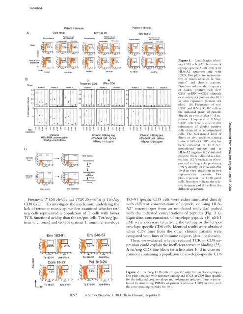
Figure 1. Identification of tet/<br />
neg <strong>CD8</strong> cells. (A) Detection of<br />
epitope-specific <strong>CD8</strong> cells <strong>with</strong><br />
HLA-A2 tetramers and <strong>with</strong><br />
ICCS. Dot plots are representative<br />
of results obtained in “immune”<br />
and chronic patients.<br />
Numbers indicate the frequency<br />
of double positive cells (tet/<br />
<strong>CD8</strong> or IFN-/<strong>CD8</strong> ) directly<br />
ex vivo (top dot plots) or after 10 d<br />
in vitro expansion (bottom dot<br />
plots). (B) Frequency of tet/<br />
<strong>CD8</strong> and IFN-/<strong>CD8</strong> cells in<br />
the indicated group of patients<br />
directly ex vivo or after 10 d expansion.<br />
Frequency of IFN-/<br />
<strong>CD8</strong> cells were calculated after<br />
subtraction of double positive<br />
cells obtained in nonstimulated<br />
cells. The background level of<br />
direct ex vivo tetramer staining<br />
(value 0.03% of <strong>CD8</strong> cells) has<br />
been calculated in HLA-A2 <br />
noninfected subjects and in<br />
HLA-A2 negative HBV-infected<br />
patients; this is indicated as a dotted<br />
line. (C) Visualization of tet/<br />
pos and tet/neg cells producing<br />
IFN- directly ex vivo and after<br />
10 d in vitro expansion in two<br />
representative patients. Dot<br />
plots represent live <strong>CD8</strong> gated<br />
cells. Numbers indicate the relative<br />
frequency of the cells in the<br />
different quadrants.<br />
Downloaded from www.jem.org on June 16, 2008<br />
Functional T Cell Avidity and TCR Expression of Tet/Neg<br />
<strong>CD8</strong> <strong>Cells</strong>. To investigate the mechanism underlying the<br />
lack of tetramer reactivity, we first examined whether tet/<br />
neg cells represented a population of T cells <strong>with</strong> lower<br />
TCR functional avidity than the tet/pos cells. Tet/neg (patient<br />
7, chronic) and tet/pos (patient 1, immune) envelope<br />
183–91-specific <strong>CD8</strong> cells were either stimulated directly<br />
<strong>with</strong> different concentrations of peptide, or using HLA-<br />
A2 macrophages from an uninfected individual pulsed<br />
<strong>with</strong> the indicated concentrations of peptides (Fig. 3 a).<br />
Equivalent concentrations of envelope peptide (10 nM/1<br />
nM) were necessary to activate the tet/neg or the tet/pos<br />
envelope specific <strong>CD8</strong> cells. Identical results were obtained<br />
when <strong>CD8</strong> lines from the other chronic patients were<br />
compared <strong>with</strong> lines of immune subjects (data not shown).<br />
Then, we evaluated whether reduced TCR or <strong>CD8</strong> expression<br />
could explain the inefficient tetramer binding (23).<br />
A tet/neg <strong>CD8</strong> line (short term line after 10 d in vitro expansion)<br />
containing a population of envelope-specific <strong>CD8</strong><br />
1092 Tetramer Negative <strong>CD8</strong> <strong>Cells</strong> in Chronic Hepatitis B<br />
Figure 2. Tet/neg <strong>CD8</strong> cells are specific only for envelope epitopes.<br />
Dot plots obtained <strong>with</strong> tetramer staining and ICCS of <strong>CD8</strong> lines specific<br />
for the indicated core, envelope and polymerase epitopes. Lines were selected<br />
by stimulating PBMCs of patient 5 (chronic HBV) in vitro <strong>with</strong><br />
the corresponding peptides for 10 d.