Examination and processing of human semen - libdoc.who.int ...
Examination and processing of human semen - libdoc.who.int ...
Examination and processing of human semen - libdoc.who.int ...
You also want an ePaper? Increase the reach of your titles
YUMPU automatically turns print PDFs into web optimized ePapers that Google loves.
APPENDIX 3 Microscopy 235<br />
Note: If the microscope is trinocular (i.e. has a third ocular to which a camera can be attached for<br />
photography or video-recording), there will be a light-deflection knob, which is generally located to<br />
the right <strong>of</strong> the eyepieces. This knob is likely to have three settings: one to allow all the light to go to<br />
the eyepieces, one to allow all light to go the camera, <strong>and</strong> a third that deflects half <strong>of</strong> the light to the<br />
eyepieces <strong>and</strong> half to the camera.<br />
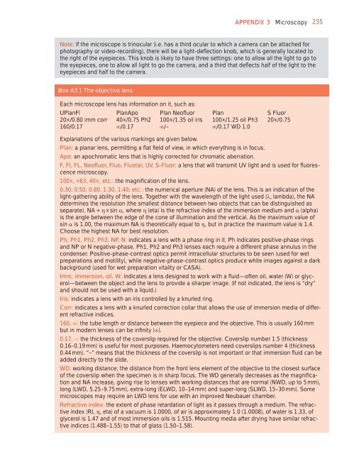
Box A3.1 The objective lens<br />
Each microscope lens has information on it, such as:<br />
UPlanFl PlanApo Plan Ne<strong>of</strong>luor Plan S Fluor<br />
20×/0.80 imm corr 40×/0.75 Ph2 100×/1.35 oil iris 100×/1.25 oil Ph3 20×/0.75<br />
160/0.17 /0.17 /– /0.17 WD 1.0<br />
Explanations <strong>of</strong> the various markings are given below.<br />
Plan: a planar lens, permitting a flat field <strong>of</strong> view, in which everything is in focus.<br />
Apo: an apochromatic lens that is highly corrected for chromatic aberration.<br />
F, Fl, FL, Ne<strong>of</strong>luor, Fluo, Fluotar, UV, S-Fluor: a lens that will transmit UV light <strong>and</strong> is used for fluorescence<br />
microscopy.<br />
100×, ×63, 40×, etc.: the magnification <strong>of</strong> the lens.<br />
0.30, 0.50, 0.80, 1.30, 1.40, etc.: the numerical aperture (NA) <strong>of</strong> the lens. This is an indication <strong>of</strong> the<br />
light-gathering ability <strong>of</strong> the lens. Together with the wavelength <strong>of</strong> the light used (lambda), the NA<br />
determines the resolution (the smallest distance between two objects that can be distinguished as<br />
separate). NA = × sin , where (eta) is the refractive index <strong>of</strong> the immersion medium <strong>and</strong> (alpha)<br />
is the angle between the edge <strong>of</strong> the cone <strong>of</strong> illumination <strong>and</strong> the vertical. As the maximum value <strong>of</strong><br />
sin is 1.00, the maximum NA is theoretically equal to , but in practice the maximum value is 1.4.<br />
Choose the highest NA for best resolution.<br />
Ph, Ph1, Ph2, Ph3, NP, N: indicates a lens with a phase ring in it. Ph indicates positive-phase rings<br />
<strong>and</strong> NP or N negative-phase. Ph1, Ph2 <strong>and</strong> Ph3 lenses each require a different phase annulus in the<br />
condenser. Positive-phase-contrast optics permit <strong>int</strong>racellular structures to be seen (used for wet<br />
preparations <strong>and</strong> motility), while negative-phase-contrast optics produce white images against a dark<br />
background (used for wet preparation vitality or CASA).<br />
Imm, immersion, oil, W: indicates a lens designed to work with a fluid—<strong>of</strong>ten oil, water (W) or glycerol—between<br />
the object <strong>and</strong> the lens to provide a sharper image. (If not indicated, the lens is “dry”<br />
<strong>and</strong> should not be used with a liquid.)<br />
Iris: indicates a lens with an iris controlled by a knurled ring.<br />
Corr: indicates a lens with a knurled correction collar that allows the use <strong>of</strong> immersion media <strong>of</strong> different<br />
refractive indices.<br />
160, : the tube length or distance between the eyepiece <strong>and</strong> the objective. This is usually 160 mm<br />
but in modern lenses can be infinity ().<br />
0.17, –: the thickness <strong>of</strong> the coverslip required for the objective. Coverslip number 1.5 (thickness<br />
0.16–0.19 mm) is useful for most purposes. Haemocytometers need coverslips number 4 (thickness<br />
0.44 mm). “–” means that the thickness <strong>of</strong> the coverslip is not important or that immersion fluid can be<br />
added directly to the slide.<br />
WD: working distance; the distance from the front lens element <strong>of</strong> the objective to the closest surface<br />
<strong>of</strong> the coverslip when the specimen is in sharp focus. The WD generally decreases as the magnification<br />
<strong>and</strong> NA increase, giving rise to lenses with working distances that are normal (NWD, up to 5 mm),<br />
long (LWD, 5.25–9.75 mm), extra-long (ELWD, 10–14 mm) <strong>and</strong> super-long (SLWD, 15–30 mm). Some<br />
microscopes may require an LWD lens for use with an improved Neubauer chamber.<br />
Refractive index: the extent <strong>of</strong> phase retardation <strong>of</strong> light as it passes through a medium. The refractive<br />
index (RI, , eta) <strong>of</strong> a vacuum is 1.0000, <strong>of</strong> air is approximately 1.0 (1.0008), <strong>of</strong> water is 1.33, <strong>of</strong><br />
glycerol is 1.47 <strong>and</strong> <strong>of</strong> most immersion oils is 1.515. Mounting media after drying have similar refractive<br />
indices (1.488–1.55) to that <strong>of</strong> glass (1.50–1.58).


