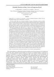
Prenatal Development of Metacarpus and Metatarsus of Cattle
Prenatal Development of Metacarpus and Metatarsus of Cattle
Prenatal Development of Metacarpus and Metatarsus of Cattle

Create successful ePaper yourself
Turn your PDF publications into a flip-book with our unique Google optimized e-Paper software.
408<br />
Materials <strong>and</strong> Methods<br />
Earlier studies <strong>of</strong> the ontogeny <strong>of</strong> the autopodium skeleton employed the method <strong>of</strong> preparing microscopic slides<br />
<strong>and</strong> their subsequent staining (see Rosenberg 1873; Mettam 1894), which is time-consuming <strong>and</strong> requires much<br />
material. The development <strong>of</strong> x-ray technique naturally lead to the introduction <strong>of</strong> this technique into the present<br />
branch <strong>of</strong> science (see Küpfer <strong>and</strong> Schinz 1923; Regli 1963; âerven˘ et al. l987 <strong>and</strong> many others). Another<br />
method used here was the imaging <strong>of</strong> the ossified sections <strong>of</strong> whole bones or their macroscopic sections stained<br />
with alizarin red. Having obtained good experience in imaging the advancing ossification as well as in staining<br />
whole fetuses, we have employed this method in the present study.<br />
The present paper is based on fixed fetuses from the collections <strong>of</strong> the Department <strong>of</strong> Anatomy, Histology <strong>and</strong><br />
Embryology, Faculty <strong>of</strong> Veterinary Medicine <strong>of</strong> the Veterinary <strong>and</strong> Pharmaceutical University in Brno. By their<br />
CRL <strong>and</strong> external characters the fetuses were classified in comparable developmental stages (Comparable Stages,<br />
CS), <strong>and</strong> their ontogenetic age (Estimated Age, EA) on days <strong>of</strong> ontogeny (DO) was determined using the following<br />
table (see ·tûrba 1987, 1995 for particulars).<br />
Stage Characteristics CRL, mm EA – DO<br />
7 Fusion <strong>of</strong> eyelids, umbilical 50 – 70 56 – 65<br />
hernia reposited)<br />
8 Numerous skinfolds present 80 – 120 75 – 90<br />
9 Tactile hairs on upper lip 130 – 250 95 – 120<br />
10 First fine hairs on body 250 – 380 140 – 215<br />
11 Eyelids separated 390 – 470 160 – 173<br />
12 Dense long hairs on body 560 – 850 210 – 270<br />
Autopodia (left) removed from fixed fetuses were longitudinally cut with a scalpel along the lateral-medial<br />
plane, so that each bone (or bone rudiment) was divided into a dorsal <strong>and</strong> a palmar half. The bones <strong>of</strong> smallersized<br />
fetuses were stained with alizarin red <strong>and</strong> subsequently cleared, using the modification <strong>of</strong> âerven˘<br />
(1971). In fetuses 210 DO <strong>and</strong> older the differentiation method <strong>of</strong> staining bone sections with alizarin red was<br />
used (âerven˘ 1979). The dimensions <strong>of</strong> the ossified sections <strong>of</strong> bone rudiments were measured with<br />
a calliper rule.<br />
Results<br />
The following facts were observed on the fetal autopodia stained with alizarin red <strong>and</strong><br />
subsequently cleared (Plate I, Figs 3, 4):<br />
Fetus CRL = 68 mm, age about 60 DO, W = 14 g, CS 7 (No. 21/1): No signs <strong>of</strong> ossification<br />
were safely demonstrated in the cartilaginous rudiments <strong>of</strong> metapodia.<br />
Fetus CRL = 120 mm, age about 80 DO, W = 76 g, CS 8 (No. 68/4): The cartilaginous<br />
rudiments <strong>of</strong> metapodia show ossifying diaphyses, with a distinct connective tissue between<br />
them. The ossified sections <strong>of</strong> os metacarpale III et IV are 9 mm long, both diaphyses being<br />
3 mm wide (Fig. 1). The ossified sections <strong>of</strong> <strong>of</strong> metacarpale III et IV are also 9 mm long, the<br />
diaphyses are 2 mm wide. No medullar cavities can be discerned. The distal epiphyses are<br />
still cartilaginous; ossification centres are already visible in digital phalanxes.<br />
Fetus CRL = 173 mm, age about 95–100 DO, W = 268 g, CS 9 (No. 29/2): Rudimentary<br />
diaphyses undergo ossification. The ossified sections <strong>of</strong> ossa metacarpalia III et IV are 12<br />
mm long <strong>and</strong> 4 mm wide; those <strong>of</strong> ossa metatarsalia III et IV are 13 mm long <strong>and</strong> 3 mm wide.<br />
The ossified sections are independent, separated by a thin connective strip; no medullar<br />
cavities were observed. The distal epiphyses are still cartilaginous.<br />
Fetus CRL = 222 mm, age about 112 DO, W = 564 g, CS 9 (No. 122/10): The ossified<br />
sections <strong>of</strong> diaphyses <strong>of</strong> metacarpals III <strong>and</strong> IV are 18 mm long <strong>and</strong> 5 mm wide, those <strong>of</strong><br />
metatarsals III <strong>and</strong> IV are 20 mm long <strong>and</strong> 4 mm wide. The bones are still independent,<br />
separated by a thin connective strip; the diaphyses show developing medullar cavities. The<br />
whole epiphyses are still cartilaginous all over.<br />
Fetus CRL = 270 mm, age about 125 DO, W = 1 092 g, CS 10-11? (No. 120/10): The<br />
ossified sections <strong>of</strong> diaphyses <strong>of</strong> os metacarpale III et IV are 29 mm long <strong>and</strong> 7 mm wide,<br />
those <strong>of</strong> os metacarpale III et IV are 30 mm long <strong>and</strong> 6 mm wide. They are still separated by<br />
a thin, continuous connective strip. There are distinct medullar cavities. Ossification centres