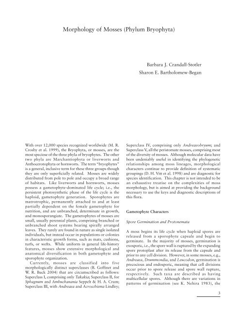
Morphology of Mosses (Phylum Bryophyta)
Morphology of Mosses (Phylum Bryophyta)
Morphology of Mosses (Phylum Bryophyta)

Create successful ePaper yourself
Turn your PDF publications into a flip-book with our unique Google optimized e-Paper software.
<strong>Morphology</strong> <strong>of</strong> <strong>Mosses</strong> (<strong>Phylum</strong> <strong>Bryophyta</strong>)<br />
Barbara J. Crandall-Stotler<br />
Sharon E. Bartholomew-Began<br />
With over 12,000 species recognized worldwide (M. R.<br />
Crosby et al. 1999), the <strong>Bryophyta</strong>, or mosses, are the<br />
most speciose <strong>of</strong> the three phyla <strong>of</strong> bryophytes. The other<br />
two phyla are Marchantiophyta or liverworts and<br />
Anthocerotophyta or hornworts. The term “bryophytes”<br />
is a general, inclusive term for these three groups though<br />
they are only superficially related. <strong>Mosses</strong> are widely<br />
distributed from pole to pole and occupy a broad range<br />
<strong>of</strong> habitats. Like liverworts and hornworts, mosses<br />
possess a gametophyte-dominated life cycle; i.e., the<br />
persistent photosynthetic phase <strong>of</strong> the life cycle is the<br />
haploid, gametophyte generation. Sporophytes are<br />
matrotrophic, permanently attached to and at least<br />
partially dependent on the female gametophyte for<br />
nutrition, and are unbranched, determinate in growth,<br />
and monosporangiate. The gametophytes <strong>of</strong> mosses are<br />
small, usually perennial plants, comprising branched or<br />
unbranched shoot systems bearing spirally arranged<br />
leaves. They rarely are found in nature as single isolated<br />
individuals, but instead occur in populations or colonies<br />
in characteristic growth forms, such as mats, cushions,<br />
turfs, or wefts. While uniform in general life-history<br />
features, mosses show extensive morphological and<br />
anatomical diversification in both gametophyte and<br />
sporophyte organization.<br />
Currently, mosses are classified into five<br />
morphologically distinct superclasses (B. G<strong>of</strong>finet and<br />
W. R. Buck 2004) that are circumscribed as follows:<br />
Superclass I, comprising only Takakia; Superclass II, for<br />
Sphagnum and Ambuchanania Seppelt & H. A. Crum;<br />
Superclass III, with Andreaea and Acroschisma Lindley;<br />
Superclass IV, comprising only Andreaeobryum; and<br />
Superclass V, all the peristomate mosses, comprising most<br />
<strong>of</strong> the diversity <strong>of</strong> mosses. Although molecular data have<br />
been undeniably useful in identifying the phylogenetic<br />
relationships among moss lineages, morphological<br />
characters continue to provide definition <strong>of</strong> systematic<br />
groupings (D. H. Vitt et al. 1998) and are diagnostic for<br />
species identification. This chapter is not intended to be<br />
an exhaustive treatise on the complexities <strong>of</strong> moss<br />
morphology, but is aimed at providing the background<br />
necessary to use the keys and diagnostic descriptions <strong>of</strong><br />
this flora.<br />
Gametophyte Characters<br />
Spore Germination and Protonemata<br />
A moss begins its life cycle when haploid spores are<br />
released from a sporophyte capsule and begin to<br />
germinate. In the majority <strong>of</strong> mosses, germination is<br />
exosporic, i.e., the spore wall is ruptured by the expanding<br />
spore protoplast after its release from the capsule and<br />
prior to any cell division. However, in some mosses, e.g.,<br />
Andreaea, Drummondia, and Leucodon, germination is<br />
precocious and endosporic, meaning that cell divisions<br />
occur prior to spore release and spore wall rupture,<br />
respectively. Such taxa are described as having<br />
multicellular spores. Although there are variations in<br />
patterns <strong>of</strong> germination (see K. Nehira 1983), the<br />
3
4 MORPHOLOGY<br />
fundamental product in most mosses is a highly branched<br />
filamentous, uniseriate protonema. Cell specialization<br />
occurs within the protonema to form two types <strong>of</strong><br />
filaments, a horizontal system <strong>of</strong> reddish brown,<br />
anchoring filaments, called the caulonema, and upright,<br />
green filaments, the chloronema. Each protonema can<br />
spread over several centimeters, forming a fuzzy green<br />
film over its substrate. Fragments <strong>of</strong> chloronema, cut<br />
out by the formation <strong>of</strong> specialized abscission cells<br />
(tmema), may further disperse the protonema. Usually<br />
this protonemal stage is short-lived, but in a few taxa,<br />
e.g., Buxbaumia, it persists as the vegetative phase <strong>of</strong> the<br />
plant.<br />
As the protonema grows, target cells usually on the<br />
caulonema generate bud initials that will ultimately divide<br />
by sequential oblique divisions to form bud apical cells.<br />
This initiates the growth <strong>of</strong> the leafy gametophore or<br />
shoot stage <strong>of</strong> the moss. Usually, numerous shoots<br />
develop from each protonema so that, in fact, a single<br />
mat or cushion <strong>of</strong> shoots can be the product <strong>of</strong> a single<br />
spore.<br />
Shoot <strong>Morphology</strong> and Habit<br />
The leafy shoot continues to grow from mitotic activity<br />
<strong>of</strong> its obovoidal to fusiform apical cell and surrounding<br />
meristem. Divisions occurring in the apical cell form<br />
spirally arranged derivatives, each <strong>of</strong> which will give rise<br />
to a single leaf and a portion <strong>of</strong> the stem. The angle <strong>of</strong><br />
divergence between successive derivatives is responsible<br />
for the spatial arrangement <strong>of</strong> the leaves or phyllotaxy<br />
<strong>of</strong> the shoot. In a few mosses, mature leaves are clearly<br />
ranked; e.g., the leaves <strong>of</strong> Fissidens and Bryoxiphium are<br />
in two rows, a 1 /2 phyllotaxy, and Fontinalis and Tetraphis<br />
have leaves aligned in three rows, a 1 /3 phyllotaxy. In<br />
most mosses, however, the leaves are spirally distributed,<br />
with 2 /5 and 3 /8 phyllotaxies being most common (W. Frey<br />
1971; B. Crandall-Stotler 1984). Branches, which<br />
develop from superficial “bud” initials sequestered in the<br />
leaf axils, mimic the development <strong>of</strong> the protonemal buds<br />
(J. Berthier 1972) and are generally heteroblastic, meaning<br />
that leaves at the base <strong>of</strong> a branch display juvenile<br />
morphology as compared to the later-formed leaves.<br />
Especially in pleurocarpic mosses, chloronema- and<br />
caulonema-like filaments, termed pseudoparaphyllia and<br />
macronemata, respectively, are found at the branch base,<br />
further suggesting its budlike attributes (Berthier).<br />
The peristomate or true mosses (Superclass V) have<br />
traditionally been divided into two broad morphological<br />
groups, namely, acrocarps and pleurocarps, based on the<br />
position <strong>of</strong> the perichaetia and subsequent sporophytes<br />
(Fig. 1). Acrocarps are characterized by erect or ascending<br />
shoot systems that are either unbranched or only<br />
sparingly branched. Branching is typically sympodial<br />
with the branches morphologically comparable to the<br />
determinant main shoot from which they arise.<br />
Perichaetia are differentiated at the tip <strong>of</strong> the main or<br />
primary shoot and terminate its growth, so further plant<br />
growth occurs only if a branch is produced below the<br />
perichaetium; such branches are called subfloral<br />
innovations. Pleurocarps are generally characterized by<br />
creeping shoot systems, with extensive lateral branching.<br />
In such systems, the indeterminant main stem may be<br />
morphologically distinct from the secondary and tertiary<br />
level branches that arise from it (C. La Farge 1996).<br />
Perichaetia in pleurocarps are produced at the tips <strong>of</strong><br />
very short, basally swollen lateral branches that are<br />
morphologically distinct from the vegetative branches.<br />
Because <strong>of</strong> the extremely reduced size <strong>of</strong> the perichaetial<br />
branches, the sporophytes appear to arise from scattered<br />
positions all along the primary stem. Cladocarpic mosses<br />
produce perichaetia at the tips <strong>of</strong> unspecialized lateral<br />
branches that display the same heteroblastic leaf series<br />
as the vegetative branches. Such branches are themselves<br />
capable <strong>of</strong> branching, and these mosses are neither<br />
acrocarpic nor pleurocarpic (La Farge). Although<br />
acrocarps, pleurocarps, and cladocarps tend to have<br />
different branching architectures, it is the morphology<br />
<strong>of</strong> the perichaetium-bearing module that defines the<br />
groups, not branching habit (La Farge).<br />
Pleurocarps form a natural, monophyletic lineage <strong>of</strong><br />
true mosses (B. G<strong>of</strong>finet and W. R. Buck 2004), but<br />
cladocarpy has evolved in several different lineages.<br />
Acrocarpy, which appears to be the plesiomorphic<br />
condition, also characterizes the Takakiopsida,<br />
Andreaeopsida, and Andreaeobryopsida. The main stems<br />
<strong>of</strong> Sphagnum (Superclass II) display a furcate or<br />
dichotomous branch architecture (H. A. Crum 1984).<br />
Along the main stems, fascicles <strong>of</strong> branches are produced<br />
in every fourth leaf (H. Leitgeb 1869), with three or more<br />
branches per fascicle. At least two branches in each<br />
fascicle hang downwards and are appressed to the stem,<br />
while one to three are divergent. Despite their distinctive<br />
fascicled arrangements, all branch development in<br />
Sphagnum is like that <strong>of</strong> other mosses, with each branch<br />
arising from a single axillary bud initial (Leitgeb). At<br />
the apex <strong>of</strong> the main shoot, the abundant developing<br />
fascicles are tightly clustered into a dense tuft called the<br />
capitulum. Archegonia terminate special, short branches<br />
in the capitulum.<br />
Rhizoids<br />
Except for Takakia and Sphagnum, mosses are anchored<br />
to their substrates by filamentous, <strong>of</strong>ten branched, reddish<br />
brown rhizoids. As in caulonemata, the rhizoids are<br />
multicellular with oblique cross walls; their walls are<br />
smooth or roughened with papillae. Rhizoids can be<br />
restricted to the base <strong>of</strong> the stem, especially in acrocarps,
MORPHOLOGY 5<br />
FIGURE 1. General morpohological features <strong>of</strong> mosses. The peristome illustrated for the acrocarpic moss<br />
Funaria is <strong>of</strong> the diplolepidous, opposite type, with endostome segments directly located to the inside <strong>of</strong> the<br />
exostome teeth. In this genus the exostome teeth remain attached to a small part <strong>of</strong> the columella, and the<br />
divisural or median line is not easily seen. The terms acrocarpic, cladocarpic and pleurocarpic refer respectively<br />
to perichaetia and the subsequent sporophytes borne at the apex <strong>of</strong> the main shoot, or on a normal branch, or on<br />
a modified lateral branch.<br />
or arise all along the stem where it is in contact with the<br />
substrate. In some taxa, e.g., Tomentypnum, densely<br />
packed rhizoids form a feltlike tomentum on the stem.<br />
Most rhizoids are slender and only sparingly branched<br />
(micronematal type) but others are larger in diameter and<br />
extensively branched (macronematal type). The former<br />
arise from any <strong>of</strong> the epidermal cells <strong>of</strong> the stem, but the<br />
latter type is associated only with branch primordia as<br />
discussed earlier. Rhizoids are not major sites <strong>of</strong> water<br />
and nutrient uptake, but can enhance capillary movement<br />
<strong>of</strong> water along the outer surface <strong>of</strong> the stem (M. C. F.<br />
Proctor 1984). They function primarily as anchoring<br />
structures and in some taxa, e.g., Leptobryum and<br />
Pyramidula, form asexual propagules that aid in the<br />
spread <strong>of</strong> the colony over unoccupied substrate.<br />
Stem Anatomy<br />
In many mosses, the stem is anatomically complex,<br />
consisting <strong>of</strong> a differentiated epidermal layer, a cortex,<br />
and a central strand <strong>of</strong> thin-walled, hydrolyzed waterconducting<br />
cells, called hydroids (Fig. 2). The epidermal<br />
cells are typically elongate in surface view and have thick,<br />
pigmented walls and small lumina. Various types <strong>of</strong> waxy<br />
deposits, analogous to cuticle or epicuticular waxes, may<br />
cover the surface <strong>of</strong> the epidermal cell wall, in both stems<br />
and leaves, especially in endohydric mosses (M. C. F.<br />
Proctor 1979). When these waxes are abundantly<br />
produced, the plants appear glaucous or iridescent to the<br />
naked eye. Although a thick-walled epidermis is<br />
common, in a variety <strong>of</strong> taxa, e.g., elements <strong>of</strong> the<br />
Pottiaceae as well as taxa <strong>of</strong> very wet habitats like<br />
Hygrohypnum, epidermal cells are thin-walled and<br />
swollen, forming a unistratose hyalodermis (Fig. 1). The<br />
cortex is generally divided into two zones. The outer<br />
zone, or sclerodermis, just to the inside <strong>of</strong> the epidermis,<br />
is comprised <strong>of</strong> stereids. These are elongated, thick-walled<br />
cells, generally with living protoplasts, that function in<br />
mechanical support, analogous to collenchyma in<br />
vascular plants. The inner zone <strong>of</strong> the cortex, or central<br />
cylinder, contains photosynthetic parenchyma cells that
6 MORPHOLOGY<br />
FIGURE 2. Stem anatomy, paraphyllia, pseudoparaphyllia and variations in leaf morphology and arrangement.<br />
become starch-filled and hyaline toward the stem interior.<br />
Some <strong>of</strong> the cells <strong>of</strong> the inner cortex are conducting<br />
parenchyma that are responsible for the transport <strong>of</strong><br />
assimilates. The central strand, absent in some taxa, is<br />
always a solid core <strong>of</strong> nonlignified, water-conducting<br />
hydroids. Hydroid structure varies among taxa<br />
(C. Hébant 1977), from very elongate cells with thickened<br />
lateral walls and partially hydrolyzed end walls in the<br />
Polytrichopsida to the moderately elongate, small<br />
diameter, thin-walled hydroids <strong>of</strong> most taxa. In addition,<br />
the Polytrichopsida have a central strand <strong>of</strong> hydroids, or<br />
hydrome, surrounded by a dissected or medullated ring<br />
<strong>of</strong> highly differentiated, phloemlike sieve cells, called<br />
leptoids. These more specialized cells function in the<br />
conduction <strong>of</strong> photoassimilates in concert with the<br />
elements <strong>of</strong> conducting parenchyma; they are <strong>of</strong><br />
widespread occurrence in immature sporophyte setae, but<br />
in the gametophyte generation are restricted to the<br />
Polytrichopsida.<br />
In some taxa, groups <strong>of</strong> hydroids extend from the leaf<br />
costa into the parenchymatous zone <strong>of</strong> the cortex (Fig.<br />
2). These small clusters <strong>of</strong> hydroids may remain only in<br />
the cortex as false leaf traces, e.g., Plagiomnium, or may<br />
extend through the cortex to connect with the central<br />
strand as true leaf traces, as occurs in the Polytrichopsida.<br />
There is substantial variation in stem organization that<br />
is systematically useful, such as number <strong>of</strong> cell layers in<br />
the sclerodermis, depth <strong>of</strong> the photosynthetic zone, and<br />
presence or absence <strong>of</strong> the central strand and/or leaf traces<br />
(I. Kawai 1989). Sphagnum possesses a unique stem<br />
anatomy in which the epidermis consists <strong>of</strong> one to five<br />
layers <strong>of</strong> large cells that are dead at maturity; in some<br />
species these cells bear fibril thickening bands, and on<br />
the divergent branches they may be modified to form<br />
retort cells. The living cells to the inside <strong>of</strong> the epidermis<br />
are differentiated into an outer zone <strong>of</strong> elongate cells with<br />
thick, <strong>of</strong>ten pigmented walls (the woody cylinder) and<br />
an inner core <strong>of</strong> large, thin-walled cells.<br />
Variation also occurs in the formation <strong>of</strong> external<br />
structures that are associated with stems. The stem<br />
surface may be smooth, or occasionally papillate, or<br />
especially in pleurocarps, be covered with green,<br />
filamentous, or sometimes foliose paraphyllia (Fig. 2).<br />
These small outgrowths presumably photosynthesize and<br />
probably enhance water conductivity by capillary action<br />
along the stem. Pseudoparaphyllia are additional<br />
structures found only in pleurocarps. Although<br />
structurally similar to paraphyllia, pseudoparaphyllia are<br />
formed only at the bases <strong>of</strong> branches. They likely serve<br />
to protect the branch primordia with which they are<br />
associated.<br />
Leaves<br />
The considerable variation that occurs in the arrangement<br />
and structure <strong>of</strong> moss leaves provides some <strong>of</strong> the most
MORPHOLOGY 7<br />
FIGURE 3. Variation in leaf morphology, anatomy and habit.<br />
useful characters for species identification (Figs. 2, 3).<br />
Leaves typically arise from all sides <strong>of</strong> the stem, most<br />
commonly exhibiting a spiral phyllotaxy, but distichous<br />
and tristichous arrangements can also be found. The<br />
mature leaves <strong>of</strong> a given shoot are usually all similar in<br />
size and shape, i.e., isophyllous, but there are taxa that<br />
are anisophyllous, with either dorsal or ventral leaves<br />
decidedly smaller than the lateral leaves (Fig. 3). Except<br />
for a few taxa like Fissidens, leaves are attached to the<br />
stem along broad transverse lines. They are generally<br />
oriented so their apices diverge radially outward from<br />
the stem. In erect mosses, this insertion exposes the<br />
adaxial surface <strong>of</strong> the leaf, that surface directed toward<br />
the stem, to light from above and directs the abaxial<br />
surface down toward the substrate. In prostrate mosses,<br />
in contrast, the adaxial surface is directed toward the<br />
substrate, and the abaxial surface is exposed. For this<br />
reason, in many keys and descriptions the abaxial surface<br />
is defined as the dorsal side <strong>of</strong> the leaf, and the adaxial<br />
surface, as the ventral side (Fig. 1). Sometimes surface<br />
features are different on the two sides <strong>of</strong> the leaf, e.g.,<br />
one surface may be smooth and the other papillose. In<br />
some taxa, all <strong>of</strong> the apices <strong>of</strong> the leaves curve to the<br />
same side in a secund arrangement (Fig. 3). In others,<br />
they may be squarrose, meaning the apices diverge<br />
outward, then bend abruptly downward; or julaceous,<br />
meaning they are strongly concave on the shoot; or<br />
complanate, meaning the adaxial surface <strong>of</strong> the leaf lies<br />
against the stem to give the shoot a flattened appearance.<br />
Moss leaves are undivided and are typically lanceolate<br />
to ovate, except for Takakia, which has unique leaves<br />
that are two to four times divided into cylindrical lobes.<br />
The fundamental moss leaf consists <strong>of</strong> a unistratose<br />
lamina and a multistratose costa or midrib (Fig. 3).<br />
However, in a few families, the entire leaf is multistratose,<br />
and in many others, leaf margins are multistratose, or<br />
otherwise differentiated from the rest <strong>of</strong> the lamina (Fig.<br />
4). For example, <strong>of</strong>ten the margin bears a border <strong>of</strong><br />
hyaline, elongated cells with thickened walls. Such<br />
marginal borders, termed limbidia (singular limbidium),<br />
are thought to provide additional support to the lamina.<br />
Leaf margins may be plane, recurved, or incurved and<br />
are <strong>of</strong>ten toothed, with the teeth varying from pointed<br />
cellular protuberances to large, several-celled projections.<br />
The areolation or cellular network <strong>of</strong> the leaf lamina<br />
also provides useful characters in moss systematics (Fig.<br />
4). Laminal cells in acrocarps are <strong>of</strong>ten short and<br />
isodiametric, particularly in the distal portion <strong>of</strong> the leaf,<br />
while the typical pleurocarp bears laminal cells that are<br />
long and thin; however, all cell shapes, from linear to
8 MORPHOLOGY<br />
FIGURE 4. Variation in leaf morphology and types <strong>of</strong> asexual diaspores. In Sphagnum, leaf shape and cell<br />
pattern differ between the main stem and branches. In Tetrodontium protonema produce leaflike flaps and in<br />
Schistostega dimorphic shoots arise from the light-reflecting protonema. Gemmae are quite variable in size,<br />
shape and location on the plant, while leafy or budlike propagula <strong>of</strong>ten occur in the axils <strong>of</strong> leaves near the shoot<br />
apex, as in Platygyrium.<br />
oblate, can be found in either group. Usually, cell shapes<br />
and sizes are not uniform throughout the leaf. In<br />
particular, the cells near the leaf base are comparatively<br />
longer and broader than those <strong>of</strong> the leaf middle, and<br />
still smaller cells occur at the leaf apex. The basal cells<br />
<strong>of</strong> the leaf margin, known as alar cells, are also frequently<br />
differentiated in size, shape or color from the other cells<br />
<strong>of</strong> the lamina (Fig. 1). Sometimes this zone <strong>of</strong><br />
differentiated cells extends from the margin all the way<br />
to the costa. Hyaline alar cells that are inflated and thinwalled<br />
may control leaf orientation in response to<br />
hygroscopic conditions, causing the leaf to collapse<br />
downward during dry conditions. Laminal cells are<br />
smooth or variously ornamented, commonly bearing a<br />
variety <strong>of</strong> wall projections called papillae (Fig. 3). Papillae<br />
are heterogeneous in form, size, and distribution. They<br />
may be solid or hollow, simple or branched, single or<br />
multiple, on both exposed cell surfaces or only on the<br />
abaxial surface, over the cell lumen or just at the distal<br />
(occasionally proximal or both) end <strong>of</strong> the cell (prorose<br />
or prorulose). Various types <strong>of</strong> epicuticular wax deposits<br />
may add to this ornamentation (M. C. F. Proctor 1979).<br />
The costa or midrib <strong>of</strong> the leaf provides yet another<br />
suite <strong>of</strong> taxonomically useful characters (Fig. 3). Leaves<br />
<strong>of</strong> acrocarps usually have a clearly defined, single costa<br />
that extends from the base <strong>of</strong> a leaf to shortly before its<br />
apex, or to its apex (percurrent) or even beyond its apex<br />
(excurrent). In a few acrocarps and many pleurocarps,<br />
the costa is short, generally confined to the leaf base,<br />
and double, or absent (Fig. 1). Costa anatomy is variable<br />
with several distinctive patterns (I. Kawai 1968). The<br />
costa may be homogeneously composed <strong>of</strong> thick-walled<br />
stereids, or have adaxial or abaxial epidermal cells larger<br />
than the internal cells. Often, a row <strong>of</strong> large, thin-walled<br />
cells, referred to as guide cells, or deuters, bisect the costa,<br />
extending between the two laminar segments;<br />
occasionally, there will be a small strand <strong>of</strong> hydroids<br />
between the guide cells and one <strong>of</strong> the stereid bands,<br />
especially in those acrocarpic taxa in which leaf traces<br />
occur. As suggested by the abundance <strong>of</strong> stereids in most<br />
costae, one <strong>of</strong> the primary functions <strong>of</strong> the costa is to<br />
provide support for the unistratose lamina, but it can<br />
also function in transport. The guide cells <strong>of</strong> the costa<br />
are conducting elements involved in the symplastic
MORPHOLOGY 9<br />
transport <strong>of</strong> photoassimilates from the leaf lamina into<br />
the stem cortex (C. Hébant 1977), and the hydroid<br />
strands move water upward from the stem into the distal<br />
portions <strong>of</strong> the leaf.<br />
In the Polytrichopsida, as well as a few other taxa,<br />
unistratose, photosynthetic lamellae arise from the<br />
adaxial (ventral) surface <strong>of</strong> the costa. They extend as<br />
parallel sheets <strong>of</strong> cells from just beyond the sheathing<br />
leaf base <strong>of</strong>ten to the apex <strong>of</strong> the costa. In some taxa,<br />
the lamina is incurved over the lamellae, thereby forming<br />
an almost mesophyll-like tissue. Variations in the number<br />
and height <strong>of</strong> lamellae as well as lamellar cell<br />
morphologies provide taxonomically informative<br />
characters in this group. Leaves <strong>of</strong> the Fissidentaceae<br />
are also distinct from those <strong>of</strong> other mosses, consisting<br />
<strong>of</strong> two basal, clasping vaginant laminae and two vertically<br />
oriented, distal laminae, designated as the ventral<br />
(superior) lamina and dorsal (inferior) lamina. The two<br />
distal laminae arise from the adaxial (ventral) and abaxial<br />
(dorsal) surfaces <strong>of</strong> the costa, respectively, while the<br />
vaginant laminae extend laterally from the costa as in<br />
other mosses. Still another modification in leaf<br />
organization occurs in Sphagnum and several other taxa,<br />
e.g., Leucobryum and Leucoloma, in which the leaf is<br />
composed <strong>of</strong> two types <strong>of</strong> cells, very small, chlorophyllose<br />
cells (chlorocysts) and empty, hyaline cells (hyalocysts in<br />
Sphagnum or leucocysts in other taxa). In Leucobryum,<br />
the leaf is multistratose, with the chlorocysts embedded<br />
beween the two layers <strong>of</strong> leucocysts. In Sphagnum the<br />
chlorocysts form a reticulum within a unistratose leaf;<br />
variations in pore distribution in the hyalocysts and<br />
arrangement <strong>of</strong> chlorocysts in relation to hyalocysts<br />
provide important taxonomic characters in this genus.<br />
Juvenile moss leaves possess one to several uniseriate<br />
hairs in their axils. These are persistent in some acrocarps,<br />
but are usually physiologically active and secrete mucilage<br />
only near the shoot apex. They <strong>of</strong>ten deteriorate as the<br />
leaf matures. Variation in the number <strong>of</strong> hairs per leaf<br />
and the number and length <strong>of</strong> cells per hair, nonetheless,<br />
has proven useful in some moss groups (L. Hedenäs<br />
1989).<br />
Asexual Diaspores<br />
It has been estimated that up to 15% <strong>of</strong> moss taxa<br />
produce some type <strong>of</strong> asexual diaspore (H. A. Crum<br />
2001). These include caducous leaves, stems, and<br />
rhizoids, as well as morphologically specialized brood<br />
bodies. Fragments <strong>of</strong> gametophytes, if dispersed to a<br />
suitable substrate, produce protonemata from which new<br />
shoots will develop. Brood bodies are <strong>of</strong> two major types,<br />
gemmae, which are small, unicellular or more commonly,<br />
multicellular structures comprised <strong>of</strong> undifferentiated<br />
cells, and propagula, which are small, easily detached<br />
plantlets or buds (Fig. 4). Gemmae can be filiform,<br />
discoid, or cylindrical, and <strong>of</strong>ten possess pigmented and<br />
thickened outer cell walls. They can be produced in<br />
special splash cups, as in Tetraphis, or near the tip <strong>of</strong><br />
elongate shoot apices (e.g., Aulacomnium), in the axils<br />
<strong>of</strong> leaves (e.g., some species <strong>of</strong> Didymodon), or on the<br />
laminar or costal surfaces (e.g., Orthotrichum), as well<br />
as on rhizoids and protonemata (e.g., Zygodon).<br />
Rhizoidal gemmae that are somewhat buried are<br />
commonly referred to as tubers. Propagula, sometimes<br />
termed bulbils, may be clustered at the tips <strong>of</strong> the shoots<br />
as in Platygyrium, or occur singly in the axils <strong>of</strong> leaves as<br />
in Leptobryum.<br />
Sexual Reproduction<br />
Gametangia are typically clustered with interspersed,<br />
sterile hairs, called paraphyses, at shoot or branch apices.<br />
Androecia, or male inflorescences, contain numerous,<br />
elongate, ovoid to cylindrical antheridia (over 100 in some<br />
taxa) and paraphyses, surrounded by perigonial leaves,<br />
and may be either budlike or disciform. Gynoecia, or<br />
female inflorescences, contain groups <strong>of</strong> long-necked,<br />
stalked archegonia, paraphyses, and surrounding<br />
perichaetial leaves, and are only budlike, never disciform.<br />
In rare cases, e.g., Takakia, organized androecia and<br />
gynoecia are lacking and the gametangia simply occur in<br />
the axils <strong>of</strong> unmodified leaves. Both the organization<br />
and arrangement <strong>of</strong> androecia and gynoecia are <strong>of</strong><br />
taxonomic importance.<br />
<strong>Mosses</strong> are either dioicous, i.e., bearing androecia and<br />
gynoecia on separate gametophytes, or monoicous,<br />
bearing both sexes on a single gametophyte. There are<br />
several different kinds <strong>of</strong> monoicous arrangements,<br />
depending on the relative positions <strong>of</strong> the antheridia and<br />
archegonia. In autoicous arrangements, there are separate<br />
androecia and gynoecia on the same plant, <strong>of</strong>ten on<br />
separate branches (cladautoicous), while in both<br />
synoicous and paroicous arrangements antheridia and<br />
archegonia occur in a single inflorescence, either<br />
intermixed within the same cluster (synoicous), or in<br />
separate clusters in different leaf axils (paroicous). In<br />
plants described as heteroicous, more than one form <strong>of</strong><br />
monoicy occurs on the same plant. In a few dioicous<br />
mosses, e.g., Dicranum, male plants are highly reduced<br />
and actually grow as epiphytes upon the leaves or stems<br />
<strong>of</strong> the much larger female plants. Although<br />
physiologically dioicous, the production <strong>of</strong> dwarf males<br />
to some degree mimics the male/female relationship <strong>of</strong><br />
autoicous taxa, so this is sometimes called a<br />
pseudautoicous condition. When male plants arise from<br />
the same protonema as female plants, it mimics dioicy,<br />
and this feature is termed rhizautoicy.<br />
Numerous biflagellated sperm are produced by mitosis<br />
inside the antheridia while a single egg develops in the<br />
base <strong>of</strong> each archegonium. When the sperm mature, the
10 MORPHOLOGY<br />
antheridia swell and open at their apices. Drops <strong>of</strong> rain<br />
water or morning dew disperse the sperm to a gynoecium,<br />
perhaps aided by microarthropods (N. Cronberg et al.<br />
2006). Slimy mucilage secretions in the archegonial necks<br />
help pull the sperm downward to the egg. Fertilization<br />
<strong>of</strong> an egg initiates the diploid sporophyte phase.<br />
Sporophyte Characters<br />
Embryo Development<br />
Concomitant with growth <strong>of</strong> the embryonic sporophyte,<br />
cell divisions in the surrounding archegonial center, basal<br />
archegonial stalk, and subtending gametophyte shoot<br />
produce an enclosing epigonium. Early in development,<br />
a nutrient transfer zone, or placenta, is differentiated<br />
between the conical foot <strong>of</strong> the sporophyte and the basal<br />
part <strong>of</strong> the epigonium. Both organic nutrients and water<br />
move from the gametophyte into the sporophyte across<br />
the placenta. A stemlike seta is differentiated above the<br />
foot and embryonic capsule initials are cleaved from an<br />
apical cell at the apex <strong>of</strong> the sporophyte. The<br />
establishment <strong>of</strong> a seta meristem just below the<br />
differentiating capsule elongates the seta, tearing the<br />
epigonium. The basal part <strong>of</strong> the epigonium, which still<br />
encloses the lower part <strong>of</strong> the seta and foot, is now<br />
referred to as the vaginula, and the upper part that<br />
remains over the tip <strong>of</strong> the sporophyte is the calyptra. In<br />
taxa in which the seta is lacking or much reduced, such<br />
as Sphagnum and Andreaea, the enlarging capsule<br />
ruptures the epigonium. In a few mosses, e.g.,<br />
Bryobartramia, the epigonium remains intact, never<br />
separating into a vaginula and calyptra (H. A. Crum<br />
2001, fig. 25A). Variation in the timing <strong>of</strong> epigonial<br />
tearing can affect calyptra morphology; for example,<br />
calyptrae torn away late in development are elongate, as<br />
in Encalypta, while those separated early are short and<br />
thin, as in Archidium.<br />
The calyptra remains seated over the developing<br />
capsule until spore maturation is complete. Calyptrae<br />
are <strong>of</strong> two types based on form. The cucullate type is slit<br />
up one side and sits like a hood over the capsule, and the<br />
mitrate type is conic and undivided or equally lobed at<br />
the base. Variations in calyptra shape, size, areolation,<br />
and surface ornamentation provide taxonomically useful<br />
characters, as detailed by P. Janzen (1916).<br />
Seta Anatomy<br />
The seta and capsule continue to develop after the<br />
emergence <strong>of</strong> the sporophyte from the epigonium, the<br />
seta from a generalized, apical meristem and the capsule<br />
by patterned divisions in earlier formed capsule initials.<br />
Usually only one sporophyte matures per gynoecium, but<br />
in some taxa, e.g., Dicranum, more than one can develop,<br />
resulting in polysety, or multiple setae emerging from a<br />
single perichaetium.<br />
Anatomically, the seta resembles the stem <strong>of</strong> the<br />
gametophyte in being differentiated into epidermis, cortex<br />
<strong>of</strong> outer stereid and inner parenchymatous zones, and a<br />
central conducting strand. There is always a waxy,<br />
cuticlelike covering associated with the epidermis.<br />
Central strands can be well-developed even in taxa that<br />
lack such strands in their gametophytes, and residual<br />
leptoids are reported to occur in some taxa, e.g., Funaria,<br />
Splachnum, and Meesia, although there is disagreement<br />
as to the exact nature <strong>of</strong> these cells (D. Schulz and<br />
C. Wiencke 1976; C. Hébant 1977). Prior to capsule<br />
ripening, the seta is typically chlorophyllose, but at<br />
maturity it becomes reddish or yellowish brown, due to<br />
pigments deposited in the thickened walls <strong>of</strong> epidermal<br />
cells and stereids. It is not uncommon for the interior<br />
cells <strong>of</strong> the seta, including those <strong>of</strong> the central strand, to<br />
deteriorate in late stages <strong>of</strong> spore maturation, producing<br />
a hollow seta (W. Lorch 1931; Hébant). In most mosses<br />
the seta surface is smooth, but in some taxa it is roughened<br />
with papillae or bristly outgrowths. Often, the epidermal<br />
cell rows are helically aligned; this arrangement as well<br />
as differential wall thickness on different sides <strong>of</strong> the seta<br />
allows the seta to twist as it dries, and untwist when<br />
rehydrated. The direction and pattern <strong>of</strong> the twist is<br />
taxonomically useful in some groups. The extent <strong>of</strong> seta<br />
emergence above the perichaetium varies among taxa.<br />
In the Sphagnopsida and Andreaeopsida the seta is absent,<br />
and the capsule is instead elevated above the perichaetium<br />
by elongation <strong>of</strong> the gametophytic cells subtending the<br />
foot. This extended gametophytic tissue is called a<br />
pseudopodium.<br />
Capsule Anatomy<br />
<strong>Mosses</strong> are monosporangiate, meaning that each<br />
sporophyte produces only a single sporangium, or<br />
capsule. Very early in development, periclinal divisions<br />
in the apically produced capsule initials separate an inner,<br />
endothecial zone from an outer ring <strong>of</strong> amphithecial cells.<br />
With the exception <strong>of</strong> Sphagnum, a columella and sporeproducing<br />
archesporium are formed from the<br />
endothecium, and outer parts <strong>of</strong> the capsule, including<br />
the peristome, develop from patterned divisions <strong>of</strong> the<br />
amphithecium. Details <strong>of</strong> these later stages in capsule<br />
ontogeny have been documented by several workers,<br />
including, among others, F. Kienitz-Gerl<strong>of</strong>f (1878),<br />
J. C. French and D. J. Paolillo (1975), S. R. Edwards<br />
(1984), and A. J. Shaw et al. (1989).<br />
Variations in the morphology <strong>of</strong> the mature capsule<br />
provide a wealth <strong>of</strong> systematically important characters<br />
(Fig. 5). Capsules vary in shape from spheroid to ovoid,
MORPHOLOGY 11<br />
FIGURE 5. Variation in capsule dehiscence and peristomes. In Scouleria the columella remains attached to the<br />
operculum and is elevated above the urn when the capsule opens. Interpretations are equivocal regarding the<br />
peristome <strong>of</strong> Buxbaumia; the endostome is arthrodontous, but the true nature <strong>of</strong> the external rings <strong>of</strong> teeth and<br />
the prostome (also known as the parastome) is not clear.<br />
obovoid, pyriform, turbinate, ellipsoid, or long-cylindric,<br />
and can be either symmetric, e.g., Orthotrichum, or<br />
asymmetric, e.g., Funaria. They may be erect, inclined,<br />
nodding (cernuous), or even pendent. They are typically<br />
terete in transverse section, but can also be 4-angled, e.g.,<br />
members <strong>of</strong> the Polytrichopsida, or longitudinally<br />
furrowed, with 8 or 16 ribs. The exothecium, or<br />
epidermis, <strong>of</strong> the capsule bears a shiny cuticlelike layer,<br />
and is usually smooth-surfaced, although surface<br />
ornamentations occur in a few genera. Exothecial<br />
characters, such as cell size, shape, and arrangement can<br />
be <strong>of</strong> systematic value. Immature capsules are green, but<br />
when mature are various shades <strong>of</strong> yellow, red, or brown.<br />
Distinctive types <strong>of</strong> capsule anatomy characterize the<br />
five major groups or superclasses <strong>of</strong> mosses. In the<br />
majority <strong>of</strong> true mosses (Superclass V), capsules consist<br />
<strong>of</strong> three anatomically distinct zones, namely, the basal<br />
neck, the median spore-containing urn or theca, and the<br />
distal operculum. Capsules with a dehiscent operculum<br />
are stegocarpous. Capsules that lack an operculum and<br />
dehisce irregularly due to generalized breakdown <strong>of</strong> the<br />
capsule walls are cleistocarpous. In Takakia (Superclass<br />
I), Andreaea (Superclass III), and Andreaeobryum<br />
(Superclass IV), there is no differentiation <strong>of</strong> a neck or<br />
operculum; their capsules open along incomplete<br />
longitudinal sutures, with the capsule staying intact above<br />
and below the slits (Fig. 5). There is one spiral suture in<br />
Takakia and four vertical sutures in Andreaea and<br />
Andreaeobryum. The globose capsule <strong>of</strong> Sphagnum<br />
(Superclass II) possesses a small, lidlike operculum, but<br />
lacks a neck; in contrast to true mosses its spore mass, or<br />
archesporium, overarches a dome-shaped columella, an<br />
arrangement that is also characteristic <strong>of</strong> Andreaea and<br />
Andreaeobryum. In Sphagnum, contraction <strong>of</strong> the<br />
capsule wall releases the operculum, explosively<br />
dispersing the spores.<br />
In some mosses, the neck tapers gradually into the<br />
seta, but in others, it is markedly differentiated, as a<br />
swollen hypophysis (or apophysis), the most notable <strong>of</strong><br />
which is the highly inflated, parasol-like hypophysis in<br />
the Splachnaceae. Stomatal complexes, i.e., stoma and<br />
their associated guard cells, are frequent within the<br />
exothecium <strong>of</strong> the neck; these may be superficial or<br />
sunken (immersed) in different species <strong>of</strong> the same genus,<br />
e.g., Orthotrichum, and can be either open or closed.<br />
The guard cells are usually completely divided, as in<br />
vascular plants, but in some taxa, like Funaria, they are<br />
only partially divided so that the stoma appears as a
12 MORPHOLOGY<br />
slitlike opening in a single cell. In Sphagnum, pairs <strong>of</strong><br />
guard cells occur scattered throughout the exothecium,<br />
but there are no stomata associated with them. Variations<br />
in number, form, and distribution <strong>of</strong> stomata are<br />
taxonomically informative (J. A. Paton and J. V. Pearce<br />
1957).<br />
Internally, the central strand <strong>of</strong> the seta extends up<br />
into the neck; this strand is surrounded by a<br />
chlorophyllose zone <strong>of</strong> either somewhat spongy<br />
parenchyma or highly differentiated aerenchyma, into<br />
which the stoma open. This is a major site <strong>of</strong><br />
photosynthesis in the developing capsule, providing up<br />
to 50% <strong>of</strong> the photoassimilate necessary for capsule<br />
growth (M. C. F. Proctor 1977).<br />
A solid column <strong>of</strong> parenchymatous cells forms the<br />
central columella that extends from the neck through<br />
the urn to the operculum <strong>of</strong> the capsule. The <strong>of</strong>ten<br />
elongate spore sac encircles the columella. To the outside<br />
<strong>of</strong> the spore sac, the wall <strong>of</strong> the capsule is differentiated<br />
into an inner aerenchyma <strong>of</strong> air spaces transversed by<br />
chlorophyllose filaments, an outer zone <strong>of</strong> hyaline cells<br />
and the thick-walled exothecium. In Polytrichopsida, a<br />
filamentous aerenchyma can also be formed between the<br />
spore sac and the columella.<br />
The operculum <strong>of</strong> stegocarpous mosses is attached to<br />
the distal end <strong>of</strong> the urn by an annulus, i.e., a ring <strong>of</strong><br />
differentiated cells that tear to release the operculum<br />
during capsule dehiscence. Annulus anatomy varies<br />
among taxa, in both the number <strong>of</strong> cell layers and the<br />
structure <strong>of</strong> the cells comprising it. In its simplest form,<br />
the annulus consists <strong>of</strong> a ring <strong>of</strong> small, quadrate cells,<br />
<strong>of</strong>ten several cells high, that differ in wall thickness from<br />
the neighboring exothecial and opercular cells. This type<br />
<strong>of</strong> annulus is either persistent or falls <strong>of</strong>f in fragments<br />
when the capsule is moistened due to differential swelling<br />
<strong>of</strong> its cells compared to the neighboring cells. This is the<br />
common mode <strong>of</strong> capsule dehiscence in pleurocarps, but<br />
also occurs in other taxa, e.g., Orthotrichum. In many<br />
acrocarps, e.g., Funaria and Bryum, the annulus consists<br />
<strong>of</strong> an internal ring <strong>of</strong> enlarged cells with hydrophilic walls<br />
and an outer ring <strong>of</strong> small cells with thickened,<br />
hydrophobic walls (Fig. 1). During dehiscence, the<br />
internal cells swell with the uptake <strong>of</strong> water, but the outer<br />
cells remain unchanged. This differential swelling causes<br />
the annulus to coil away in a single piece from the<br />
overlapping cells <strong>of</strong> the operculum; this is termed a<br />
revoluble annulus. In either case, removal <strong>of</strong> the<br />
operculum, or capsule lid, exposes the mouth <strong>of</strong> the urn<br />
and its spore mass for dispersal.<br />
The Peristome<br />
In the majority <strong>of</strong> stegocarpous mosses, spore dispersal<br />
is mediated by the peristome, a circular system <strong>of</strong> teeth<br />
that is inserted on the mouth <strong>of</strong> the urn, to the inside <strong>of</strong><br />
the operculum. The developmental history and<br />
architecture <strong>of</strong> the peristome provide a suite <strong>of</strong> important<br />
systematic characters (Fig. 5). Peristomes are <strong>of</strong> two<br />
fundamentally different types, nematodontous, which are<br />
found only in Polytrichopsida and Tetraphidopsida, and<br />
arthrodontous. In a nematodontous peristome, the teeth<br />
are constructed <strong>of</strong> bundles <strong>of</strong> whole, dead cells.<br />
Commonly in the Polytrichopsida, 32 or 64 (rarely 16)<br />
short lingulate teeth, comprised <strong>of</strong> up to four layers <strong>of</strong><br />
vertically elongate, very thick-walled cells, are attached<br />
by their inner surface to a membranous expansion <strong>of</strong> the<br />
columella called the epiphragm. The release <strong>of</strong> the<br />
operculum exposes small slits between the teeth through<br />
which the spores are slowly released. In the<br />
Tetraphidopsida, there are four erect, wedge-shaped<br />
peristome teeth, each <strong>of</strong> which represents a quadrant <strong>of</strong><br />
the peristomial cell layers.<br />
In contrast to the cellular peristomes <strong>of</strong> these taxa,<br />
arthrodontous peristomes, found in the rest <strong>of</strong><br />
stegocarpous mosses, consist at maturity only <strong>of</strong> remnants<br />
<strong>of</strong> paired, periclinal cell walls. As reviewed by several<br />
authors (e.g., S. R. Edwards 1984; A. J. Shaw and<br />
H. Robinson 1984; W. R. Buck and B. G<strong>of</strong>finet 2000),<br />
arthrodontous peristomes differentiate from the three<br />
innermost layers <strong>of</strong> the amphithecium formed by<br />
fundamental square divisions (K. Goebel 1900–1905, vol.<br />
2) in the apex <strong>of</strong> the embryonic capsule. Following<br />
H. L. Blomquist and L. L. Robertson (1941), these are<br />
termed the outer peristomial (OPL), primary peristomial<br />
(PPL), and inner peristomial layers (IPL). The number<br />
<strong>of</strong> cells in the peristomial layers in a 1 /8 slice <strong>of</strong> a transverse<br />
section is expressed as the peristomial formula (Edwards<br />
1979); thus, a peristomial formula <strong>of</strong> 4:2:3 describes a<br />
capsule with 32 OPL, 16 PPL, and 24 IPL cells. The<br />
number and arrangement <strong>of</strong> cells in the peristomial layers<br />
cannot always be determined with certainty in mature<br />
capsules, so peristomial formulae are generally not<br />
included in taxonomic descriptions.<br />
Arthrodontous peristomes are <strong>of</strong> two major types,<br />
namely, haplolepidous and diplolepidous (Fig. 5). The<br />
haplolepidous peristome consists <strong>of</strong> a single ring <strong>of</strong> 16<br />
teeth that are formed by cell wall deposition on the paired<br />
walls <strong>of</strong> the PPL and IPL. The peristomial formula is<br />
always 0(4):2:3, with a single column <strong>of</strong> PPL cells forming<br />
the outer (dorsal) surface <strong>of</strong> each tooth, and unequal parts<br />
<strong>of</strong> two IPL cells forming the inner (ventral) surface.<br />
Consequently, the outer surface <strong>of</strong> the tooth, which may<br />
be variously ornamented with horizontal striae,<br />
trabeculae, or papillae, lacks median or divisural lines<br />
(= vertical cell walls). The teeth can be forked at their<br />
apices, as in the Dicranaceae, or be fused at the base into<br />
an elongate tube, or basal membrane, or be divided into<br />
32 long narrow, filaments, e.g., the Pottiaceae.<br />
Development from the OPL is highly reduced or absent,<br />
forming at best prostomial bumps at the base <strong>of</strong> the<br />
peristome (S. R. Edwards 1984).
MORPHOLOGY 13<br />
Diplolepidous peristomes have the same number <strong>of</strong><br />
cells in the OPL and PPL as haplolepidous peristomes,<br />
but display substantial variation in the IPL numbers, with<br />
peristomial formulae ranging from 4:2:4 to 4:2:14. Two<br />
sets <strong>of</strong> teeth are differentiated, the exostome, or outer<br />
peristome, formed by deposition on the paired walls <strong>of</strong><br />
the OPL and PPL, and the endostome, formed at the<br />
PPL–IPL wall junctures. The exostome typically consists<br />
<strong>of</strong> 16 teeth, equal to the number <strong>of</strong> cells in the PPL, while<br />
the outer surface <strong>of</strong> each tooth bears a divisural line that<br />
marks the two columns <strong>of</strong> cells <strong>of</strong> the OPL. The teeth<br />
may be joined together in pairs, or secondarily divided,<br />
and are <strong>of</strong>ten highly ornamented, especially on the outer<br />
surface (A. J. Shaw 1985). The architecture <strong>of</strong> the<br />
endostome is likewise variable, with different patterns<br />
<strong>of</strong> surface ornamentation on outer and inner surfaces<br />
(Shaw and J. R. Rohrer 1984). In a diplolepidousalternate<br />
peristome (D. H. Vitt 1984) <strong>of</strong> the bryoid or<br />
hypnoid type, the endostome comprises a basal, <strong>of</strong>ten<br />
keeled membrane, topped by 16 broad, perforate<br />
segments that alternate with the exostome teeth. One to<br />
four uniseriate cilia occur between the segments, opposite<br />
the exostome teeth. In some taxa, the endostome<br />
segments are highly reduced or absent, and the inner<br />
peristome consists only <strong>of</strong> cilia (Fig. 5). In contrast, in<br />
the diplolepidous-opposite peristome <strong>of</strong> the Funariales,<br />
there is no basal membrane, the endostome segments<br />
occur opposite the exostome teeth, and there are no cilia<br />
(Fig. 1). In some taxa, e.g., Orthotrichum, a short,<br />
rudimentary system <strong>of</strong> processes, called a prostome or<br />
preperistome, is formed just to the outside <strong>of</strong> the outer<br />
teeth.<br />
Movements <strong>of</strong> the exostome teeth <strong>of</strong> diplolepidous<br />
taxa as well as the single ring <strong>of</strong> teeth <strong>of</strong> haplolepidous<br />
taxa are due to the differential composition <strong>of</strong> the wall<br />
deposits on the outer versus the inner surfaces <strong>of</strong> the teeth.<br />
Specifically, one surface readily absorbs water and<br />
elongates, while the other does not. This differential<br />
response to water absorption causes the teeth to bend<br />
when moistened. In many taxa the teeth close over the<br />
mouth <strong>of</strong> the capsule when moistened, so spores are<br />
released only when the air is dry, but in others they bend<br />
outward when wet, allowing spore release in moist<br />
conditions (D. M. J. Mueller and A. J. Neumann 1988).<br />
With drying, the teeth return to their original stance. This<br />
process can be repeated several times, resulting in the<br />
gradual release <strong>of</strong> the spores from the capsule.<br />
Arrest <strong>of</strong> peristome development can result in the loss<br />
<strong>of</strong> segments, cilia, teeth, the entire endostome or<br />
exostome, or the whole peristome. Stegocarpous mosses<br />
that lack a peristome, e.g., Physcomitrium, are termed<br />
gymnostomous. Although they lack a peristome at<br />
capsule maturity, such mosses, nonetheless, display<br />
characteristic peristomial layers in their developing<br />
capsules, and can be aligned with peristomate taxa using<br />
their peristomial formulae.<br />
Spores<br />
Most mosses are isosporous, meaning that spore sizes<br />
are unimodal, with variation ranging around one<br />
arithmetic mean (G. S. Mogensen 1983). Some dioicous<br />
mosses that produce dwarf males, however, are<br />
anisosporous (D. H. Vitt 1968). In this case, half <strong>of</strong> the<br />
spores in any capsule are significantly smaller than the<br />
other half, that is, spore sizes are bimodal within a single<br />
capsule (H. P. Ramsay 1979). Culture studies have<br />
documented that in many taxa the small spores germinate<br />
later than the large spores and give rise to dwarf males<br />
(M. Ernst-Schwarzenbach 1944). Bimodality <strong>of</strong> spore<br />
sizes does not, however, always correlate with sexual<br />
dimorphism. In some instances, the small spores are<br />
consistently abortive, a condition termed pseudoanisospory<br />
(Mogensen 1978). Mogensen hypothesized<br />
that a lethal combination <strong>of</strong> alleles from two genes is<br />
responsible for the abortive spores and that this condition<br />
leads to balanced polymorphism in the taxon.<br />
Pseudoanisopory is more common than true anisospory<br />
and occurs in both dioicous and monoicous taxa.<br />
Spores in the majority <strong>of</strong> mosses are dispersed as single<br />
cells, but precociously germinated multicellular spores<br />
occur in some xerophytes or epiphytes, such as<br />
Drummondia. Spores are typically spheroidal, but may<br />
also be ovoid, reniform, or tetrahedral. They are <strong>of</strong>ten<br />
small, less than 20 μm in diameter, with a finely papillose<br />
ornamentation, but much larger, more highly ornamented<br />
spores can also occur, primarily in cleistocarpic taxa.<br />
Ornamentation <strong>of</strong> the outer spore wall comes primarily<br />
from the perine, which is formed from deposits <strong>of</strong><br />
globular materials produced within the spore sac<br />
(G. S. Mogensen 1983), although in some taxa, e.g.,<br />
Polytrichum and Astoma, the exine also contributes to<br />
the sculpturing (J. W. McClymont and D. A. Larson<br />
1964). In most cases the globular deposits <strong>of</strong> perine<br />
appear to be rather randomly deposited over the spore<br />
surface as granulose papillae, e.g., Cinclidium (Mogensen<br />
1981), but in others the deposits seem to be laid down in<br />
a regular pattern, perhaps controlled by a predetermined<br />
network on the spore surface, e.g., Funaria<br />
(H. V. Neidhart 1979). Variations in spore wall<br />
ornamentation have been little used in moss systematics,<br />
with the exception <strong>of</strong> a few groups that have been studied<br />
using SEM, e.g., Polytrichopsida (Gary L. Smith 1974),<br />
Encalyptaceae (D. H. Vitt and C. D. Hamilton 1974),<br />
and Pottiaceae (K. Saito and T. Hirohama 1974;<br />
J. S. Carrión et al. 1990).