Targeting focal adhesions: Helicobacter pylori-host communication ...
Targeting focal adhesions: Helicobacter pylori-host communication ...
Targeting focal adhesions: Helicobacter pylori-host communication ...

Create successful ePaper yourself
Turn your PDF publications into a flip-book with our unique Google optimized e-Paper software.
Cell Communication and Signaling 2008, 6:2<br />
http://www.biosignaling.com/content/6/1/2<br />
prising that the number of CagA-interacting proteins is<br />
steadily increasing. These proteins are involved in diverse<br />
cellular signal transduction pathways targeting cell proliferation,<br />
cellular junctions and <strong>adhesions</strong> [9-11].<br />
A hallmark of cultured H. <strong>pylori</strong>-infected epithelial cells is<br />
the development of the so-called hummingbird phenotype,<br />
the characteristic formation of which is dependent<br />
on CagA PY (Fig. 1) [13]. This phenotype might influence<br />
the immune response, wound healing, metastasis or invasive<br />
growth of cancer cells in vivo [14,15]. The H. <strong>pylori</strong>induced<br />
hummingbird phenotype is reminiscent of<br />
growth factor-induced cell scattering, which consists of<br />
several processes, namely (i) cell movement, driven by<br />
rearrangement of the cytoskeleton and (ii) the assembly/<br />
disassembly of cell-matrix contacts [16]. However, the<br />
way in which H. <strong>pylori</strong> regulates these cellular processes is<br />
even less understood. Here, we review the recent progress<br />
in the study of H. <strong>pylori</strong> <strong>communication</strong> with intercellular<br />
signal transduction pathways controlling the coordinated<br />
action of cytoskeletal-dependent migration, cell morphology<br />
and cell-matrix adhesion, all of which might contribute<br />
to the pathogenesis of H. <strong>pylori</strong>.<br />
Cellular aspects of cell migration<br />
Cell migration comprises several temporally and spatially<br />
coordinated events, including elongation of the leading<br />
edge, adhesion of this protrusion to the matrix, movement<br />
of the cell body and release of the trailing edge of the<br />
cell [14,15]. Important structures are <strong>focal</strong> <strong>adhesions</strong><br />
(FAs) and the actin cytoskeleton, which are strictly regulated<br />
during cell migration.<br />
The elongation Figure H. <strong>pylori</strong>-mediated 1 and migration hummingbird phenotype involves cell<br />
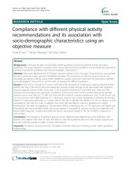
The H. <strong>pylori</strong>-mediated hummingbird phenotype<br />
involves cell elongation and migration. While noninfected<br />
gastric epithelial AGS cells (mock) show a round<br />
morphology, infection with H. <strong>pylori</strong> wild type (wt) induced<br />
loss of cell-to-cell contacts, cell elongation and migration.<br />
The elongated cell morphology in response to H. <strong>pylori</strong> is<br />
dependent on the injection of CagA, since AGS cells infected<br />
with a cagA-deficient H. <strong>pylori</strong> mutant (ΔcagA) do not elongate.<br />
FAs are comprised of α and β integrin heterodimers that<br />
form a bridge between the intracellular actin cytoskeleton<br />
and the extracellular matrix (ECM) [17]. While the extracellular<br />
domain of integrins binds directly to ECM proteins,<br />
the cytoplasmic tail is linked to the actin<br />
cytoskeleton via a steadily increasing number of signaling<br />
and adapter proteins, such as <strong>focal</strong> adhesion kinase (FAK),<br />
vinculin, talin and paxillin. [17]. Initially, integrins were<br />
presumed to act simply as cell adhesion receptors, but it<br />
has become clear that they also play crucial roles in the<br />
<strong>communication</strong> of cellular signal transduction pathways<br />
leading to adhesion and rearrangement of the actin<br />
cytoskeleton [14,18]. The integrity and stability of proteins<br />
located in the FA site are mainly regulated by tyrosine<br />
phosphorylation, which is controlled by classical<br />
"outside in" and "inside out" signaling cascades [19].<br />
However, cell migration does not only require a coordinated<br />
assembly and disassembly of FAs to move the cell<br />
body on the ECM, but also needs active actin polymerization<br />
along the plasma membrane to contract the cell cortex.<br />
The Rho family of small GTPases, including the well<br />
studied members Cdc42, Rac1 and RhoA, is a key regulator<br />
of the cytoskeleton (Fig. 2A). Generation of cortical<br />
tension and a rounded cell morphology are critically controlled<br />
by FAK, Src and the small GTPase RhoA, while<br />
Rac1 and Cdc42 direct actin assembly to generate lamellipodia<br />
and elongation [20,21].<br />
Besides Rho-GTPase-controlled actin rearrangements, cell<br />
morphology might also be influenced by deregulated FAbased<br />
cell adhesion resulting in the development of high<br />
traction forces on both cell poles and in a drastic cell elongation<br />
(Fig. 2A). Indeed, video microscopy has revealed<br />
that cells infected with H. <strong>pylori</strong> fail to release their back<br />
ends during cell migration, probably through the maintenance<br />
of vinculin-containing FA complexes at their distal<br />
tips [22]. This observation indicates that H. <strong>pylori</strong> causes a<br />
FA-dependent retraction defect of motile cells leading to<br />
the elongated cell morphology, the molecular mechanism<br />
of which will be discussed below.<br />
H. <strong>pylori</strong> injects CagA across FAs and regulates the<br />
elongation of infected epithelial cells<br />
Like the growth factor-induced scattering phenotype, the<br />
hummingbird phenotype involves an altered cell morphology<br />
and migration, which are regulated by H. <strong>pylori</strong>.<br />
Injection of CagA is strongly associated with the development<br />
of an elongated cell morphology, while the process<br />
of migration requires a functional T4SS system, but not<br />
the CagA protein itself [23-25]. These observations led to<br />
the speculation that either another H. <strong>pylori</strong> factor translocates<br />
through the T4SS into <strong>host</strong> cells or that the T4SS<br />
pilus directly interacts with an unknown cell surface<br />
receptor to stimulate specific signal transduction pathways<br />
leading to the motogenic response of H. <strong>pylori</strong>-colonized<br />
epithelial cells [26]. Indeed, peptidoglycan has been<br />
shown to translocate into the <strong>host</strong> cytoplasm via the T4SS<br />
Page 2 of 7<br />
(page number not for citation purposes)