Appendicular skeleton and dermal armour of the Late Cretaceous ...
Appendicular skeleton and dermal armour of the Late Cretaceous ...
Appendicular skeleton and dermal armour of the Late Cretaceous ...

You also want an ePaper? Increase the reach of your titles
YUMPU automatically turns print PDFs into web optimized ePapers that Google loves.
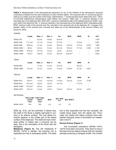
DÍEZ DÍAZ, PEREDA SUBERBIOLA, & SANZ: TITANOSAUR SKELETON<br />
TABLE 5. Measurements <strong>of</strong> <strong>the</strong> best-preserved specimens (in cm) <strong>of</strong> <strong>the</strong> hindlimb <strong>of</strong> <strong>the</strong> titanosaurian sauropod<br />
Lirainosaurus astibiae from <strong>the</strong> <strong>Late</strong> <strong>Cretaceous</strong> <strong>of</strong> Laño (nor<strong>the</strong>rn Spain). The measures with a ≈ symbol are approximate<br />
as iron oxides cover <strong>the</strong> surface <strong>of</strong> <strong>the</strong> bones. Abbreviations: **: eroded proximal <strong>and</strong>/or distal ends; ECC = femoral<br />
mid-shaft width/femoral anteroposterior width (Wilson <strong>and</strong> Carrano, 1999); Max. : maximum diameter in <strong>the</strong><br />
narrowest part <strong>of</strong> <strong>the</strong> diaphysis; Max. MLW D/Prx: maximum mediolateral width <strong>of</strong> <strong>the</strong> distal/proximal end; MDW: maximum<br />
width <strong>of</strong> <strong>the</strong> distal end; Min. : minimum diameter in <strong>the</strong> narrowest part <strong>of</strong> <strong>the</strong> diaphysis; MLW: mediolateral width;<br />
MPW: maximum width <strong>of</strong> <strong>the</strong> proximal end; Per: perimeter in <strong>the</strong> narrowest part <strong>of</strong> <strong>the</strong> diaphysis; RI = average <strong>of</strong> <strong>the</strong><br />
greatest widths <strong>of</strong> <strong>the</strong> proximal end, mid-shaft <strong>and</strong> distal end <strong>of</strong> <strong>the</strong> element/length <strong>of</strong> <strong>the</strong> element (Wilson <strong>and</strong><br />
Upchurch, 2003).<br />
FEMORA<br />
Length Max. Min. Per. MPW MDW RI ECC<br />
MCNA 1817 - 9,2 cm 4,8 cm 24,8 cm - - - 1.916<br />
MCNA 3160 71,1 cm** 11,1 cm 4,3 cm 27,7 cm - 17,5 cm - 2.581<br />
MCNA 7468 68,2 cm 9,1 cm 5,4 cm 25,2 cm 18,3 cm 17,6 cm 0.219 1.685<br />
MCNA 14465 68 cm 9,2 cm 4,3 cm 24,1 cm 15,9 cm 17,7 cm 0.209 2.139<br />
MCNA 14466 81,6 cm 12,1 cm 5,3 cm 33 cm 22,5 cm - - 2.283<br />
MCNA 14468 75 cm** 13,8 cm 5,2 cm 35,2 cm 23,6 cm** - - 2.654<br />
TIBIAE<br />
Length Max. Min. Per. MPW MDW RI<br />
MCNA 2203 - 7,7 cm 4,1 cm 21,4 cm - 10,2 cm -<br />
MCNA 13860 41,2 cm 5,7 cm 3,3 cm 17,6 cm 13,9 cm 9,7 cm 0.237<br />
FIBULAE<br />
Length Max. Min. Per. MPW MDW RI<br />
MCNA 7472 55 cm** ≈ 5 cm ≈ 3,5 cm ≈ 15 cm 10,2 cm 7,3 cm 0.136<br />
MCNA 9410 51,4 cm** 4,6 cm 3,8 cm 14,1 cm - ≈ 7,3 cm -<br />
MCNA 14469 47,5 cm** 6,6 cm 3,6 cm - - - -<br />
MCNA 14471 42,3 cm 4,4 cm 2,8 cm 13,5 cm 10,3 cm 6,5 cm 0.167<br />
METATARSAL<br />
Total Length<br />
(medial<br />
view)<br />
Total Length<br />
(lateral<br />
view)<br />
MLW<br />
Max. MLW<br />
Prx<br />
Max. MLW D<br />
MCNA 14474 19.4 19.8 4.9 1.8 1.9<br />
2008, fig. 15.E), can be confirmed. In lateral view,<br />
<strong>the</strong> shaft <strong>of</strong> <strong>the</strong> fibula is slightly sigmoidal to concave<br />
in its anterior surface. The oval lateral trochanter<br />
appears in <strong>the</strong> middle part <strong>of</strong> <strong>the</strong> lateral<br />
surface <strong>of</strong> <strong>the</strong> diaphysis. The distal end has a triangular<br />
pr<strong>of</strong>ile. In medial view, a concavity can be<br />
seen; this is <strong>the</strong> articulation <strong>of</strong> <strong>the</strong> fibula with <strong>the</strong><br />
astragalus.<br />
Metatarsal (Figure 6). The left metatarsal III<br />
(MCNA 14474) is slender; <strong>the</strong> proximal end is<br />
exp<strong>and</strong>ed <strong>and</strong> has a rectangular pr<strong>of</strong>ile. The distal<br />
end is less exp<strong>and</strong>ed <strong>and</strong> has two condyles, <strong>the</strong><br />
medial being larger than <strong>the</strong> lateral one. Proximally,<br />
<strong>the</strong> medial <strong>and</strong> lateral surfaces have welldefined<br />
triangular areas <strong>of</strong> articulation for metatarsals<br />
II <strong>and</strong> IV.<br />
Dermal Armour (Figure 7)<br />
Two incomplete osteoderms (MCNA 14473,<br />
14474) have been recovered. They seem to have a<br />
flat base <strong>and</strong> two lateral surfaces that join dorsally,<br />
perhaps producing a triangular pr<strong>of</strong>ile, one <strong>of</strong> <strong>the</strong>m<br />
10