MAxILLARY SINUS ELEvATION BY LATERAL WINDOW ...
MAxILLARY SINUS ELEvATION BY LATERAL WINDOW ...
MAxILLARY SINUS ELEvATION BY LATERAL WINDOW ...

Create successful ePaper yourself
Turn your PDF publications into a flip-book with our unique Google optimized e-Paper software.
Journal of evidence-based dental practice Special Issue—Periodontal and Implant Treatment<br />
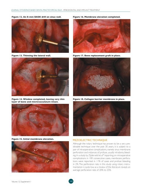
Figure 12. An 8-mm DASK drill on sinus wall.<br />
Figure 16. Membrane elevation completed.<br />
Figure 13. Thinning the lateral wall.<br />
Figure 17. Bone replacement graft in place.<br />
Figure 14. Window completed, leaving very thin<br />
layer of bone and microvasculature intact.<br />
Figure 18. Collagen barrier membrane in place.<br />
Figure 15. Initial membrane elevation.<br />
Piezoelectric Technique<br />
Although the rotary technique has proven to be a very predictable<br />
technique over the past 30 years, it is subject to a<br />
pair of intraoperative complications, namely sinus membrane<br />
perforation and instances of profuse, usually intrabony, bleeding.<br />
In a study by Zijderveld et al 49 reporting on intraoperative<br />
complications in 100 consecutive cases, membrane perforations<br />
were reported in 11% of cases and profuse bleeding<br />
in 2%. The perforation rate in this study using rotary instrumentation<br />
is quite low, as a review of the literature reveals an<br />
average perforation rate of 20% to 25%.<br />
Volume 12, Supplement 1<br />
168