Maxillary sinus vascular anatomy and its relation to sinus lift surgery
Maxillary sinus vascular anatomy and its relation to sinus lift surgery
Maxillary sinus vascular anatomy and its relation to sinus lift surgery

Create successful ePaper yourself
Turn your PDF publications into a flip-book with our unique Google optimized e-Paper software.
Rosano et al Haemorrhage risk during <strong>sinus</strong> <strong>surgery</strong><br />
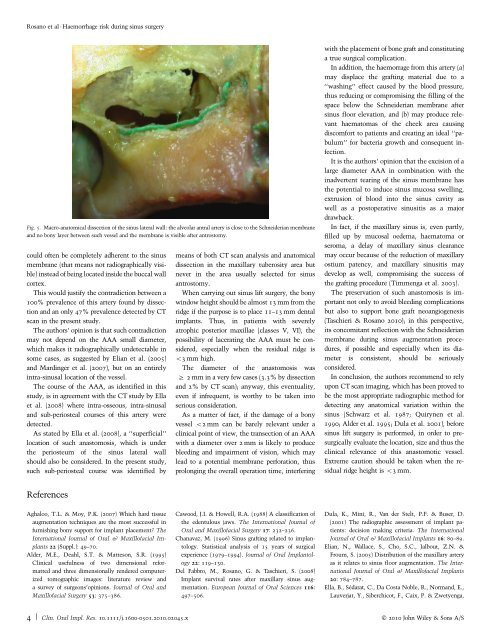
Fig. 5. Macro-ana<strong>to</strong>mical dissection of the <strong>sinus</strong> lateral wall: the alveolar antral artery is close <strong>to</strong> the Schneiderian membrane<br />
<strong>and</strong> no bony layer between such vessel <strong>and</strong> the membrane is visible after antros<strong>to</strong>my.<br />
could often be completely adherent <strong>to</strong> the <strong>sinus</strong><br />
membrane (that means not radiographically visible)<br />
instead of being located inside the buccal wall<br />
cortex.<br />
This would justify the contradiction between a<br />
100% prevalence of this artery found by dissection<br />
<strong>and</strong> an only 47% prevalence detected by CT<br />
scan in the present study.<br />
The authors’ opinion is that such contradiction<br />
may not depend on the AAA small diameter,<br />
which makes it radiographically undetectable in<br />
some cases, as suggested by Elian et al. (2005)<br />
<strong>and</strong> Mardinger et al. (2007), but on an entirely<br />
intra-<strong>sinus</strong>al location of the vessel.<br />
The course of the AAA, as identified in this<br />
study, is in agreement with the CT study by Ella<br />
et al. (2008) where intra-osseous, intra-<strong>sinus</strong>al<br />
<strong>and</strong> sub-periosteal courses of this artery were<br />
detected.<br />
As stated by Ella et al. (2008), a ‘‘superficial’’<br />
location of such anas<strong>to</strong>mosis, which is under<br />
the periosteum of the <strong>sinus</strong> lateral wall<br />
should also be considered. In the present study,<br />
such sub-periosteal course was identified by<br />
means of both CT scan analysis <strong>and</strong> ana<strong>to</strong>mical<br />
dissection in the maxillary tuberosity area but<br />
never in the area usually selected for <strong>sinus</strong><br />
antros<strong>to</strong>my.<br />
When carrying out <strong>sinus</strong> <strong>lift</strong> <strong>surgery</strong>, the bony<br />
window height should be almost 13 mm from the<br />
ridge if the purpose is <strong>to</strong> place 11–13 mm dental<br />
implants. Thus, in patients with severely<br />
atrophic posterior maxillae (classes V, VI), the<br />
possibility of lacerating the AAA must be considered,<br />
especially when the residual ridge is<br />
o3mmhigh.<br />
The diameter of the anas<strong>to</strong>mosis was<br />
2 mm in a very few cases (3.3% by dissection<br />
<strong>and</strong> 2% by CT scan); anyway, this eventuality,<br />
even if infrequent, is worthy <strong>to</strong> be taken in<strong>to</strong><br />
serious consideration.<br />
As a matter of fact, if the damage of a bony<br />
vessel o2 mm can be barely relevant under a<br />
clinical point of view, the transection of an AAA<br />
with a diameter over 2 mm is likely <strong>to</strong> produce<br />
bleeding <strong>and</strong> impairment of vision, which may<br />
lead <strong>to</strong> a potential membrane perforation, thus<br />
prolonging the overall operation time, interfering<br />
with the placement of bone graft <strong>and</strong> constituting<br />
atruesurgicalcomplication.<br />
In addition, the haemorrage from this artery (a)<br />
may displace the grafting material due <strong>to</strong> a<br />
‘‘washing’’ effect caused by the blood pressure,<br />
thus reducing or compromising the filling of the<br />
space below the Schneiderian membrane after<br />
<strong>sinus</strong> floor elevation, <strong>and</strong> (b) may produce relevant<br />
haema<strong>to</strong>mas of the cheek area causing<br />
discomfort <strong>to</strong> patients <strong>and</strong> creating an ideal ‘‘pabulum’’<br />
for bacteria growth <strong>and</strong> consequent infection.<br />
It is the authors’ opinion that the excision of a<br />
large diameter AAA in combination with the<br />
inadvertent tearing of the <strong>sinus</strong> membrane has<br />
the potential <strong>to</strong> induce <strong>sinus</strong> mucosa swelling,<br />
extrusion of blood in<strong>to</strong> the <strong>sinus</strong> cavity as<br />
well as a pos<strong>to</strong>perative <strong>sinus</strong>itis as a major<br />
drawback.<br />
In fact, if the maxillary <strong>sinus</strong> is, even partly,<br />
filled up by mucosal oedema, haema<strong>to</strong>ma or<br />
seroma, a delay of maxillary <strong>sinus</strong> clearance<br />
may occur because of the reduction of maxillary<br />
ostium patency, <strong>and</strong> maxillary <strong>sinus</strong>itis may<br />
develop as well, compromising the success of<br />
the grafting procedure (Timmenga et al. 2003).<br />
Thepreservationofsuchanas<strong>to</strong>mosisisimportant<br />
not only <strong>to</strong> avoid bleeding complications<br />
but also <strong>to</strong> support bone graft neoangiogenesis<br />
(Taschieri & Rosano 2010); in this perspective,<br />
<strong>its</strong> concomitant reflection with the Schneiderian<br />
membrane during <strong>sinus</strong> augmentation procedures,<br />
if possible <strong>and</strong> especially when <strong>its</strong> diameter<br />
is consistent, should be seriously<br />
considered.<br />
In conclusion, the authors recommend <strong>to</strong> rely<br />
upon CT scan imaging, which has been proved <strong>to</strong><br />
be the most appropriate radiographic method for<br />
detecting any ana<strong>to</strong>mical variation within the<br />
<strong>sinus</strong> (Schwarz et al. 1987; Quirynen et al.<br />
1990; Alder et al. 1995; Dula et al. 2001), before<br />
<strong>sinus</strong> <strong>lift</strong> <strong>surgery</strong> is performed, in order <strong>to</strong> presurgically<br />
evaluate the location, size <strong>and</strong> thus the<br />
clinical relevance of this anas<strong>to</strong>motic vessel.<br />
Extreme caution should be taken when the residual<br />
ridge height is o3mm.<br />
References<br />
Aghaloo, T.L. & Moy, P.K. (2007) Which hard tissue<br />
augmentation techniques are the most successful in<br />
furnishing bony support for implant placement? The<br />
International Journal of Oral & Maxillofacial Implants<br />
22 (Suppl.): 49–70.<br />
Alder, M.E., Deahl, S.T. & Matteson, S.R. (1995)<br />
Clinical usefulness of two dimensional reformatted<br />
<strong>and</strong> three dimensionally rendered computerized<br />
<strong>to</strong>mographic images: literature review <strong>and</strong><br />
a survey of surgeons’opinions. Journal of Oral <strong>and</strong><br />
Maxillofacial Surgery 53: 375–386.<br />
Cawood, J.I. & Howell, R.A. (1988) A classification of<br />
the edentulous jaws. The International Journal of<br />
Oral <strong>and</strong> Maxillofacial Surgery 17: 232–236.<br />
Chanavaz, M. (1996) Sinus grafting related <strong>to</strong> implan<strong>to</strong>logy.<br />
Statistical analysis of 15 years of surgical<br />
experience (1979–1994). Journal of Oral Implan<strong>to</strong>logy<br />
22: 119–130.<br />
Del Fabbro, M., Rosano, G. & Taschieri, S. (2008)<br />
Implant survival rates after maxillary <strong>sinus</strong> augmentation.<br />
European Journal of Oral Sciences 116:<br />
497–506.<br />
Dula, K., Mini, R., Van der Stelt, P.F. & Buser, D.<br />
(2001) The radiographic assessment of implant patients:<br />
decision making criteria. The International<br />
Journal of Oral & Maxillofacial Implants 16: 80–89.<br />
Elian, N., Wallace, S., Cho, S.C., Jalbout, Z.N. &<br />
Froum, S. (2005) Distribution of the maxillary artery<br />
as it relates <strong>to</strong> <strong>sinus</strong> floor augmentation. The International<br />
Journal of Oral & Maxillofacial Implants<br />
20: 784–787.<br />
Ella, B., Sédarat, C., Da Costa Noble, R., Norm<strong>and</strong>, E.,<br />
Lauverjat, Y., Siberchicot, F., Caix, P. & Zwetyenga,<br />
4 | Clin. Oral Impl. Res. 10.1111/j.1600-0501.2010.02045.x c 2010 John Wiley & Sons A/S