AVIDITY TEST FOR IgG ANTIBODIES
AVIDITY TEST FOR IgG ANTIBODIES
AVIDITY TEST FOR IgG ANTIBODIES

Create successful ePaper yourself
Turn your PDF publications into a flip-book with our unique Google optimized e-Paper software.
322 ZDRAV VESTN 2001; 70<br />
or other stages of infection. One of these tests could be the<br />
avidity test of specific <strong>IgG</strong> antibodies or Western blot.<br />
Avidity tells about the strength of antibodies’ binding to multivalent<br />
antigen. Avidity can be measured after short incubation<br />
of the antigen-antibody complex with 6 M urea (5). High<br />
avidity antibodies persist in binding to the antigen, while low<br />
avidity antibodies are destroyed after the addition of urea.<br />
In this study we’ve measured the avidity of <strong>IgG</strong> antibodies<br />
(anti-EA, anti-VCA, anti-EBNA) in sera of patients previously<br />
examined by ELISA to define their immune status to EBV.<br />
The results of »in house« avidity test were compared to the<br />
results obtained by commercially available test kit, oriented to<br />
measure avidity of <strong>IgG</strong> anti-EBV.<br />
Material and methods<br />
Sera of patients. In this study 74 sera of patients previously<br />
examined for EBV immune status (4, 7) were included for<br />
measuring <strong>IgG</strong> antibodies’ avidity. According to their anti-EBV<br />
status sera were divided into four groups: patients’ sera with<br />
acute EBV infection (n = 22), recent infection (n = 18), past<br />
infection (n = 11) and with virus reactivation (n = 23).<br />
»In house« avidity test. All sera were repetedly tested by ELISA.<br />
After the incubation of sera with specific antigens, the plates<br />
were washed; in each well 100 µl of 6 M urea was added for 3<br />
mins at room temperature. Urea was removed, the plates were<br />
washed again and ELISA was continued as described elsewhere<br />
(5, 7).<br />
Avidity test with commercial kit – Enzygnost Anti-EBV <strong>IgG</strong>;<br />
Avidity Reagent for Enzygnost (Dade Behring, Marburg, Germany).<br />
This test was performed following instructions of the<br />
manufacturer (8).<br />
Measurement of avidity. Avidity of <strong>IgG</strong> antibodies is expressed<br />
by the avidity index (AI). In »in house« avidity test AI was obtained<br />
when optical density (OD) of the sample, treated with<br />
urea, was devided by OD of the same sample which was not<br />
treated with urea. When AI is 0.50–1.00, it means high avidity<br />
of <strong>IgG</strong> antibodies and this is characteristic for past infection.<br />
When AI is < 0.25, it means low avidity of <strong>IgG</strong> antibodies and<br />
this reflects acute infection. AI values of 0.25–0.50 are equivocal<br />
(5).<br />
By the use of commercial test kit, AI was determined in the<br />
same way as in »in house« avidity test; the only difference was<br />
that the quocient was multiplied by 100. So AI was expressed<br />
in percentages. Cut off value was 40% (8).<br />
Statistics. The results were statistically evaluated in different<br />
EBV infections by χ 2 test.<br />
Results<br />
»In house« avidity test<br />
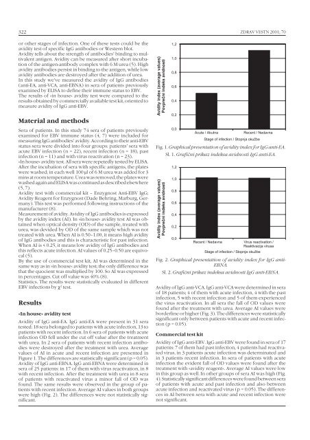
Avidity of <strong>IgG</strong> anti-EA. <strong>IgG</strong> anti-EA were present in 31 sera<br />
tested: 18 sera belonged to patients with acute infection, 13 to<br />
patients with recent infection. In 6 sera of patients with acute<br />
infection OD fell under the cut off value after the treatment<br />
with urea. In 2 sera of patients with recent infection antibodies<br />
were destroyed after the treatment with urea. Average<br />
values of AI in acute and recent infection are presented in<br />
Figure 1. The differences are statistically significant (p = 0.05).<br />
Avidity of <strong>IgG</strong> anti-EBNA. <strong>IgG</strong> anti-EBNA were determined in<br />
sera of 25 patients: in 17 of them with virus reactivation, in 8<br />
with recent infection. After the treatment with urea in 8 sera<br />
of patients with reactivated virus a minor fall of OD was<br />
found. The same results were observed in the group of patients<br />
with recent infection. Average AI values in both groups<br />
were high (Fig. 2). The differences were not statistically significant.<br />
Avidity index (average values)<br />
Povpreèni indeks avidnosti<br />
1,2<br />
1,0<br />
0,8<br />
0,6<br />
0,4<br />
0,2<br />
0,0<br />
Acute / Akutna<br />
Recent / Nedavna<br />
Stage of infection / Stopnja okužbe<br />
Fig. 1. Graphical presentation of avidity index for <strong>IgG</strong> anti-EA.<br />
Sl. 1. Grafični prikaz indeksa avidnosti <strong>IgG</strong> anti-EA.<br />
Avidity index (average values)<br />
Povpreèni indeks avidnosti<br />
1,2<br />
1,0<br />
0,8<br />
0,6<br />
0,4<br />
0,2<br />
0,0<br />
Recent / Nedavna<br />
Virus reactivation /<br />
Reaktivacija virusa<br />
Stage of infection / Stopnja okužbe<br />
Fig. 2. Graphical presentation of avidity index for <strong>IgG</strong> anti-<br />
EBNA.<br />
Sl. 2. Grafični prikaz indeksa avidnosti <strong>IgG</strong> anti-EBNA.<br />
Avidity of <strong>IgG</strong> anti-VCA. <strong>IgG</strong> anti-VCA were determined in sera<br />
of 18 patients; 4 of them with acute infection, 4 with the past<br />
infection, 5 with recent infection and 5 of them experienced<br />
the virus reactivation. In all sera the fall of OD values were<br />
found after the treatment with urea. Average AI values were<br />
borderline or higher (Fig. 3). The differences were statistically<br />
significant only between patients with acute and recent infection<br />
(p = 0.05).<br />
Commercial test kit<br />
Avidity of <strong>IgG</strong> anti-EBV. <strong>IgG</strong> anti-EBV were found in sera of 17<br />
patients: 7 of them had past infection, 4 patients had reactivated<br />
virus, in 3 patients acute infection was determinated and<br />
in 3 patients recent infection. In sera of patients with acute<br />
infection the evident fall of OD values were found after the<br />
treatment with »avidity reagent«. Average AI values were low<br />
in this group as well. In other groups of sera AI was high (Fig.<br />
4). Statistically significant differences were found between sera<br />
of patients with acute and past infection and also between<br />
acute infection and reactivated virus (p = 0.05). The differences<br />
in AI between sera with acute and recent infection were<br />
not significant.