

2. spongiotic dermatitis with a mixed - Our Dermatology Online Journal
2. spongiotic dermatitis with a mixed - Our Dermatology Online Journal
2. spongiotic dermatitis with a mixed - Our Dermatology Online Journal

Create successful ePaper yourself
Turn your PDF publications into a flip-book with our unique Google optimized e-Paper software.
Prior studies have attempted to characterize the<br />
inflammatory infiltrates present in <strong>spongiotic</strong> <strong>dermatitis</strong><br />
[9-10]. A predominant T lymphocyte population has<br />
been reported [9-10]. In this study, our findings were in<br />
agreement <strong>with</strong> several authors because we also detected<br />
a large population of T lymphocytes. However, we also<br />
found several CD1a, factor XIIIa, HAM-56 and CD68<br />
positive cells. Consistent <strong>with</strong> our findings, other authors<br />
have also reported that immunoglobulin D may play a<br />
pathophysiologic role [11]. Interestingly, we found<br />
robust B and T lymphocyte activity, and a prominent<br />
antigen presenting cell (APC) population.<br />
Further, the reactive cell and cytokine combination has<br />
not been previously highlighted in <strong>spongiotic</strong><br />
dermatitides. The APCs participate in the initiation of the<br />
inflammatory process in various immune-mediated<br />
dermatoses, via activation of antigen specific T<br />
lymphocytes. The skin contains several different subsets<br />
of APCs. Non-professional APCs do not constitutively<br />
express the MHC class II proteins required for<br />
interaction <strong>with</strong> naive T cells; these are expressed only<br />
upon stimulation of the non-professional APC by certain<br />
cytokines such as gamma interferon (IFN-γ).<br />
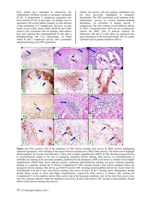
Figure 1.a CD1a positive cells in the epidermis by IHC (brown staining, blue arrow), b. H&E sections highlighting<br />
epidermal spongiosis, <strong>with</strong> widening of the spaces between keratinocytes (100X) (blue arrows). The black arrows highlight<br />
dermal papillary tip vascular microthrombi. c. Same as b, at higher magnification (400X). d. IHC showing strong positivity<br />
to myeloid/histoid antigen in the area of <strong>spongiotic</strong> epidermis (brown staining; blue arrows), in contradistinction to<br />
markedly less staining at the specimen periphery, unaffected by the spongiosis (100X) (red arrow). e. Similar to d, at higher<br />
magnification (400X) Blue arrow indicates positive epidermal myeloid/histoid staining; red arrow indicates punctuate<br />
staining in a papillary dermal tip. f. Positive Complement/C3 IHC staining around a hair follicle periphery below the<br />
spongiosis (brown staining, blue arrow). g. HAM-56 positive IHC straining around papillary dermal tip vessels where the<br />
microthrombi in b and c were seen (brown staining, blue arrow). h and i H & E staining shows inflammation around<br />
dermal blood vessels at lower and higher magnifications, respectively (blue arrows). j. Positive IHC staining for<br />
Complement/C3 in the papillary dermis (blue arrow) and at the basement membrane zone of the skin (blue arrow); also,<br />
note fine, punctate deposits <strong>with</strong>in the epidermis (red arrow). k and l. IgD positive IHC staining in deep papillary dermal<br />
blood vessels (brown staining, blue arrows).<br />
54<br />
© N Dermatol <strong>Online</strong> <strong>2.</strong>2011


