Antibacterial Activity of Thai Medicinal Plant Extracts on the Skin ...
Antibacterial Activity of Thai Medicinal Plant Extracts on the Skin ...
Antibacterial Activity of Thai Medicinal Plant Extracts on the Skin ...

Create successful ePaper yourself
Turn your PDF publications into a flip-book with our unique Google optimized e-Paper software.
evarporated to dryness in vacuo using rotary evaporator at 45°C. For c<strong>on</strong>tinuous<br />
extracti<strong>on</strong>, porti<strong>on</strong>s <str<strong>on</strong>g>of</str<strong>on</strong>g> <strong>the</strong> powdered plant materials were separately extracted until<br />
exhausted by soxhlet apparatus using n-hexane, chlor<str<strong>on</strong>g>of</str<strong>on</strong>g>orm and 95% ethanol,<br />
respectively. The extract soluti<strong>on</strong>s were filtered and <strong>the</strong>n <strong>the</strong> solvents were removed by<br />
evaporati<strong>on</strong> at 45°C to yield <strong>the</strong> n-hexane extract, chlor<str<strong>on</strong>g>of</str<strong>on</strong>g>orm extract and ethanol extract<br />
<str<strong>on</strong>g>of</str<strong>on</strong>g> four kinds <str<strong>on</strong>g>of</str<strong>on</strong>g> plant materials.<br />
For T. chebula, extracti<strong>on</strong> was d<strong>on</strong>e by c<strong>on</strong>tinuous extracti<strong>on</strong> and <strong>the</strong> solvent<br />
extracti<strong>on</strong> method adopted from Sato et al. (1997). C<strong>on</strong>tinuous extracti<strong>on</strong> was d<strong>on</strong>e by<br />
using methanol as solvent. For solvent extracti<strong>on</strong> method (Sato et al., 1997), <strong>the</strong> powder<br />
<str<strong>on</strong>g>of</str<strong>on</strong>g> T. chebula was extracted three times with 50% ethanol under reflux for 2 hours. The<br />
extract soluti<strong>on</strong>s were combined, c<strong>on</strong>centrated in vacuo to evaporate all ethanol and <strong>the</strong>n<br />
extracted three times with diethyl e<strong>the</strong>r and n-butanol, respectively. The combined <str<strong>on</strong>g>of</str<strong>on</strong>g><br />
each extract soluti<strong>on</strong>s was evaporated or freeze dried to dryness (Table 1).<br />
Test Organisms<br />
The test organisms used, S. aureus (ATCC 25923) and E. coli (ATCC 25922),<br />
were obtained from <strong>the</strong> Department <str<strong>on</strong>g>of</str<strong>on</strong>g> Microbiology, Faculty <str<strong>on</strong>g>of</str<strong>on</strong>g> Medicine, Chiang Mai<br />
University.<br />
The inoculum size <str<strong>on</strong>g>of</str<strong>on</strong>g> each test strain was standardized according to <strong>the</strong> Committee<br />
for Clinical Laboratory Standard (NCCLS, 1993). The test bacterial strain was inoculated<br />
into Mueller Hint<strong>on</strong> broth, MHB (Oxoid) medium and incubated for 3-6 hrs at 35°C until<br />
<strong>the</strong> culture attained a turbidity <str<strong>on</strong>g>of</str<strong>on</strong>g> 0.5 McFarland unit. The final inoculum was adjusted to<br />
5 x 10 5 cfu/ml.<br />
Screening for <str<strong>on</strong>g>Antibacterial</str<strong>on</strong>g> <str<strong>on</strong>g>Activity</str<strong>on</strong>g><br />
The extracts were evaluated for antibacterial activity by agar well diffusi<strong>on</strong><br />
method. A molten 6 ml MHA was poured in a sterile petri dish. After agar hardening, four<br />
cups <str<strong>on</strong>g>of</str<strong>on</strong>g> 12 mm were placed. 0.1 ml <str<strong>on</strong>g>of</str<strong>on</strong>g> <strong>the</strong> standard suspensi<strong>on</strong> (5 x 10 5 cfu/ml) <str<strong>on</strong>g>of</str<strong>on</strong>g> each<br />
test bacterial strain was inoculated to molten 14 ml MHA (about 50°C) and <strong>the</strong>n <strong>the</strong><br />
mixture was poured in <strong>the</strong> prepared petri dish. After agar hardening and cup removing,<br />
each well was applied with 0.1 ml <str<strong>on</strong>g>of</str<strong>on</strong>g> each 0.1% extract. The solvents were used as<br />
c<strong>on</strong>trol. The applied plates were incubated at 35°C for 18-24 hrs and <strong>the</strong>n inhibiti<strong>on</strong> z<strong>on</strong>e<br />
diameters (IZD) were measured.<br />
The minimal inhibitory c<strong>on</strong>centrati<strong>on</strong> (MIC) and minimal bactericidal<br />
c<strong>on</strong>centrati<strong>on</strong> (MBC) were determined for <strong>the</strong> extracts that have inhibiti<strong>on</strong> z<strong>on</strong>e diameter<br />
more than 20 mm. The MIC was determined by macrobroth diluti<strong>on</strong> method and<br />
compared with commercial antibiotic Grammicin (gentamicin sulfate) as positive c<strong>on</strong>trol.<br />
Each extract was rec<strong>on</strong>stitued with its solvent, <strong>the</strong>n two-fold serial diluti<strong>on</strong>s <str<strong>on</strong>g>of</str<strong>on</strong>g> <strong>the</strong><br />
extracts and c<strong>on</strong>trol drug were prepared in tubes with MHB as diluent. Each diluti<strong>on</strong> was<br />
seeded with test organism to <strong>the</strong> standard c<strong>on</strong>centrati<strong>on</strong> (5 x 10 5 cfu/ml). The MIC was<br />
taken as <strong>the</strong> last diluti<strong>on</strong> showing no noticeable growth (turbidity).<br />
The MBC determinati<strong>on</strong> was determined by subculturing 10 µl <str<strong>on</strong>g>of</str<strong>on</strong>g> <strong>the</strong> culture<br />
medium from each tube (in macrobroth MIC assay) showing no apparent growth <strong>on</strong> MHA<br />
and incubated at 35°C for 18-24 hrs. The MBC was read as <strong>the</strong> least c<strong>on</strong>centrati<strong>on</strong><br />
showing <strong>the</strong> growth <str<strong>on</strong>g>of</str<strong>on</strong>g> not more than five col<strong>on</strong>ies <str<strong>on</strong>g>of</str<strong>on</strong>g> each organism (99.9% were dead).<br />
Phytochemical Screening<br />
The high potential fracti<strong>on</strong>, PG-1, was preliminary studied for phytochemical<br />
c<strong>on</strong>stituents using standard method.<br />
RESULTS AND DISCUSSION<br />
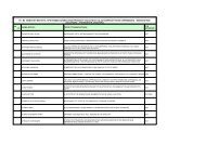
In this present study, five <str<strong>on</strong>g>Thai</str<strong>on</strong>g> medicinal plants were separately extracted with<br />
various methods to yield 21 kinds <str<strong>on</strong>g>of</str<strong>on</strong>g> extracts. All extracts were evaluated for <strong>the</strong>ir<br />
antibacterial activity against S. aureus and E. coli, representing gram positive and gram<br />
negative bacteria. The results are shown in Table 1 and 2. Three out <str<strong>on</strong>g>of</str<strong>on</strong>g> 21 kinds <str<strong>on</strong>g>of</str<strong>on</strong>g><br />
154