Toxoplasmosis presenting as a swelling in the axillary tail of the ...
Toxoplasmosis presenting as a swelling in the axillary tail of the ...
Toxoplasmosis presenting as a swelling in the axillary tail of the ...

Create successful ePaper yourself
Turn your PDF publications into a flip-book with our unique Google optimized e-Paper software.
Siriwardana et al. Journal <strong>of</strong> Medical C<strong>as</strong>e Reports 2011, 5:348<br />
http://www.jmedicalc<strong>as</strong>ereports.com/content/5/1/348<br />
Page 2 <strong>of</strong> 4<br />
diagnosis w<strong>as</strong> considered to be a malignant lesion <strong>in</strong> <strong>the</strong><br />
left bre<strong>as</strong>t with met<strong>as</strong>tatic <strong>in</strong>volvement <strong>of</strong> an <strong>axillary</strong><br />
lymph node.<br />
She underwent ultr<strong>as</strong>ound and mammographic exam<strong>in</strong>ations<br />
<strong>of</strong> her bre<strong>as</strong>ts. The mammogram showed a<br />
smooth-outl<strong>in</strong>ed, s<strong>of</strong>t-density lesion <strong>in</strong> her left bre<strong>as</strong>t<br />
with no microcalcifications and a few small lymph<br />
nodes <strong>in</strong> her left <strong>axillary</strong> <strong>tail</strong>. Ultr<strong>as</strong>ound revealed that<br />
<strong>the</strong> palpable lump <strong>in</strong> <strong>the</strong> lateral part <strong>of</strong> her left bre<strong>as</strong>t<br />
w<strong>as</strong> a 2 cm solid lesion with reduced echogenicity. The<br />
o<strong>the</strong>r nodule, <strong>in</strong> <strong>the</strong> upper part <strong>of</strong> <strong>the</strong> left axilla, w<strong>as</strong><br />
also solid (1 cm) and suggestive <strong>of</strong> a lymph node (M4<br />
U4; that is, suspicious abnormality accord<strong>in</strong>g to <strong>the</strong><br />
Bre<strong>as</strong>t Imag<strong>in</strong>g Report<strong>in</strong>g and Data System, or BIRADS).<br />
The radiological appearance w<strong>as</strong> highly suggestive <strong>of</strong> a<br />
lymphoma. Then she underwent targeted f<strong>in</strong>e-needle<br />
<strong>as</strong>piration cytology (FNAC) <strong>of</strong> <strong>the</strong> <strong>axillary</strong> lesion and<br />
core needle biopsy <strong>of</strong> <strong>the</strong> bre<strong>as</strong>t lesion. The FNAC w<strong>as</strong><br />
<strong>in</strong>determ<strong>in</strong>ate (C3) but showed numerous monotonous<br />
lymphocytes <strong>in</strong> a background conta<strong>in</strong><strong>in</strong>g lymphogranular<br />
bodies suggestive <strong>of</strong> granulomatous <strong>in</strong>flammation<br />
such <strong>as</strong> toxopl<strong>as</strong>mosis. There were no malignant cells.<br />
The core biopsy showed a small aggregate <strong>of</strong> epi<strong>the</strong>leoid<br />
histiocytes and mult<strong>in</strong>uclear giant cells <strong>in</strong> keep<strong>in</strong>g with<br />
granulomatous <strong>in</strong>flammation. There w<strong>as</strong> no evidence <strong>of</strong><br />
a malignancy.<br />
Her c<strong>as</strong>e w<strong>as</strong> discussed at <strong>the</strong> multidiscipl<strong>in</strong>ary meet<strong>in</strong>g,<br />
and <strong>the</strong> team recommended a wide local excision <strong>of</strong><br />
<strong>the</strong> bre<strong>as</strong>t lesion with palpable <strong>axillary</strong> lymph node<br />
biopsy. The results <strong>of</strong> a histological exam<strong>in</strong>ation (Figures<br />
1 and 2) <strong>of</strong> <strong>the</strong> resected specimens <strong>of</strong> bre<strong>as</strong>t and<br />
<strong>axillary</strong> lesions were suggestive <strong>of</strong> an <strong>in</strong>tramammary and<br />
<strong>axillary</strong> lymph node m<strong>as</strong>s with marked follicular<br />
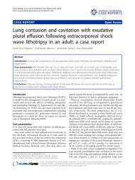
Figure 1 A microscopic exam<strong>in</strong>ation <strong>of</strong> <strong>the</strong> specimens <strong>of</strong><br />
bre<strong>as</strong>t (<strong>axillary</strong> <strong>tail</strong>) lump and <strong>axillary</strong> lymph node shows<br />
marked follicular hyperpl<strong>as</strong>ia with prom<strong>in</strong>ent small granulom<strong>as</strong><br />
composed almost entirely <strong>of</strong> epi<strong>the</strong>lioid cells.<br />
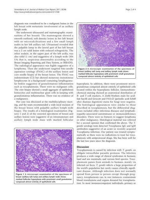
Figure 2 A microscopic exam<strong>in</strong>ation <strong>of</strong> <strong>the</strong> specimens <strong>of</strong><br />
bre<strong>as</strong>t (<strong>axillary</strong> <strong>tail</strong>) lump and <strong>axillary</strong> lymph node shows<br />
marked follicular hyperpl<strong>as</strong>ia with prom<strong>in</strong>ent small granulom<strong>as</strong><br />
composed almost entirely <strong>of</strong> epi<strong>the</strong>lioid cells.<br />
hyperpl<strong>as</strong>ia. In addition, <strong>the</strong>re were prom<strong>in</strong>ent microgranulom<strong>as</strong><br />
composed almost entirely <strong>of</strong> epi<strong>the</strong>lioid cells<br />
located with<strong>in</strong> <strong>the</strong> hyperpl<strong>as</strong>tic follicles. Immunohistochemical<br />
sta<strong>in</strong><strong>in</strong>g showed an anatomical distribution <strong>of</strong><br />
B- and T-cell markers. A Ziehl-Neelsen sta<strong>in</strong> for acidf<strong>as</strong>t<br />
bacilli and Grocott and PAS+D (periodic acid-Schiff<br />
after di<strong>as</strong>t<strong>as</strong>e digestion) sta<strong>in</strong>s for fungi were negative.<br />
The histological appearances were similar to those<br />
described <strong>in</strong> toxopl<strong>as</strong>mosis, but <strong>the</strong> differential diagnoses<br />
<strong>in</strong>cluded o<strong>the</strong>r <strong>in</strong>fectious dise<strong>as</strong>es and lymphadenopathy-<strong>as</strong>sociated<br />
autoimmune or immunodeficiency<br />
disorders. There were no features to suggest lymphoma<br />
or o<strong>the</strong>r malignancy. Histological material w<strong>as</strong> referred<br />
for a second op<strong>in</strong>ion that confirmed <strong>the</strong> above. The T.<br />
gondii serology tests detected Toxopl<strong>as</strong>ma IgG and IgM<br />
antibodies suggestive <strong>of</strong> an acute or recently acquired<br />
Toxopl<strong>as</strong>ma <strong>in</strong>fection. Our patient w<strong>as</strong> treated symptomatically<br />
<strong>as</strong> <strong>the</strong>re were no <strong>in</strong>dications to treat her toxoplamosis<br />
with antiprotozoal drugs. She h<strong>as</strong> been well for<br />
<strong>the</strong> l<strong>as</strong>t two years s<strong>in</strong>ce <strong>the</strong> diagnosis.<br />
Discussion<br />
<strong>Toxopl<strong>as</strong>mosis</strong> is caused by <strong>in</strong>fection with T. gondii, an<br />
obligate <strong>in</strong>tracellular par<strong>as</strong>itic protozoa. The <strong>in</strong>fection<br />
produces a wide range <strong>of</strong> cl<strong>in</strong>ical syndromes <strong>in</strong> humans,<br />
land and sea mammals, and various bird species. <strong>Toxopl<strong>as</strong>mosis</strong><br />
p<strong>as</strong>ses from animals to humans, ma<strong>in</strong>ly via<br />
<strong>in</strong>fected cat feces. T. gondii <strong>in</strong>fects a large proportion <strong>of</strong><br />
<strong>the</strong> world’s population but rarely causes cl<strong>in</strong>ically significant<br />
dise<strong>as</strong>e. Although <strong>in</strong>fection does not normally<br />
spread from person to person except through pregnancy,<br />
toxopl<strong>as</strong>mosis can, <strong>in</strong> rare <strong>in</strong>stances, contam<strong>in</strong>ate<br />
blood transfusions and organs donated for transplantation.<br />
In most immunocompetent <strong>in</strong>dividuals, primary or