PDF - Journal of Medical Case Reports
PDF - Journal of Medical Case Reports
PDF - Journal of Medical Case Reports

Create successful ePaper yourself
Turn your PDF publications into a flip-book with our unique Google optimized e-Paper software.
Klimek et al. <strong>Journal</strong> <strong>of</strong> <strong>Medical</strong> <strong>Case</strong> <strong>Reports</strong> 2012, 6:419 Page 2 <strong>of</strong> 3<br />
http://www.jmedicalcasereports.com/content/6/1/419<br />
Blood and urine tests were normal. Transabdominal ultrasound<br />
with a low-filled bladder detected an 8cm × 6.5cm<br />
hypoechoic mass in the right iliac fossa without perfusion,<br />
interpreted as a uterine malformation, a hematocolpos or<br />
an ovarian torsion.<br />
Due to the risk <strong>of</strong> an underlying ovarian torsion,<br />
the patient was taken to theater for vaginoscopy<br />
under anesthesia and, in the absence <strong>of</strong> any pathologic<br />
findings, for exploratory laparoscopy and, if necessary,<br />
laparotomy. Vaginoscopy showed a normal<br />
vaginal vault and a single cervix. Ovarian torsion<br />
could not be excluded by laparoscopy due to rightsided<br />
peritubal adhesions. Following a Pfannenstiel<br />
laparotomy, a bicornuate uterus with a blood- filled<br />
right uterine horn was diagnosed. The ovaries and<br />
tubes were normal. The right uterine hematometra<br />
was transabdominally drained.<br />
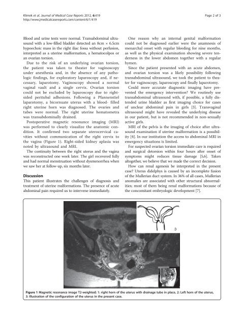
Postoperative magnetic resonance imaging (MRI)<br />
was performed to clearly visualize the anatomic condition.<br />
It confirmed two separate uterocervical cavities<br />
without communication <strong>of</strong> the right cervix to<br />
the vagina (Figure 1). Right-sided kidney aplasia was<br />
notedbyultrasoundandMRI.<br />
The continuity between the right uterus and the vagina<br />
was reconstructed one week later. The girl recovered fully<br />
and had normal menstruation without dysmenorrhea when<br />
we saw her at follow-up, six months later.<br />
Discussion<br />
This patient illustrates the challenges <strong>of</strong> diagnosis and<br />
treatment <strong>of</strong> uterine malformations. The presence <strong>of</strong> acute<br />
abdominal pain required us to intervene immediately.<br />
One reason why an internal genital malformation<br />
could not be diagnosed earlier were the anamnesis <strong>of</strong><br />
menarchal onset with regular bleeding for nine months,<br />
as well as the physical examination showing severe tenderness<br />
in the lower abdomen together with a regular<br />
hymen.<br />
Since the patient presented with an acute abdomen,<br />
and ovarian torsion was a likely possibility following<br />
transabdominal ultrasound, we took the patient to theater<br />
for vaginoscopy, laparoscopy and finally laparotomy.<br />
Could more accurate diagnostic imaging have prevented<br />
the emergency intervention? We routinely use<br />
transabdominal ultrasound with, if possible, a fully distended<br />
urine bladder as first imaging choice for cases<br />
<strong>of</strong> unclear abdominal pain in girls [3]. Transvaginal<br />
ultrasound might have revealed the underlying disease<br />
in our patient, but is not recommended in non-sexually<br />
active girls.<br />
MRI <strong>of</strong> the pelvis is the imaging <strong>of</strong> choice after ultrasound<br />
examination if uterine malformation is a possibility<br />
[4]. In our institution the access to abdominal MRI in<br />
emergency situations is limited.<br />
For suspected ovarian torsion immediate care is required<br />
and surgical detorsion within four hours after onset <strong>of</strong><br />
symptoms might reduces tissue damage [5,6]. Taken<br />
altogether, we believe that we made the correct decision.<br />
How can renal agenesis be interpreted in the present<br />
case? Uterus didelphys is caused by an incomplete fusion<br />
<strong>of</strong> the Mullerian duct system. In 36% <strong>of</strong> all cases, Mullerian<br />
anomalies are associated with other structural abnormalities;<br />
most <strong>of</strong> them being renal malformations because <strong>of</strong><br />
the concomitant embryologic development [7].<br />
3<br />
Figure 1 Magnetic resonance image T2-weighted; 1: right horn <strong>of</strong> the uterus with drainage tube in place, 2: Left horn <strong>of</strong> the uterus,<br />
3: Illustration <strong>of</strong> the configuration <strong>of</strong> the uterus in the present case.