Retinal Detachment - Kontaktlinse Instituttet
Retinal Detachment - Kontaktlinse Instituttet
Retinal Detachment - Kontaktlinse Instituttet

You also want an ePaper? Increase the reach of your titles
YUMPU automatically turns print PDFs into web optimized ePapers that Google loves.
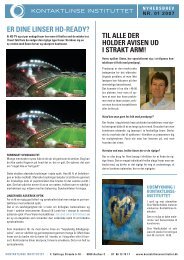
Differential Diagnosis of RD<br />
Based on Objective Findings<br />
• Lattice degeneration<br />
• Retinoschisis<br />
• White Without Pressure (WWP)<br />
• Posterior vitreous detachment (PVD)<br />
• Other peripheral retinal disorders<br />
Nicolitz Eye Consultants © 2011<br />
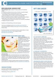
PVD is probably the most common<br />
differential diagnosis in RD evaluations<br />
Weiss’s<br />
Ring<br />
Nicolitz Eye Consultants © 2011<br />
Objective Findings Associated<br />
with <strong>Retinal</strong> <strong>Detachment</strong><br />
• Binocular indirect ophthalmoscopy (BIO) cont.<br />
o “Schaffer’s sign”<br />
• Presence of vitreous pigment cells;<br />
• 99% accurate for confirmed Dx of RD<br />
o Holes, tears, breaks<br />
o Carefully evaluate...<br />
• Lattice degeneration areas<br />
• Retinoschisis<br />
• “White without pressure”<br />
• Posterior vitreous detachment *<br />
o Scleral depression technique<br />
Nicolitz Eye Consultants © 2011<br />
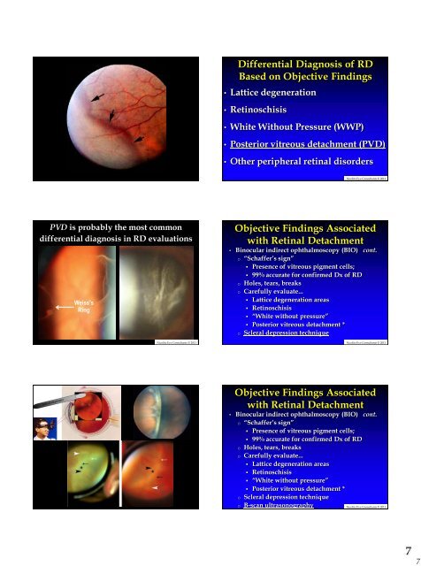
Objective Findings Associated<br />
with <strong>Retinal</strong> <strong>Detachment</strong><br />
• Binocular indirect ophthalmoscopy (BIO) cont.<br />
o “Schaffer’s sign”<br />
• Presence of vitreous pigment cells;<br />
• 99% accurate for confirmed Dx of RD<br />
o Holes, tears, breaks<br />
o Carefully evaluate...<br />
• Lattice degeneration areas<br />
• Retinoschisis<br />
• “White without pressure”<br />
• Posterior vitreous detachment *<br />
o Scleral depression technique<br />
o B-scan ultrasonography<br />
Nicolitz Eye Consultants © 2011<br />
7<br />
7