PROVINCIAL GUIDELINES FOR VASCULAR ... - BC Renal Agency
PROVINCIAL GUIDELINES FOR VASCULAR ... - BC Renal Agency
PROVINCIAL GUIDELINES FOR VASCULAR ... - BC Renal Agency

You also want an ePaper? Increase the reach of your titles
YUMPU automatically turns print PDFs into web optimized ePapers that Google loves.
<strong>PROVINCIAL</strong> <strong>GUIDELINES</strong> <strong>FOR</strong> <strong>VASCULAR</strong> ACCESS<br />
<strong>FOR</strong> PATIENTS WITH HD AS PRIMARY MODALITY<br />
Enclosed please find the Provincial Vascular Access Guidelines, developed and<br />
supported by the Provincial Vascular Access Services Team and <strong>BC</strong>PRA.<br />
Signed and approved on behalf of the Provincial Vascular Access Services Team:<br />
Chair, Provincial Vascular Access Services Team: Joanne Cozac<br />
Chair, Vascular Access Surgeons Group, Dr Jerry Chen:<br />
Chair, Vascular Access Radiology Group, Dr Phil Harrison:<br />
Executive Director, <strong>BC</strong> Provincial <strong>Renal</strong> <strong>Agency</strong>, Dr Adeera<br />
Levin:
<strong>PROVINCIAL</strong> <strong>GUIDELINES</strong> <strong>FOR</strong> <strong>VASCULAR</strong> ACCESS<br />
<strong>FOR</strong> PATIENTS WITH HD AS PRIMARY MODALITY<br />
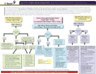
A) CREATION<br />
1. Referral for creation of VA should be done when GFR < 25 ml/min and change in<br />
GFR is > 5 ml/min/year.<br />
2. It is recommended that patients be assessed pre-operatively by vascular surgeon<br />
and/or nephrologist whether radiological assessment of their vasculature by either<br />
ultrasound or venography is required prior to creation of vascular access.<br />
3. VA of choice are AVF followed by AVG and lastly tunneled catheters in pts who<br />
do not have any suitable vessels for AVF/AVG creation.<br />
4. Tunneled catheters should be placed if hemodialysis catheter is to remain in situ<br />
for > 1-3 months.<br />
5. Catheters should be placed preferably in internal jugular veins as the first site.<br />
6. Tunneled cuffed catheters should not be placed on the same side as a maturing<br />
AV access, if possible.<br />
7. Real-time ultrasound guided insertion is recommended to reduce insertion-related<br />
complications (Guideline 5D).<br />
8. Fluoroscopy is recommended for insertion of all cuffed dialysis catheters and the<br />
catheter tip should be adjusted to the level of the caval/atrial junction or into the<br />
right atrium to ensure optimal blood flow (Guideline 5C).<br />
9. Femoral hemodialysis catheters may be placed when urgent vascular access is<br />
required, using long ( ≥ 20 cm) catheters and a more suitable access (IJ catheter)<br />
should be established as soon as possible (within one week).<br />
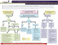
B) MONITORING<br />
a) Maturation of created access:<br />
1. At 2 weeks after creation: AVF and grafts (if applicable) should be assessed by<br />
trained individual (vascular access or kidney clinic nurse +/- nephrologist +/-<br />
vascular surgeon)<br />
a. If absent thrill or bruit, the patient should be urgently referred back to the<br />
vascular surgeon<br />
2. At 6 weeks after creations: AVF and grafts (if applicable) should be assessed by<br />
the vascular access team for maturation failure<br />
a. If inadequate maturation, appropriate investigations (fistulogram) and<br />
interventions should be initiated<br />
3. At 4-6 weeks prior to anticipated initiation of hemodialysis: AVF and grafts<br />
should be assessed by vascular access team<br />
a. If access inadequate for cannulation, appropriate investigations and<br />
interventions should be initiated
) Established Vascular Access:<br />
1. AVF and AVG should be monitored on a q 4-6 weekly schedule by the preferable<br />
methods in following order:<br />
a. Access flow measurements<br />
b. Dynamic or venous pressure measurement<br />
c. Access recirculation using 2 needle, 3 sample urea method if other<br />
technology not available<br />
2. Catheter function can be followed using recirculation values preferably using<br />
dilution method (transonic machine) q 4-6 weekly<br />
3. Evaluation and assessment should be preferably done by identified vascular<br />
access team:<br />
a. Vascular access nurse<br />
b. Nephrologist<br />
c. Vascular surgeon<br />
d. Radiologist<br />
4. Investigation and treatment by venography (fistulogram) is recommended<br />
a. Within 1-2 weeks for:<br />
i. Absolute access flows of < 500 ml/min in AVF and < 650ml/min<br />
in AVG<br />
ii. Decrease in access flow of > 20% from baseline values<br />
iii. Inability to achieve a blood pump speed on dialysis of ≥ 300<br />
ml/min by week 3 of initiating hemodialysis or 2 occasions in 2 week period should be<br />
investigated with hemodialysis catheter dye study to rule our persistent<br />
thrombus, fibrin sheath or malposition<br />
b. If evidence of fibrin sheath around catheter, trial of catheter stripping<br />
under radiology may be attempted, if possible. If not, catheter will need to<br />
be replaced.<br />
6. Persistent catheter dysfunction or the development of new facial swelling in any<br />
patient should be investigated with venography to rule our central vein stenosis.
C) INTERVENTION<br />
1. Treatment of stenosis of the fistula/graft or central veins should first be attempted<br />
by angioplasty unless otherwise directed by the radiologist or vascular surgeon.<br />
2. Angioplasty should be done in a timely manner:<br />
a. Usually within 2 weeks<br />
b. Within 2 days if<br />
i. absolute access flow < 300 ml/min or<br />
ii. drop from baseline of > 50% or<br />
iii. clinical indication ( severe bleeding or unable to properly dialyze<br />
pts)<br />
3. Surgical revisions of stenosed fistula/graft should be done on a more urgent basis,<br />
as per surgical priority scale.