View 2005 Report - RSC - Australian National University
View 2005 Report - RSC - Australian National University
View 2005 Report - RSC - Australian National University

You also want an ePaper? Increase the reach of your titles
YUMPU automatically turns print PDFs into web optimized ePapers that Google loves.
BIOLOGICAL CHEMISTRY<br />
least in some cases. In collaborative work with the Biomolecular NMR group, we determined the molecular structure of<br />
the folded domain of the τ subunit that distinguishes it from γ (Figure 1). Although this domain contains the regions of τ<br />
that interact with the α subunit of Pol III and DnaB, the major sites of interaction are not in its structured region, but in<br />
the flexible regions that flank it. In other work, we showed that ψ interacts with γ through its intrinsically unstructured<br />
N-terminal region, and NMR and crystallographic studies of the structure and function of the ε proofreading exonuclease<br />
subunit have continued in collaboration with Professors Gottfried Otting and David Ollis, Drs Max Keniry and Gary Schenk.<br />
(With P D Carr, M J Headlam, S Jergic, M John, M A Keniry, K V Loscha, A J Oakley, D L Ollis, G Otting, K Ozawa, A-Y Park,<br />
P Prosselkov, P M Schaeffer, X-C Su, N K Williams, P S C Wu, and E Liepinsh [Latvian U, Riga], G Schenk [U Queensland])<br />
Replication Termination<br />
In the final stage of replication, forks encounter the terminator protein Tus in complex<br />
with Ter-site DNA, and are arrested in a polar manner – a replisome approaching<br />
from the one face of the Tus-Ter complex can progress, while another approaching<br />
from the other face is blocked. We showed that this process works like a molecular<br />
mousetrap that is set by DNA strand separation by DnaB, and sprung to create a<br />
structure that is kinetically trapped by unusually stable binding of the Ter DNA to<br />
Tus. This year, we confirmed the mechanism that determines polarity by solving<br />
the structure of the arrested “Tus-Ter Lock” complex (Figure 2). (With M Mulcair,<br />
A J Oakley, P M Schaeffer, and T M Hill [U North Dakota], C Neylon [U Southampton])<br />
New Protein Technologies<br />
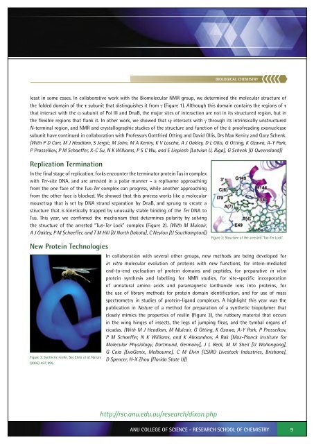
Figure 3: Synthetic resilin. See Elvin et al. Nature<br />
(<strong>2005</strong>) 437, 999.<br />
Figure 2: Structure of the arrested “Tus-Ter Lock”.<br />
In collaboration with several other groups, new methods are being developed for<br />
in vitro molecular evolution of proteins with new functions, for intein-mediated<br />
end-to-end cyclisation of protein domains and peptides, for preparative in vitro<br />
protein synthesis and labelling for NMR studies, for site-specific incorporation<br />
of unnatural amino acids and paramagnetic lanthanide ions into proteins, for<br />
the use of library methods for protein domain identification, and for use of mass<br />
spectrometry in studies of protein-ligand complexes. A highlight this year was the<br />
publication in Nature of a method for preparation of a synthetic biopolymer that<br />
closely mimics the properties of resilin (Figure 3), the rubbery material that occurs<br />
in the wing hinges of insects, the legs of jumping fleas, and the tymbal organs of<br />
cicadas. (With M J Headlam, M Mulcair, G Otting, K Ozawa, A-Y Park, P Prosselkov,<br />
P M Schaeffer, N K Williams, and K Alexandrov, A Rak [Max-Planck Institute for<br />
Molecular Physiology, Dortmund, Germany], J L Beck, M M Sheil [U Wollongong],<br />
G Coia [EvoGenix, Melbourne], C M Elvin [CSIRO Livestock Industries, Brisbane],<br />
D Spencer, H-X Zhou [Florida State U])<br />
http://rsc.anu.edu.au/research/dixon.php<br />
ANU COLLEGE OF SCIENCE - RESEARCH SCHOOL OF CHEMISTRY<br />
9