

DERMATOLOGY EPONYMS â SIGN â LEXICON â (H) - Our ...
DERMATOLOGY EPONYMS â SIGN â LEXICON â (H) - Our ...
DERMATOLOGY EPONYMS â SIGN â LEXICON â (H) - Our ...

Create successful ePaper yourself
Turn your PDF publications into a flip-book with our unique Google optimized e-Paper software.
HUTCHINSON’S INCISORS <strong>SIGN</strong><br />
There are depressions or notching of the incisal edges of the<br />
labial surfaces of the permanent incisors. A sign of congenital<br />
syphilis (Fig. 30-32) [42]. Also called Hutchinson’s teeth<br />
sign and Screwdriver sign.<br />
HUTCHINSON’S TEETH <strong>SIGN</strong><br />
see Hutchinson’s Incisors sign<br />
HUTCHINSON’S TRIO <strong>SIGN</strong><br />
The precence of interstitial keratitis, notched teeth, and<br />
otitis occurring together. A sign of inherited syphilis [43].<br />
The Triad is characterized by three signs: 1) deformation of<br />
teeth as a result of direct influence pale спирохеты on tooth<br />
rudiments of a fruit or on the bodies regulating growth of<br />
teeth. Changes concern the top central cutters, is more rare<br />
— lateral and central bottom (a barrel-like form, semi-lunar<br />
defects of cutting edge); 2) parenchymatous keratity; 3) the<br />
progressing relative deafness arising owing to a degeneration<br />
of a preddvernoulitkovy nerve, lying in a stony part of a<br />
temporal bone (syphilitic лабиринтит). The triad belongs to<br />
symptoms of late congenital syphilis. At one patient two can<br />
be observed only or one of signs, meet all three less often.<br />
The triad is described for the first time by J. Hutshinson in<br />
1858.<br />
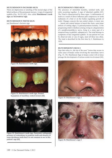
HUTCHINSON’S <strong>SIGN</strong> 2<br />
Sign that refers to „the tip of the nose” lesion that occurs in<br />
some cases of herpes zoster involving the nasociliary nerve<br />
(Fig. 33, 34). Hutchinson Sign in herpes zoster will at times<br />
presage the development of serious ocular involvement [44].<br />
Figure 30. Hutchinson’s teeth sign<br />
Figure 31. Hutchinson’s teeth sign. Enamel<br />
hypoplasia of maxillary central incisors [42]<br />
Figure 33. Hutchinson’s sign 2<br />
Figure 32. Hutchinson’s teeth sign. Panoramic radiograph:<br />
presence of restorations in posterior teeth and absence of<br />
some deciduous teeth that it is not common to see in this<br />
patient’s age [42]<br />
Figure 34. Hutchinson’s sign 2<br />
140 © <strong>Our</strong> Dermatol Online 1.2013


