

download full issue - Our Dermatology Online Journal
download full issue - Our Dermatology Online Journal
download full issue - Our Dermatology Online Journal

You also want an ePaper? Increase the reach of your titles
YUMPU automatically turns print PDFs into web optimized ePapers that Google loves.
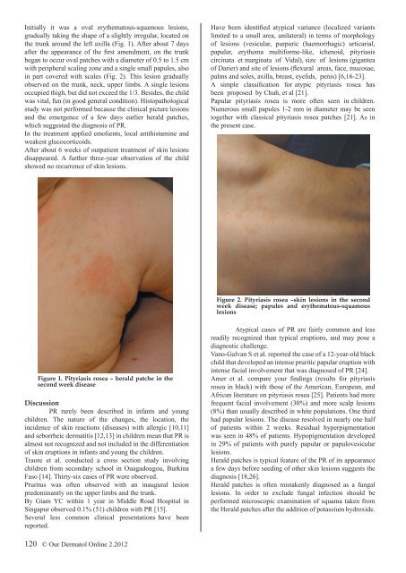
Initially it was a oval erythematous-squamous lesions,<br />
gradually taking the shape of a slightly irregular, located on<br />
the trunk around the left axilla (Fig. 1). After about 7 days<br />
after the appearance of the first amendment, on the trunk<br />
began to occur oval patches with a diameter of 0.5 to 1.5 cm<br />
with peripheral scaling zone and a single small papules, also<br />
in part covered with scales (Fig. 2). This lesion gradually<br />
observed on the trunk, neck, upper limbs. A single lesions<br />
occupied thigh, but did not exceed the 1/3. Besides, the child<br />
was vital, fun (in good general condition). Histopathological<br />
study was not performed because the clinical picture lesions<br />
and the emergence of a few days earlier herald patches,<br />
which suggested the diagnosis of PR.<br />
In the treatment applied emolients, local antihistamine and<br />
weakest glucocorticoids.<br />
After about 6 weeks of outpatient treatment of skin lesions<br />
disappeared. A further three-year observation of the child<br />
showed no recurrence of skin lesions.<br />
Have been identified atypical variance (localized variants<br />
limited to a small area, unilateral) in terms of morphology<br />
of lesions (vesicular, purpuric (haemorrhagic) urticarial,<br />
papular, erythema multiforme-like, ichenoid, pityriasis<br />
circinata et marginata of Vidal), size of lesions (gigantea<br />
of Darier) and site of lesions (flexural areas, face, mucosae,<br />
palms and soles, axilla, breast, eyelids, penis) [6,16-23].<br />
A simple classification for atypic pityriasis rosea has<br />
been proposed by Chuh, et al [21].<br />
Papular pityriasis rosea is more often seen in children.<br />
Numerous small papules 1-2 mm in diameter may be seen<br />
together with classical pityriasis rosea patches [21]. As in<br />
the present case.<br />
Figure 2. Pityriasis rosea –skin lesions in the second<br />
week disease; papules and erythematous-squamous<br />
lesions<br />
Figure 1. Pityriasis rosea – herald patche in the<br />
second week disease<br />
Discussion<br />
PR rarely been described in infants and young<br />
children. The nature of the changes, the location, the<br />
incidence of skin reactions (diseases) with allergic [10,11]<br />
and seborrheic dermatitis [12,13] in children mean that PR is<br />
almost not recognized and not included in the differentiation<br />
of skin eruptions in infants and young the children.<br />
Traore et al. conducted a cross section study involving<br />
children from secondary school in Ouagadougou, Burkina<br />
Faso [14]. Thirty-six cases of PR were observed.<br />
Pruritus was often observed with an inaugural lesion<br />
predominantly on the upper limbs and the trunk.<br />
By Giam YC within 1 year in Middle Road Hospital in<br />
Singapur observed 0.1% (51) children with PR [15].<br />
Several less common clinical presentations have been<br />
reported.<br />
Atypical cases of PR are fairly common and less<br />
readily recognized than typical eruptions, and may pose a<br />
diagnostic challenge.<br />
Vano-Galvan S et al. reported the case of a 12-year-old black<br />
child that developed an intense pruritic papular eruption with<br />
intense facial involvement that was diagnosed of PR [24].<br />
Amer et al. compare your findings (results for pityriasis<br />
rosea in black) with those of the American, European, and<br />
African literature on pityriasis rosea [25]. Patients had more<br />
frequent facial involvement (30%) and more scalp lesions<br />
(8%) than usually described in white populations. One third<br />
had papular lesions. The disease resolved in nearly one half<br />
of patients within 2 weeks. Residual hyperpigmentation<br />
was seen in 48% of patients. Hypopigmentation developed<br />
in 29% of patients with purely papular or papulovesicular<br />
lesions.<br />
Herald patches is typical feature of the PR of its appearance<br />
a few days before seeding of other skin lesions suggests the<br />
diagnosis [18,26].<br />
Herald patches is often mistakenly diagnosed as a fungal<br />
lesions. In order to exclude fungal infection should be<br />
performed microscopic examination of squama taken from<br />
the Herald patches after the addition of potassium hydroxide.<br />
120 © <strong>Our</strong> Dermatol <strong>Online</strong> 2.2012


