3B Scientific - Microscopy Catalog
3bscientific.com
3bscientific.com

Create successful ePaper yourself
Turn your PDF publications into a flip-book with our unique Google optimized e-Paper software.
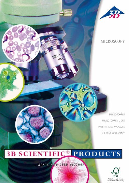
MICROSCOPY<br />
MICROSCOPES<br />
MICROSCOPE SLIDES<br />
MULTIMEDIA-PACKAGES<br />
<strong>3B</strong> MICROanatomy<br />
...going one step further<br />
Printed on paper from a<br />
sustainable forest company
Dear Customer,<br />
We have dedicated this catalogue to taking an in-depth look at working with a microscope. First<br />
of all, because we would particularly like to demonstrate to you our know-how in this important<br />
part of your biology class. Secondly, because we have some important news for you:<br />
We have slashed the minimum order quantity by 60 % for individual slides. You can order<br />
individual slides from ten items per order so that you can complete your collection more quickly<br />
and easily.<br />
Enjoy the variety!<br />
Kind regards<br />
Petra von Corvin<br />
Product Manager<br />
Petra von Corvin<br />
Product Manager<br />
K13<br />
Page 31<br />
Microslides<br />
Page 18<br />
W30600<br />
Page 4<br />
CONTENT<br />
MICROSCOPES 1<br />
Accessories 12<br />
Camreras for Microscopes 14<br />
MICROSCOPE SLIDES<br />
School Sets 18<br />
Series for Secondary schools 19<br />
Individual Slides 24<br />
Multimedia Packages 26<br />
DISSECTING KITS 27<br />
Storage Boxes, Microscope slides,<br />
Forceps, Needles 28<br />
<strong>3B</strong> MICROanatomy 30<br />
Anatomical Models 32<br />
Please also ask for our complete<br />
range of catalogues<br />
Biology<br />
Committed to quality<br />
<strong>3B</strong> <strong>Scientific</strong> provides you with good quality at fair prices. Our sophisticated quality<br />
management complies with the ISO 9001:2008 standards and the Worlddidac<br />
Quality Charter and is regularly approved by independent experts.<br />
That’s something you can rely on.<br />
Phsics<br />
Experiments<br />
Copyright © 2010, <strong>3B</strong> <strong>Scientific</strong> GmbH, Hamburg. Unauthorized reproduction and publication of this material is strictly forbidden.
Information About Microscopes<br />
Wide field eyepieces<br />
Seidentopf head<br />
Tube<br />
Revolving quadruple nosepiece<br />
with objectives<br />
Stand<br />
Object guide<br />
Object stage<br />
Coaxial adjustment for<br />
coarse and fine focusing<br />
Abbé condenser<br />
Illumination<br />
x-y cross-table<br />
adjustment<br />
Mains switch<br />
Base<br />
Course Microscope<br />
Course microscopes are robust, low cost microscopes with basic optical<br />
features that are ideally suited for lessons in school or for beginners in<br />
microscopy.<br />
Barrel<br />
The barrel is the tube in which the oculars can be placed.<br />
Monocular barrel: for observation with a single eye.<br />
Binocular barrel: for stereo observation. This makes the work easier<br />
and less tiring than with a monocular microscope.<br />
Trinocular barrel: for stereo observation but also allowing for addition<br />
of a camera.<br />
Ocular<br />
The ocular magnifies the real image thrown by the microscope’s objective.<br />
The diameter of the field of vision, i.e. the area of the slide that<br />
can be viewed at one time, is calculated by dividing the field number by<br />
the scaling factor. Thus for a 10x 18 mm ocular, the viewing field has a<br />
diameter of 1.8 mm.<br />
Objective Revolver<br />
The objective revolver accommodates between 3 and 5 objectives and<br />
makes it possible to change the magnification rapidly when viewing a<br />
slide.<br />
Objective<br />
An objective produces a real image of the object. The size of the image<br />
is given by the scaling factor (e.g. 10x) and the resolution is determined<br />
by the numerical aperture (e.g. 0.65). The larger the numerical aperture<br />
the more detailed the image produced.<br />
Achromatic objectives provide only a limited amount of correction for<br />
lens aberrations but this is nevertheless sufficient for most uses that<br />
arise in schools. Planar achromatic objectives eliminate image field<br />
curvature and throw an image that is uniformly focused from the centre<br />
of the field of vision to the edge.<br />
Resolution of Objectives<br />
The resolution of an objective is given by the following formula<br />
l<br />
d = 2 · A<br />
where d = distance between two points, l = wavelength of the light,<br />
A = numerical aperture<br />
Example: numerical aperture = 0.65, l = 0.55 µm, resolution<br />
d = 0.423 μm.<br />
Object Stage<br />
The object stage is the shelf upon which slides are placed for observation<br />
through a microscope. Using an x-y cross-table allows the slide to<br />
be moved by specific distances along the x and/or y axes. The scales<br />
mean that once a specific location on the slide has been found, it is<br />
easy to locate it again.<br />
Condenser<br />
The function of a condenser is to allow for careful adjustment of the<br />
aperture to ensure an optimum compromise between image contrast<br />
and resolution. As the aperture is made smaller, the contrast increases<br />
but the resolution is simultaneously reduced.<br />
Coarse and Fine Focusing<br />
Coarse and fine adjustment gears allow for optimum focusing of an<br />
image. They are mostly fitted along a common axis on either side of the<br />
column leading up from the base.<br />
Illumination<br />
Microscope slides can be illuminated by means of incandescent tungsten<br />
lamps, fluorescent tubes, LEDs or halogen lamps. Halogen lamps<br />
are best suited to the task because they provide such intense light.<br />
Fluorescent tubes and LEDs eliminate the problem of slides warming up<br />
due to the heat from the light during longer periods of observation.<br />
...going one step further 1
Microscope<br />
NEW<br />
W30690<br />
The W30690 microscope is intended for exacting analysis using bright-field transmitted light. Its ergonomic design allows<br />
for lengthy periods of use without tiring. The high quality infinite optical system guarantees excellent image quality. Such<br />
microscopes are used in general practice.<br />
W30690<br />
Product name Laboratory Microscope BS-300<br />
Stand<br />
Robust and stable all metal stand, pinion knobs attached on both sides of the stand for coarse and fine focusing with friction<br />
coupling<br />
Tube Binocular at 45° angle, rotatable through 360°<br />
Eyepieces<br />
Pair of eyepieces PL10x 20 mm with infinite optics<br />
Objectives Inverted objective revolver with plan achromatic infinite objectives 4x, 10x, 40xS und 100xS Oil<br />
Enlargement 40x – 1000x<br />
Objekttisch x-y mechanical stage, 150 mm x 140 mm, adjustment range 50 mm x 76 mm<br />
Illumination Adjustable 6 V/20 W halogen lamp, built-in transformer for 90 to 240V mains voltage<br />
Condenser Condenser NA1.25, iris diaphragm, filter holder and blue filter<br />
Accessories Complete with dust cover<br />
2 <strong>3B</strong> <strong>Scientific</strong>® <strong>Microscopy</strong>
U30722<br />
U30723<br />
High-quality mechanics and optics along with ease of operation are the stand-out features of the polarisation microscopes U30722 and U30723. Their<br />
compact and ergonomic design makes it easier to work with them. The main application for these microscopes is in biology, for instance when studying the<br />
structure of starch grains, the texture of cellulose fibres in cell walls or the position of rod-like viruses in cells (e.g. tobacco mosaic virus). They are also used<br />
in mineralogy to study rock specimens, identify minerals and investigate crystals.<br />
Product name<br />
Stand<br />
Tube<br />
Polarisation<br />
equipment<br />
Eyepieces<br />
Objectives<br />
Enlargement<br />
Object stage<br />
Illumination<br />
Condenser<br />
Dimensions<br />
Weight<br />
Supplied<br />
New<br />
U30722<br />
Monocular Polarisation Microscope<br />
Robust, all metal stand with arm permanently connected to the<br />
base. Focusing by means of separate knobs for coarse and fine<br />
adjustment located on either side of the stand and operated<br />
by rack and pinion drive with ball bearings and retaining lever,<br />
adjustable stopper for protecting the object slides and objective.<br />
Monocular inclined 30°, head rotation 360°<br />
Polariser with scale and analyser, which can be inserted into the<br />
tube.<br />
Wide field eyepiece WF 10x 18 mm<br />
Inverted objective revolver with 3 achromatic objectives 4x /<br />
0.10, 10x / 0.25, 40x / 0.65, (oil)<br />
40x – 400x<br />
Circular object stage 120 mm in diameter, which can be rotated<br />
360°, scale with Vernier and 2 specimen clips<br />
Adjustable 6 V, 20 W halogen lamp incorporated into the base,<br />
universal 85 to 265 V, 50/60 Hz power supply<br />
Abbe condenser N.A.1,25 with iris diaphragm, focused via rack<br />
and pinion drive<br />
240 mm x 190 mm x 385 mm<br />
5,5 kg<br />
Complete with dust cover<br />
U30723<br />
Binocular Polarisation Microscope<br />
Robust, all metal stand with arm permanently connected to the<br />
base. Focusing by means of separate knobs for coarse and fine<br />
adjustment located on either side of the stand and operated<br />
by rack and pinion drive with ball bearings and retaining lever,<br />
adjustable stopper for protecting the object slides and objective.<br />
Binocular Seidentopf head, 30° viewing angle, 360° rotatable<br />
head, viewing distance adjustable between 54 and 75 mm, ±5<br />
dioptric compensation for both eyepieces<br />
Polariser with scale and analyser, which can be inserted into the<br />
tube.<br />
Pair of wide field eyepieces WF 10x 18 mm Pair of wide field<br />
eyepieces WF 10x 18 mm<br />
Inverted objective revolver with 3 achromatic objectives 4x /<br />
0.10, 10x / 0.25, 40x / 0.65 (oil)<br />
40x – 400x<br />
Circular object stage 120 mm in diameter, which can be rotated<br />
360°, scale with Vernier and 2 specimen clips<br />
Adjustable 6 V, 20 W halogen lamp incorporated into the base,<br />
universal 85 to 265 V, 50/60 Hz power supply<br />
Abbe condenser N.A.1,25 with iris diaphragm, focused via rack<br />
and pinion drive<br />
240 mm x 190 mm x 425 mm<br />
6 kg<br />
Complete with dust cover<br />
microscope Polarisation Microscopes<br />
...going one step further 3
Course microscopes Biologic Microscopes<br />
W30600-115<br />
W30600-230<br />
W30610-115<br />
W30610-230<br />
W30605-115<br />
W30605-230<br />
The monocular course microscopes W30600, W30605 and W30610 are distinguished by their robust construction and ease of operation. They are equipped<br />
with three achromatic objectives as used in common practice and have a simple object stage with two clips for holding slides. They can be supplemented<br />
by means of a variety of spare parts and accessories. The LED lighting of the W30605 and W30610 makes for uniform illumination of the object and avoids<br />
the problem of heat affecting the slide when viewed for extended periods. The microscopes are equipped with rechargeable batteries and can be used<br />
without a mains connection. Digital curriculum microscope W30605 is additionally equipped with a 300 kilopixel camera. The user-friendly “Photolib”<br />
software allows for…<br />
• Full screen real time video<br />
• Image processing<br />
• Image plane processing<br />
• Noise reduction filter for image enhancement, user-defined filter<br />
Good value for money<br />
Bestseller<br />
Robust<br />
• False colour image display<br />
• 3D representation<br />
• Extensive evaluation and measurement options<br />
with<br />
camera<br />
cordless<br />
Product Name<br />
Product Name<br />
Stand<br />
Tube<br />
Eyepieces<br />
Objectives<br />
Enlargement<br />
Object stage<br />
Illumination<br />
Condenser<br />
Dimensions<br />
Weight<br />
Supplied<br />
W30600-115, W30600-230<br />
Monocular Course Microscope Model 100<br />
–<br />
–<br />
All-metal stand, arm firmly connected with base, pinion knobs<br />
attached on both sides of the stand for coarse and fine focusing<br />
Monocular inclined 45°, head rotation 360°<br />
Wide field eyepiece WF 10x 18 mm with pointer and eyepiece<br />
lock<br />
Revolving nosepiece with 3 achromatic objectives<br />
4x / 0.10, 10x / 0.25, 40x / 0.65,<br />
40x, 100x, 400x<br />
110 mm x 120 mm with 2 specimen clips<br />
115 V resp. 230 V, 20 W tungsten lamp integrated in base, with<br />
blue filter in lamp shaft and a converging lens, power supply<br />
115 V resp. 230 V 50/60 Hz<br />
Bright-field condenser N.A. 0.65, iris diaphragm and filter holder<br />
175 mm x 135 mm x 370 mm<br />
2.9 kg<br />
Complete with dust cover<br />
W30610-115, W30610-230<br />
Monocular Course Microscope Model 100, LED<br />
W30605-115, W30605-230<br />
Digital Course Microscope Model 100, LED with built-in<br />
Camera<br />
Basic apparatus as per W30600 with the following differences:<br />
W30610 / W30605<br />
Illumination: With adjustable LED lighting incorporated into<br />
the base and a focusing lens in the lighting shaft, power supplied<br />
by rechargeable battery, 115 V or 230 V, 50/60 Hz charger.<br />
W30605<br />
Camera sensor: 1/3˝ CMOS, 300 kpixel, colour prints<br />
Power supply: Via USB 2.0<br />
System Requirements: WIN95, WIN98, WIN2000 and WINXP<br />
4 <strong>3B</strong> <strong>Scientific</strong>® <strong>Microscopy</strong>
U30700-115<br />
U30700-230<br />
U30701-115<br />
U30701-230<br />
Biologic Microscopes<br />
Course microscopes U30700 and U30701 are especially robust microscopes for educational purposes. They are simple to use and their mechanical and optical<br />
quality stands out. Separate adjustment knobs for fine and coarse setting allow the microscopes to be focused quickly. The low-temperature lighting<br />
provides for uniform illumination of the object and avoids the problem of heat affecting the slide when observed for long periods. Seidentopf head and<br />
30° viewing angle for comfortable observation of the object.<br />
Course microscopes<br />
Product name<br />
Stand<br />
Tube<br />
Eyepieces<br />
Objectives<br />
Enlargement<br />
Object stage<br />
Illumination<br />
Condenser<br />
Dimensions<br />
Weight<br />
Supplied<br />
U30700-115, U30700-230<br />
Monocular Course Microscope Model 200<br />
Robust, all metal stand with arm permanently connected to the<br />
base. Focusing by means of separate knobs for coarse and fine<br />
adjustment located on either side of the stand and operated by<br />
rack and pinion drive with dovetail teeth, adjustable stopper for<br />
protecting the object stage and objective.<br />
Monocular inclined 45°, head rotation 360°<br />
Wide field eyepiece WF 10x 18 mm<br />
Revolving nosepiece with 3 achromatic objectives 4x, 10x, 40x<br />
40x, 100x, 400x<br />
127 mm x 132 mm with 2 specimen clips<br />
5 W fluorescent lamp incorporated in the base, power supply<br />
115 V resp. 230 V 50/60 Hz<br />
NA 0.65 with iris diaphragm , filter holder and blue filter<br />
220 mm x 148 mm x 356 mm<br />
4 kg<br />
Complete with dust cover<br />
U30701-115, U30701-230<br />
Binocular Course Microscope Model 200<br />
Robust, all metal stand with arm permanently connected to the<br />
base. Focusing by means of separate knobs for coarse and fine<br />
adjustment located on either side of the stand and operated by<br />
rack and pinion drive with dovetail teeth, adjustable stopper for<br />
protecting the object stage and objective.<br />
Binocular Seidentopf head, 30° viewing angle, 360° rotatable<br />
head, viewing distance adjustable between 54 and 75 mm, ±5<br />
dioptric compensation for both eyepieces<br />
Pair of wide field eyepieces WF 10x 18 mm<br />
Revolving nosepiece with 3 achromatic objectives 4x, 10x, 40x<br />
40x, 100x, 400x<br />
127 mm x 132 mm with 2 specimen clips<br />
5 W fluorescent lamp incorporated in the base, power supply<br />
115 V resp. 230 V 50/60 Hz<br />
NA 0.65 with iris diaphragm , filter holder and blue filter<br />
282 mm x 148 mm x 357 mm<br />
4.69 kg<br />
Complete with dust cover<br />
...going one step further 5
Biologic Microscopes<br />
U30705-115<br />
U30705-230<br />
U30706-115<br />
U30706-230<br />
Course microscopes for advanced users<br />
Course microscopes U30705 and U30706 are suitable for any applications that may arise in the course of advanced biology lessons. The microscopes are<br />
equipped with a cross table, a 4-way objective revolver with DIN achromatic objectives, a focusing Abbe condenser and the coaxial drive knobs are arranged<br />
as per common practice. The low-temperature lighting provides for uniform illumination of the object and avoids the problem of heat affecting the<br />
slide when viewed for extended periods. Accessories include planar and semi-planar achromatic objectives and a dark-field condenser.<br />
Product name<br />
Stand<br />
Tube<br />
Eyepieces<br />
Objectives<br />
Enlargement<br />
Object stage<br />
Illumination<br />
Condenser<br />
Dimensions<br />
Weight<br />
Supplied<br />
U30705-115, U30705-230<br />
Monocular Course Microscope Model 300<br />
Robust, all metal stand with arm permanently connected to the<br />
base. Focusing by means of separate knobs for coarse and fine<br />
adjustment located on either side of the stand and operated by<br />
rack and pinion drive with ball bearings, adjustable stopper for<br />
protecting the object slides and objective.<br />
Monocular inclined 45°, head rotation 360°<br />
Wide field eyepiece WF 10x 18 mm<br />
Revolving nosepiece with 4 achromatic objectives 4x, 10x, 40x,<br />
100x (oil)<br />
40x, 100x, 400x, 1000x<br />
x-y cross table, 125 mm x 130 mm, with object guide and coaxial<br />
adjustment knobs perpendicular to the object stage, adjustment<br />
range 70 mm x 30 mm<br />
5 W fluorescent lamp incorporated in the base, power supply<br />
115 V resp. 230 V 50/60 Hz<br />
Abbé condenser N.A.1,25 NA 0.65 with iris diaphragm , filter<br />
holder and blue filter, focused via rack and pinion drive<br />
220 mm x 154 mm x 359 mm<br />
4.5 kg<br />
Complete with dust cover<br />
U30706-115, U30706-230<br />
Binocular Course Microscope Model 300<br />
Robust, all metal stand with arm permanently connected to the<br />
base. Focusing by means of separate knobs for coarse and fine<br />
adjustment located on either side of the stand and operated by<br />
rack and pinion drive with ball bearings, adjustable stopper for<br />
protecting the object slides and objective.<br />
Binocular Seidentopf head, 30° viewing angle, 360° rotatable<br />
head, viewing distance adjustable between 54 and 75 mm, ±5<br />
dioptric compensation for both eyepieces<br />
Pair of wide field eyepieces WF 10x 18 mm<br />
Revolving nosepiece with 4 achromatic objectives 4x, 10x, 40x,<br />
100x (oil)<br />
40x, 100x, 400x, 1000x<br />
x-y cross table, 125 mm x 130 mm, with object guide and coaxial<br />
adjustment knobs perpendicular to the object stage, adjustment<br />
range 70 mm x 30 mm<br />
5 W fluorescent lamp incorporated in the base, power supply<br />
115 V resp. 230 V 50/60 Hz<br />
Abbé condenser N.A.1,25 NA 0.65 with iris diaphragm , filter<br />
holder and blue filter, focused via rack and pinion drive<br />
282 mm x 148 mm x 357 mm<br />
5.2 kg<br />
Complete with dust cover<br />
6 <strong>3B</strong> <strong>Scientific</strong>® <strong>Microscopy</strong>
U30710<br />
U30711<br />
Microscopes U30710, U30711, U30712 and U30713 are characterised by their robust design, excellent mechanical and optical quality and ease of operation.<br />
They are equipped with a large cross-stage and a 4-way objective revolver with 4 DIN achromatic objectives. U30710, U30711 and U30712 are also supplied<br />
with a second wide field WF15x eyepiece as standard, allowing for various magnifications of a slide. A halogen lamp incorporated into the base makes for<br />
bright and uniform illumination of the object. Seidentopf head and 30° viewing angle for comfortable observation of the object.<br />
Product name<br />
Stand<br />
Tube<br />
Eyepieces<br />
Objectives<br />
Enlargement<br />
Object stage<br />
Illumination<br />
Condenser<br />
Dimensions<br />
Weight<br />
Supplied<br />
U30710<br />
Monocular Microscope Model 400<br />
Robust, all metal stand with arm permanently connected to the<br />
base. Focusing by means of separate knobs for coarse and fine<br />
adjustment located on either side of the stand and operated<br />
by rack and pinion drive with ball bearings and retaining lever,<br />
adjustable stopper for protecting the object slides and objective.<br />
Focus range: 15mm<br />
Resolution of fine focusing adjustment: 0.002 mm<br />
Monocular inclined 30°, head rotation 360°<br />
Wide field eyepieces WF 10x 18 mm and WF 15x 13 mm<br />
Revolving nosepiece with 4 achromatic objectives 4x, 10x, 40x,<br />
100x (oil)<br />
40X – 1500X<br />
x-y mechanical stage, 132 mm x 145 mm, with object guide and<br />
coaxial adjustment knobs perpendicular to the object stage,<br />
adjustment range 50 mm x 76 mm<br />
Adjustable 6 V, 20 W halogen lamp incorporated into the base,<br />
universal 85 to 265 V, 50/60 Hz power supply<br />
Abbé condenser N.A.1,25 NA 0.65 with iris diaphragm , filter<br />
holder and blue filter, focused via rack and pinion drive<br />
291 mm x 214 mm x 356 mm<br />
5.6 kg<br />
Complete with dust cover<br />
U30711<br />
Binocular Microscope Model 400<br />
Robust, all metal stand with arm permanently connected to the<br />
base. Focusing by means of separate knobs for coarse and fine<br />
adjustment located on either side of the stand and operated<br />
by rack and pinion drive with ball bearings and retaining lever,<br />
adjustable stopper for protecting the object slides and objective.<br />
Focus range: 15mm<br />
Resolution of fine focusing adjustment: 0.002 mm<br />
Binocular Seidentopf head, 30° viewing angle, 360° rotatable<br />
head, viewing distance adjustable between 54 and 75 mm, ±5<br />
dioptric compensation for both eyepieces<br />
Pair of wide field eyepieces WF 10x 18 mm and WF 15x 13 mm<br />
Revolving nosepiece with 4 achromatic objectives 4x, 10x, 40x,<br />
100x (oil)<br />
40X – 1500X<br />
x-y mechanical stage, 132 mm x 145 mm, with object guide and<br />
coaxial adjustment knobs perpendicular to the object stage,<br />
adjustment range 50 mm x 76 mm<br />
Adjustable 6 V, 20 W halogen lamp incorporated into the base,<br />
universal 85 to 265 V, 50/60 Hz power supply<br />
Abbé condenser N.A.1,25 NA 0.65 with iris diaphragm , filter<br />
holder and blue filter, focused via rack and pinion drive<br />
328 mm x 214 mm x 394 mm<br />
6.1 kg<br />
Complete with dust cover<br />
Biologic Microscopes Microscopes for advanced users<br />
...going one step further 7
Biologic Microscopes<br />
U30713<br />
U30712<br />
Microscopes for advanced users<br />
Microscopes U30712 and U30713 provide for binocular or monocular viewing as well as allowing simultaneous fitting of a camera for photographic or<br />
video recording of the image.<br />
Product name<br />
Stand<br />
Tube<br />
Eyepieces<br />
Objectives<br />
Enlargement<br />
Object stage<br />
Illumination<br />
Condenser<br />
Dimensions<br />
Weight<br />
Supplied<br />
U30713<br />
Monocular Microscope Model 400 with Vertical Viewing<br />
Robust, all metal stand with arm permanently connected to the<br />
base. Focusing by means of separate knobs for coarse and fine<br />
adjustment located on either side of the stand and operated<br />
by rack and pinion drive with ball bearings and retaining lever,<br />
adjustable stopper for protecting the object slides and objective.<br />
Focus range: 15 mm<br />
Resolution of fine focusing adjustment: 0.002 mm<br />
Head with double viewing capability, one tube with 30° viewing<br />
angle, one with vertical viewing, head rotation 360°<br />
Pair of wide field eyepieces WF 10x 18 mm<br />
Revolving nosepiece with 4 achromatic objectives 4x, 10x, 40x,<br />
100x (oil)<br />
40x, 100x, 400x, 1000x<br />
x-y mechanical stage, 132 mm x 145 mm, with object guide and<br />
coaxial adjustment knobs perpendicular to the object stage,<br />
adjustment range 50 mm x 76 mm<br />
Adjustable 6 V, 20 W halogen lamp incorporated into the base,<br />
universal 85 to 265 V, 50/60 Hz power supply<br />
Abbé condenser N.A.1,25 NA 0.65 with iris diaphragm , filter<br />
holder and blue filter, focused via rack and pinion drive<br />
291 mm x 214 mm x 415 mm<br />
5.8 kg<br />
Complete with dust cover<br />
U30712<br />
Trinocular Microscope Model 400<br />
Robust, all metal stand with arm permanently connected to the<br />
base. Focusing by means of separate knobs for coarse and fine<br />
adjustment located on either side of the stand and operated<br />
by rack and pinion drive with ball bearings and retaining lever,<br />
adjustable stopper for protecting the object slides and objective.<br />
Focus range: 15 mm<br />
Resolution of fine focusing adjustment: 0.002 mm<br />
Trinocular Seidentopf head, 360° rotatable, binocular tubus<br />
with 30° viewing angle, viewing distance adjustable between 54<br />
and 75 mm, ±5 dioptric compensation for both eyepieces, one<br />
tube with vertical viewing angle<br />
Pair of wide field eyepieces WF 10x 18 mm and WF 15x 13 mm<br />
Revolving nosepiece with 4 achromatic objectives 4x, 10x, 40x,<br />
100x (oil)<br />
40x – 1500x<br />
x-y mechanical stage, 132 mm x 145 mm, with object guide and<br />
coaxial adjustment knobs perpendicular to the object stage,<br />
adjustment range 50 mm x 76 mm<br />
Adjustable 6 V, 20 W halogen lamp incorporated into the base,<br />
universal 85 to 265 V, 50/60 Hz power supply<br />
Abbé condenser N.A.1,25 NA 0.65 with iris diaphragm , filter<br />
holder and blue filter, focused via rack and pinion drive<br />
328 mm x 214 mm x 449 mm<br />
6.2 kg<br />
Complete with dust cover<br />
8 <strong>3B</strong> <strong>Scientific</strong>® <strong>Microscopy</strong>
U30720<br />
U30721<br />
Microscopes U30720 and U30721 are suitable for any applications that may arise in the course of advanced biology lessons. Their compact and ergonomic<br />
design facilitates ease of working with the microscope. They are equipped as standard with a polarisation fitting and have a large cross table, 2 pairs of<br />
wide-field eyepieces (WF 10x, WF 15x) and a four way objective revolver with planar achromatic objectives, for outstanding observation of tiny details with<br />
uniform focus from centre to edge of field of view.<br />
Product name<br />
Stand<br />
Tube<br />
Polarisation<br />
equipment<br />
Eyepieces<br />
Objectives<br />
Enlargement<br />
Object stage<br />
Illumination<br />
Condenser<br />
Dimensions<br />
Weight<br />
Supplied<br />
U30720<br />
Monocular Microscope Model 500 with<br />
Polarisation Equipment<br />
Robust, all metal stand with arm permanently connected to the<br />
base. Focusing by means of separate knobs for coarse and fine<br />
adjustment located on either side of the stand and operated<br />
by rack and pinion drive with ball bearings and retaining lever,<br />
adjustable stopper for protecting the object slides and objective.<br />
Focus range: 15 mm<br />
Resolution of fine focusing adjustment: 0.002 mm<br />
Monocular inclined 30°, head rotation 360°<br />
Polariser and analyser<br />
Wide field eyepieces WF 10x 18 mm and 15x 13 mm<br />
Inverted and angled objective revolver with 4 plan achromatic<br />
objectives 4x, 10x, 40x, 100x (oil)<br />
40x – 1500x<br />
x-y mechanical stage, 155 mm x 145 mm, with object guide and<br />
coaxial adjustment knobs perpendicular to the object stage,<br />
adjustment range 50 mm x 76 mm<br />
Adjustable 6 V, 20 W halogen lamp incorporated into the base,<br />
universal 85 to 265 V, 50/60 Hz power supply<br />
Abbé condenser N.A.1,25 NA 0.65 with iris diaphragm , filter<br />
holder and blue filter, focused via rack and pinion drive<br />
256 mm x 190 mm x 378 mm<br />
6 kg<br />
Complete with dust cover<br />
Top<br />
Quality<br />
U30721<br />
Binocular Microscope Model 500 with<br />
Polarisation Equipment<br />
Robust, all metal stand with arm permanently connected to the<br />
base. Focusing by means of separate knobs for coarse and fine<br />
adjustment located on either side of the stand and operated<br />
by rack and pinion drive with ball bearings and retaining lever,<br />
adjustable stopper for protecting the object slides and objective.<br />
Focus range: 15 mm<br />
Resolution of fine focusing adjustment: 0.002 mm<br />
Binocular Seidentopf head, 30° viewing angle, 360° rotatable<br />
head, viewing distance adjustable between 54 and 75 mm, ±5<br />
dioptric compensation for both eyepieces<br />
Polariser and analyser<br />
Pair of wide field eyepieces WF 10x 18 mm and 15x 13 mm<br />
Inverted and angled objective revolver with 4 plan achromatic<br />
objectives 4x, 10x, 40x, 100x (oil)<br />
40x – 1500x<br />
x-y mechanical stage, 155 mm x 145 mm, with object guide and<br />
coaxial adjustment knobs perpendicular to the object stage,<br />
adjustment range 50 mm x 76 mm<br />
Adjustable 6 V, 20 W halogen lamp incorporated into the base,<br />
universal 85 to 265 V, 50/60 Hz power supply<br />
Abbé condenser N.A.1,25 NA 0.65 with iris diaphragm , filter<br />
holder and blue filter, focused via rack and pinion drive<br />
306 mm x 190 mm x 407 mm<br />
6.6 kg<br />
Complete with dust cover<br />
Biologic Microscopes Microscopes for advanced users<br />
...going one step further 9
Stereo microscopes<br />
W30660-115<br />
W30660-230<br />
W30661-115<br />
W30661-230<br />
W30665-115<br />
W30665-230<br />
Cold light<br />
Stereo microscopes<br />
Cordless<br />
Stereo microscopes W30660 , W30661 and W30665 are robust microscopes that are distinguished by their ease of operation and excellent mechanical and<br />
optical quality. They can be used in numerous applications within the fields of biology and geology. They are equipped with quick change fitting that allows<br />
for rapid replacement of the objective. With the aid of accessories, a magnification of up to 120x can be achieved. Model W30660 is lit from the top,<br />
while W30661 and W30665 can be illuminated by top light, by transmitted light, or by a combination of both. The large object stage of the W30661 and<br />
W30665 also allows large objects to be observed.<br />
Stereo microscope W30665 features low temperature lighting (LED) to ensure even illumination of the object while preventing heat damage to the specimen<br />
during prolonged observations. It also eliminates the risk of burning if the lighting unit is touched inadvertently. Power is provided by rechargeable<br />
batteries so that the microscope can be used without needing to plug in a main lead<br />
Product name<br />
Stand<br />
Tube<br />
Eyepieces<br />
Objectives<br />
Enlargement<br />
Object plate<br />
W30660-115, W30660-230<br />
Stereo Microscope, 20x, Top Light<br />
Illumination<br />
Metal stand, column firmly connected<br />
with base, pinion knobs attached on<br />
both sides of the stand for coarse and<br />
fine focusing<br />
Binocular inclined 45°, interocular distance<br />
adjustable between 55 and 75 mm<br />
Exchangeable pair of wide field eyepiece<br />
WF 10x with eyepiece lock and rubber<br />
eyepiece cups, diopter compensation ±5<br />
on the left eyepiece<br />
Lens 2x with slide and quick change<br />
device<br />
20x<br />
Base with detachable object plate (plastic,<br />
black/white)<br />
60 mm Ø and 2 specimen clips<br />
W30661-115, W30661-230<br />
Stereo Microscope, 20x, Top,<br />
Transmitted and<br />
Mixed Light Illumination<br />
Metal stand, column firmly connected<br />
with base, pinion knobs attached on<br />
both sides of the stand for coarse and<br />
fine focusing<br />
Binocular inclined 45°, interocular distance<br />
adjustable between 55 and 75 mm<br />
Exchangeable pair of wide field eyepiece<br />
WF 10x with eyepiece lock and rubber<br />
eyepiece cups, diopter compensation ±5<br />
on the left eyepiece<br />
Lens 2x with slide and quick change<br />
device<br />
20x<br />
Base with detachable object plate (plastic,<br />
black/white and glass)<br />
95 mm Ø and 2 specimen clips<br />
W30665-115, W30665-230<br />
Stereo Microscope, 20x, LED<br />
Metal stand, column firmly connected<br />
with base, pinion knobs attached on<br />
both sides of the stand for coarse and<br />
fine focusing<br />
Binocular inclined 45°, interocular distance<br />
adjustable between 55 and 75 mm<br />
Exchangeable pair of wide field eyepiece<br />
WF 10x with eyepiece lock and rubber<br />
eyepiece cups, diopter compensation ±5<br />
on the left eyepiece<br />
Lens 2x with slide and quick change<br />
device<br />
20x<br />
Base with detachable object plate<br />
(plastic, black/white and glass) 95 mm<br />
dia. and 2 specimen clips<br />
Illumination<br />
Dimensions<br />
Weight<br />
Supplied<br />
Top light illumination,12 V/10 W, with<br />
toggle switch, power supply 115 V resp.<br />
230 V 50/60 Hz<br />
170 mm x 300 mm x 115 mm<br />
2.4 kg<br />
Complete with dust cover<br />
Top , transmitted and mixed light illumination,<br />
12 V/10 W lamp, toggle switch<br />
to turn ON, rotary switch to select light<br />
combination, power supply 115 V resp.<br />
230 V 50/60 Hz<br />
190 mm x 300 mm x 115 mm<br />
2.9 kg<br />
Complete with dust cover<br />
LED, top , transmitted and mixed light<br />
illumination, toggle switch to turn ON,<br />
rotary switch to select light combination,<br />
power supplied by rechargeable battery,<br />
115 V or 230 V, 50/60 Hz charger<br />
190 mm x 300 mm x 115 mm<br />
2.9 kg<br />
Complete with dust cover<br />
10 <strong>3B</strong> <strong>Scientific</strong>® <strong>Microscopy</strong>
W30662-115<br />
W30662-230<br />
W30663-115<br />
W30663-230<br />
W30664-115<br />
W30664-230<br />
Bestseller<br />
Revolving<br />
head<br />
Stereo microscopes W30662, W30663 and W30664 are robust microscopes that are distinguished by their ease of operation and excellent mechanical and<br />
optical quality. They can be used in numerous applications within the fields of biology and geology. Simply by rotating the objective from the 2x setting to<br />
4x, the overall magnification can be set to 20x or 40x. With the aid of accessories, a magnification of up to 80x can be achieved. Model W30662 is lit from<br />
the top, while W30663 and W30664 can be illuminated by top light, by transmitted light, or by a combination of both. The large object stage of W30663<br />
and W30664 also allows large objects to be observed.<br />
Stereo microscope W30664 differs from W30662 and W30663 in that its stereo head can be rotated by 360°.<br />
Stereo microscopes Stereo microscopes<br />
Product name<br />
Stand<br />
Tube<br />
Eyepieces<br />
Objectives<br />
Enlargement<br />
Object plate<br />
W30662-115, W30662-230<br />
Stereo Microscope, 40x, Top Light<br />
Illumination<br />
Metal stand, column firmly connected<br />
with base, pinion knobs attached on<br />
both sides of the stand for coarse and<br />
fine focusing<br />
Binocular inclined 45°, interocular distance<br />
adjustable between 55 and 75 mm<br />
Exchangeable pair of wide field eyepiece<br />
WF 10x with eyepiece lock and rubber<br />
eyepiece cups, diopter compensation ±5<br />
on the left eyepiece<br />
Revolving nosepiece with objective 2x<br />
/ 4x<br />
20x/40x<br />
Base with detachable object plate<br />
(plastic, black/white)<br />
60 mm Ø and 2 specimen clips<br />
W30663-115, W30663-230<br />
Stereo Microscope, 40x, Top, Transmitted<br />
and<br />
Mixed Light Illumination<br />
Metal stand, column firmly connected<br />
with base, pinion knobs attached on<br />
both sides of the stand for coarse and<br />
fine focusing<br />
Binocular inclined 45°, interocular distance<br />
adjustable between 55 and 75 mm<br />
Exchangeable pair of wide field eyepiece<br />
WF 10x with eyepiece lock and rubber<br />
eyepiece cups, diopter compensation ±5<br />
on the left eyepiece, one eyepiece with<br />
pointer<br />
Revolving nosepiece with objective 2x<br />
/ 4x<br />
20x/40x<br />
Base with detachable object plate<br />
(plastic, black/white and glass)<br />
95 mm Ø and 2 specimen clips<br />
W30664-115, W30664-230<br />
Stereo Mikroskop, 40x, Rotatable Head<br />
Metal stand, column firmly connected<br />
with base, pinion knobs attached on<br />
both sides of the stand for coarse and<br />
fine focusing<br />
Binocular inclined 45°, interocular<br />
distance adjustable between 55 and 75<br />
mm, head rotatable by 360°<br />
Exchangeable pair of wide field eyepiece<br />
WF 10x with eyepiece lock and rubber<br />
eyepiece cups, diopter compensation ±5<br />
on the left eyepiece<br />
Revolving nosepiece with objective 2x<br />
/ 4x<br />
20x/40x<br />
Base with detachable object plate<br />
(plastic, black/white and glass) 95 mm<br />
dia. and 2 specimen clips<br />
Illumination<br />
Dimensions<br />
Weight<br />
Supplied<br />
Top light illumination,12 V/10 W, with<br />
toggle switch, power supply 115 V resp.<br />
230 V 50/60 Hz<br />
170 mm x 300 mm x 115 mm<br />
2.4 kg<br />
Complete with dust cover<br />
Top , transmitted and mixed light illumination,<br />
12 V/10 W lamp, toggle switch<br />
to turn ON, rotary switch to select light<br />
combination, power supply 115 V resp.<br />
230 V 50/60 Hz<br />
190 mm x 300 mm x 115 mm<br />
2.9 kg<br />
Complete with dust cover<br />
Top , transmitted and mixed light illumination,<br />
12 V/10 W lamp, toggle switch<br />
to turn ON, rotary switch to select light<br />
combination, power supply 115 V resp.<br />
230 V 50/60 Hz<br />
190 mm x 300 mm x 115 mm<br />
2.9 kg<br />
Complete with dust cover<br />
...going one step further 11
microscopes<br />
W30613 – W30617<br />
W30640 – W30643<br />
U30748 – U30753<br />
Accessories<br />
W30618 – W306201<br />
U30730 – U30731<br />
Options and Replacements for:<br />
W30600, W30605 W30610<br />
Art. No. Designation Specification<br />
W30640<br />
Wide field eyepieces<br />
WF 10x 18 mm<br />
W30641<br />
Wide field eyepieces<br />
WF 10x 18 mm with pointer<br />
W30642<br />
Wide field eyepieces<br />
WF 15x 13 mm<br />
W30643<br />
Wide field eyepieces<br />
WF 20x 11 mm<br />
W30613<br />
Achromatic objectives 4x / 0,10<br />
W30614<br />
Achromatic objectives 10x / 0,25<br />
W30615<br />
Achromatic objectives 40x / 0,65<br />
W30616<br />
Achromatic objectives 60x / 0,85<br />
W30617<br />
Achromatic objectives 100x / 1,25<br />
W30618<br />
Abbé condenser<br />
N.A.1,25 and iris diaphragm<br />
W30619<br />
Object holder<br />
Moveable<br />
W306201 Polarization device<br />
W30621-115 Spare lamps<br />
20 W for 115 V mains supply<br />
W30621-230 Spare lamps<br />
20 W for 230 V mains supply<br />
U30700, U30701, U30705, U30706, U30710, U30711, U30712, U30713, U30720, U30721, U30722, U30723<br />
U30730<br />
Wide field eyepieces<br />
WF 10x-18 mm with pointer<br />
U30731<br />
Wide field eyepieces<br />
WF 10x-18 mm with scale<br />
U30732<br />
Wide field eyepieces<br />
WF 10x-18 mm<br />
U30733<br />
Wide field eyepieces<br />
WF 15x-13 mm<br />
U30748<br />
Achromatic objectives<br />
4x<br />
U30749<br />
Achromatic objectives<br />
10x<br />
U30750<br />
Achromatic objectives<br />
20x<br />
U30751<br />
Achromatic objectives<br />
40x<br />
U30752<br />
Achromatic objectives<br />
60x<br />
U30753<br />
Achromatic objectives<br />
100x (Oil)<br />
U30735<br />
Semiplan achromatic objectives<br />
4x<br />
U30736<br />
Semiplan achromatic objectives<br />
10x<br />
U30737<br />
Semiplan achromatic objectives<br />
40x<br />
12 <strong>3B</strong> <strong>Scientific</strong>® <strong>Microscopy</strong>
U30735 – U30738<br />
Options and Replacements for:<br />
U30739 – U30743<br />
W30674 – W30678<br />
U30746 – U30747<br />
U30745<br />
U30700, U30701, U30705, U30706, U30710, U30711, U30712, U30713, U30720, U30721, U30722, U30723<br />
Art. No. Designation Specification<br />
U30738<br />
U30739<br />
U30740<br />
U30741<br />
U30742<br />
U30743<br />
U30744<br />
U30745<br />
Semiplan achromatic objectives<br />
Plan achromatic objectives<br />
Plan achromatic objectives<br />
Plan achromatic objectives<br />
Plan achromatic objectives<br />
Plan achromatic objectives<br />
Plan achromatic objectives<br />
Micrometer slide<br />
100x (Oil)<br />
4x<br />
10x<br />
20x<br />
40x<br />
60x<br />
100x (Oil)<br />
76 mm x 26 mm 1 mm / 100 div. / 0,01 mm<br />
U30700, U30701, U30705, U30706<br />
U30755-115<br />
U30755-230<br />
Spare fluorescent lamp<br />
Spare fluorescent lamp<br />
5 W for 115 V mains supply<br />
5 W for 230 V mains supply<br />
U30710, U30711, U30712, U30713, U30720, U30721<br />
U30746<br />
U30747<br />
W30651<br />
Dark field condenser<br />
Dark field condenser (Oil)<br />
Spare lamps<br />
Halogen, 6 V, 20 W<br />
W30660, W30661, W30662, W30663, , W30664, W30665<br />
W30670<br />
W30671<br />
W30672<br />
W30673<br />
W30679<br />
W30682<br />
Wide field eyepiece, Pair<br />
Wide field eyepiece, Pair<br />
Wide field eyepiece, Pair<br />
Wide field eyepiece, Pair<br />
Eyepiece cups<br />
Spare lamps<br />
WF 5x 18 mm<br />
WF 10x 20 mm<br />
WF 15x 13 mm<br />
WF 20x 10 mm<br />
Pair<br />
12 V, 10 W<br />
W30660, W30661, W30665 , W30665<br />
W30674<br />
W30675<br />
W30676<br />
W30677<br />
W30678<br />
Achromatic Objectives<br />
Achromatic Objectives<br />
Achromatic Objectives<br />
Achromatic Objectives<br />
Achromatic Objectives<br />
1x<br />
2x<br />
3x<br />
4x<br />
6x<br />
W30670 – W30673<br />
and W30679<br />
microscopes Accessories<br />
...going one step further 13
Cameras for Microscopes cameras<br />
Microscope<br />
not included<br />
Bestseller<br />
U30100<br />
U30100<br />
Digital Camera for Microscope, 1.3 Mpixel<br />
High resolution colour digital camera for connecting directly to a PC or<br />
charged laptop via the USB interface. The camera can be mounted directly<br />
onto the eyepiece of any conventional microscope. The camera is via<br />
the USB connection, thereby making external power supply unnecessary.<br />
Separate software for image pickup and recording, display and processing.<br />
The software is characterised by being particularly user friendly and makes<br />
possible, among other things:<br />
• Full screen real time video<br />
• Still picture recording<br />
• Recording films in AVI format<br />
• Adjusting image sequence and recording time<br />
• Zoom function<br />
• Image processing (similar to conventional image processing programs)<br />
• Brightness and contrast control<br />
• Real-time image printing<br />
• Memory function (jpeg, bmp, tiff etc.)<br />
• Gradation curves<br />
• Tonal value correction<br />
• FFT function<br />
• Image plane processing<br />
• Comparison of two adjacent images<br />
• Noise reduction filter for image enhancement, user-defined filter<br />
• False colour image display<br />
• 3D representation<br />
• Extensive evaluation and measurement options<br />
NEW<br />
U30110<br />
U30110<br />
Student Digital Camera for Microscope, 1.3 Mpixel<br />
Inexpensive digital colour camera for use in class which can be placed<br />
directly on any modern microscope tube. The user friendly “MiniSee” software<br />
allows for real-time video and still pictures to be recorded and stored<br />
in all formats currently in use.<br />
U30111<br />
Student Digital Camera for Microscope Classroom Set, 1,3 Mpixel<br />
The set consists of 10 x U30110 digital cameras.<br />
U30100C8<br />
Digital Camera Classroom Set for Microscope, 1.3 Mpixel<br />
The set consists of 8 x U30100 digital cameras.<br />
U30101-230<br />
U30101-230 Video Camera for Microscope, PAL, 350 kpixel<br />
Easy-to-use colour video camera which can be directly mounted onto the<br />
eyepiece of a conventional microscope. Image display takes place on a television<br />
screen. Television connection via a cinch connector. Power supply via<br />
mains supply unit. NTSC version available on request.<br />
U30101<br />
14 <strong>3B</strong> <strong>Scientific</strong>® <strong>Microscopy</strong>
cameras<br />
Camera sensor<br />
U30105<br />
Microscope<br />
not included<br />
U30105<br />
Digital Camera for Microscope, 2 Mpixel<br />
Digital colour camera for microscopes with higher resolution than<br />
U30100. One advantage of the camera is that when the viewing field of<br />
the microscope is too dark to see with the naked eye, the camera can still<br />
provide a bright, highly detailed image. It is thus highly suited to dark-field<br />
microscopy and for microscopes equipped with fluorescent illumination.<br />
For software specification see U30100.<br />
U30100 U30105 U30110 U30101-230<br />
1/2" CMOS, 1,3 Mpixel,<br />
colour image<br />
1/2" CMOS, 2 Mpixel,<br />
colour image<br />
1/4" CMOS, 1,3 Mpixel,<br />
colour image<br />
VGA,<br />
colour image<br />
Pixel size 5,2 µm X 5,2 µm 2,8 μm X 2,8 μm –<br />
Resolution 1280 X 1024 (1,3 Mpixel) 1600 X 1200 (2 Mpixel) 1280 X 1024 (1.3 Mpixel) 628X582 (350 kpixel)<br />
Minimum illumination – < 5 lux @ F 1,4 3000 K<br />
TV system – PAL<br />
Output – AV<br />
Application<br />
Direct mounting onto the microscope eyepiece<br />
Data format BMP, TIFF, JPG, PNG, PSD etc. –<br />
Exposure<br />
Automatic<br />
Automatic,<br />
Auto white balance via push<br />
button on camera housing<br />
Shutter control Automatic –<br />
Power supply<br />
Via USB interface 2.0, USB cable 1.5 m in length<br />
Via mains power supply unit<br />
220 V, 50/60 Hz<br />
System requirements Windows 2000 / XP / Vista; USB connection 2.0 –<br />
Camera housing<br />
Cylindrical, oxidised metal<br />
housing<br />
Oxidised metal housing<br />
Cylindrical, oxidised metal housing<br />
Dimensions 98 mm x 55 mm dia approx. 110 mm x 50 mm x 50 mm approx. 27 mm x 45 mm dia approx. 84 mm x 52 mm dia approx.<br />
Weight 160 g approx. 260 g approx. 40 g approx 180 g approx.<br />
Accessories<br />
2 Adapters 30 mm dia. and 30.5 mm dia<br />
2 Adapters 30 mm dia. and<br />
30.5 mm dia., mains power<br />
supply unit<br />
Cameras for Microscopes<br />
...going one step further 15
cameras<br />
Cameras for observations and for use with microscope<br />
U421051<br />
Microscope not included<br />
For a multitude<br />
of applications<br />
U42100-230<br />
U42100-230<br />
Video Flex®<br />
High resolution, desktop colour<br />
video camera for a variety of<br />
applications. Thanks to the balland-socket<br />
bearing, video head<br />
that can pivot and swivel via its<br />
flexible gooseneck, the camera can<br />
be easily and accurately connected,<br />
e.g. to microscopes and telescopes,<br />
or directed towards visual material,<br />
running processes or items of scientific<br />
or technical interest so that<br />
they can be viewed on a monitor<br />
or TV screen. The heavy, triangular<br />
base with the integrated controls<br />
ensures the necessary stability.<br />
Audio recordings are possible with<br />
the microphone integrated in the<br />
base. The high quality optics cover<br />
a range from 6 mm to infinity,<br />
allowing for magnifications of up<br />
to 50:1. The camera has normal<br />
cinch sockets for video and audio<br />
outputs. It can be connected to a<br />
video recorder for recording or to a<br />
monitor or TV set (PAL) for viewing.<br />
Includes microscope adapter, plugin<br />
power supply, connecting leads<br />
and Euro-Scart plug.<br />
NTSC version available on request.<br />
Digital Video Flex®<br />
Robust, ultra high resolution desktop digital colour camera for direct connection to a PC or notebook via a USB interface. The design of the Digital Video<br />
Flex® corresponds largely to that of the Video Flex®. U42100-230, and differs only in terms of the optical features. Audio recordings are possible via a<br />
microphone equipped computer. An external power supply is not necessary as the camera is powered via the USB connection. Includes microscope adapter,<br />
Discovery Scope Kit, Applied Vision software and carrying case. The Applied Vision software for picture recording, reproduction and processing is<br />
characterized by its user friendliness and features:<br />
• Full-screen, real-time video<br />
• Still frame recording<br />
• Recording of films in AVI format<br />
• Time-lapse recording<br />
• Internet streaming<br />
• Can be used in local network<br />
• Zoom function<br />
• Image processing<br />
• Brightness, contrast control and positive/<br />
negative image display<br />
• Drawing tools<br />
• Organiser/memo function<br />
• Printout of real-time images<br />
• Memory function (jpeg, bmp, tiff)<br />
• Choice of background<br />
• Creation of image collages<br />
• Comparison of two adjacent images<br />
• Measurement of the distance between 2 points or<br />
the area of a circle<br />
• Exporting data to an Excel spreadsheet<br />
16 <strong>3B</strong> <strong>Scientific</strong>® <strong>Microscopy</strong>
cameras<br />
U42103<br />
Vision Viewer®<br />
Lighter version of the Digital Video Flex® U421051 with similar optical properties<br />
and for similar applications. The difference is that the video head is directly<br />
attached to the swan-neck arm (with no universal joint). Includes a microscope<br />
adapter, observation set (Discovery Scope Kit) and Applied Vision Software.<br />
U42103<br />
U421101<br />
PhysicsCAM<br />
High-resolution, hand-held camera which can be connected directly via a USB<br />
interface to a PC or notebook. For a variety of applications in natural science<br />
classes, e.g. in experiments which are difficult to observe or which take<br />
place over long periods of time. The PhysicsCAM is equipped with a flexible<br />
adapter and can therefore be mounted on equipment with varying size of<br />
eye. The Applied Vision software offers a variety of functions for displaying<br />
and processing images (see U421051).<br />
U421101<br />
U42100-230 U421051 U42103 U421101<br />
Photosensitivity 1.5 lux 20 lux 20 lux 3 lux<br />
Image digitization ¼" CCD digital CMOS digital CMOS digital CMOS<br />
Output signal video Digital / USB 2.0 Digital / USB 2.0 Digital / USB 2.0<br />
Exposure automatic adjustable via software adjustable via software adjustable via software<br />
Resolution 500 lines 1280x960 SXGA 1280x960 SXGA 640x480 VGA<br />
Live video -- up to 30 images per second up to 30 images per second up to 30 images per second<br />
TV system PAL -- -- --<br />
Audio mono -- -- --<br />
Lens 8 mm Glass 8 mm Glass, C-Mount 6 mm Glass 6 mm Glass<br />
Focal distance 6 mm to infinity 6 mm to infinity 8 mm to infinity 8 to infinity<br />
Magnification 50:1 50:1 30:1 30:1<br />
Microscope adapter 34,5 mm built-in 34,5 mm built-in 34,5 mm built-in 24 to 32 mm<br />
and 28 mm and 28 mm and 28 mm<br />
Power supply 5 V DC/800 mA via via USB via USB via USB<br />
plug-in power supply<br />
Cable A/V cable 365 cm USB connecting cable, USB connecting cable, USB connecting cable,<br />
approx. 150 cm approx. 150 cm approx. 150 cm<br />
Gooseneck 650 mm x 15 mm dia aprox. 650 mm x 15 mm dia aprox. 510 mm x 13 mm dia aprox. –<br />
Base 180x180x180 mm 180x180x180 mm 180x180x180 mm --<br />
Weight approx. 2.7 kg approx. 2.7 kg approx. 1.7 kg approx. 400 g<br />
Cameras for observations and for use with microscope<br />
...going one step further 17
Microscope Slides<br />
MICROSCOPE SLIDES<br />
Our microscope slides are made under rigorous scientific control. They are the product of experience combined with<br />
the most up to date techniques. The prerequisite for excellent preparations is good material, well preserved and fixed<br />
so that the finer structures are as life-like as possible. Microtome sections are cut from this material by highly skilled<br />
and experienced staff. They are of a thickness which will result in slides from which the maximum resolution of the<br />
structural components can be obtained. Particular attention is paid to the staining technique and in each case the<br />
selected method for a particular specimen will ensure the best possible differentiation combined with clear definition<br />
and permanency of staining. These prepared microscope slides are supplied on the best glass with fine ground edges<br />
of the size 26x76 mm (1 x 30) and are mailed in rigid boxes. Most sets are supplied with comprehensive explanatory<br />
brochures. All slides can be purchased either in complete sets and series or individually at a minimum quantity of<br />
10 mixed slides. We reserve the right to make minor alterations to the sets and compilations. The delivery time is between<br />
6 – 8 weeks.<br />
SCHOOL SERIES<br />
School Sets A, B, C, D<br />
For all those who would like to give a good overall view of the important areas with their selection of specimens, the School Biology Series<br />
is a particularly useful purchase. It comprises four individual series - A, B, C and D - that can be built onto each other. Of course, the<br />
individual series and their components can be used individually in their own right, and added to one after the other.<br />
W13336 W13436 W13336F W13336S W13336P<br />
German English French Spanish Portugese<br />
School Set A (General Biology)<br />
25 Slides<br />
Zoology: 1(e) Amoeba proteus, w.m. showing nucleus and pseudopodia<br />
2(e) Hydra, w.m. extended specimen to show foot, body,<br />
mouth, and tentacles 3(c) Lumbricus, earthworm, typical t.s. back<br />
of clitellum showing muscular wall, intestine, typhlosole, nephridia<br />
etc. 4(c) Daphnia and Cyclops, small crustaceans from fresh<br />
water 5(d) Musca domestica, house fly, head and mouth parts<br />
(proboscis) w.m 6(b) Musca domestica, leg with clinging pads (pulvilli)<br />
7(c) Apis mellifica, honey bee, anterior and posterior wing<br />
Histology of Man and Mammals: 8(c) Squamous epithelium,<br />
isolated cells from human mouth 9(d) Striated muscle, l.s. showing<br />
nuclei and striations 10(d) Compact bone, t.s. special stained<br />
for cells, lamellae, and canaliculi 11(d)Human scalp, vertical<br />
section showing l.s. of hair follicles, sebaceous glands, epidermis<br />
12(c) Human blood smear, stained for red and white corpuscles<br />
Bacteria and Cryptogams: 13(d) Bacteria from mouth, smear<br />
Gram stained showing bacilli cocci, spirilli, spirochaetes 14(c)<br />
Diatoms, strewn slide of mixed species, 15(c) Spirogyra, vegetative<br />
filaments with spiral chloroplasts 16(c) Mucor or Rhizopus,<br />
mold, w.m. of mycelium and sporangia 17(c) Moss stem with<br />
leaves w.m. Phanerogams: 18(c) Ranunculus, buttercup, typical<br />
dicot root t.s., central stele 19(c) Zeamays, corn, monocot stem<br />
with scattered bundles t.s. 20(c) Helianthus, sunflower, typical<br />
herbaceous dicot stem t.s. 21(c) Syringa, lilac, leaf t.s. showing<br />
epidermis, palisade parenchyma, spongy parenchyma, vascular<br />
bundles 22(d) Lilium, lily, anthers with pollen grains and pollen<br />
sacs t.s. 23(d) Lilium, ovary t.s. showing arrangement of ovules<br />
24(c) Allium cepa, onion, w.m. of epidermis shows simple plant<br />
cells with cell walls, nuclei, and cytoplasm 25(d) Allium cepa, l.s.<br />
of root tips showing cell divisions (mitosis) in all stages, carefully<br />
stained<br />
W13337 W13437 W13337F W13337S W13337P<br />
German English French Spanish Portugese<br />
School Set B (Supplement for A)<br />
50 preparations on the subject areas of zoology, histology and anthropology, spermatophytes. For details, please go to<br />
www.3bscientific.co.uk.<br />
W13338 W13438 W13338F W13338S W13338P<br />
German English French Spanish Portugese<br />
School Set C (Supplement for A and B)<br />
50 preparations on the subject areas of zoology, histology and anthropology, spermatophytes.<br />
For details, please go to www.3bscientific.co.uk.<br />
W13339 W13439 W13339F W13339S W13339P<br />
German English French Spanish Portugese<br />
School Set D (Supplement for A, B, C and D)<br />
50 preparations on the subject areas of histology and anthropology, zoology, cytology and genetics, pathogens and diseased organs,<br />
embryology, ecology and the environment, botany. For details, please go to www.3bscientific.co.uk.<br />
W13133 W13233 W13133F W13133S W13133P<br />
German English French Spanish Portugese<br />
Manual for School Set with 175 Drawings<br />
18 <strong>3B</strong> <strong>Scientific</strong>® <strong>Microscopy</strong>
SERIES FOR SECONDARY SCHOOLS<br />
With the series for secondary school, you will receive microscope slide collections on the most popular areas. You can place important<br />
topics “under the microscope”.<br />
W13300 W13400 W13300F W13300S W13300P<br />
German English French Spanish Portugese<br />
Series I. Cells, Tissues and Organs<br />
13 Microscope Slides<br />
1(d). Simple animal cells in sec. of salamander liver 2(d). Mitosis,<br />
l.s. from Allium root tips 3(c). Ranunculus, buttercup, t.s. of a<br />
typical dicot root 4(e). Monocot and dicot stems, two t.s. for comparison<br />
5(c). Syringa, lilac, t.s. of a typical mesophytic dicot leaf<br />
6(c). Columnar epithelium, t.s of blind gut from rabbit 7(e). Bone<br />
and hyaline cartilage, t.s. 8(d). Striated muscles of mammal, l.s.<br />
9(d). Smooth muscles of mammal, l.s. and t.s. 10(c). Lung of cat,<br />
t.s. 11(c). Human blood smear 12(d). Human body skin, l.s. 13(f).<br />
Young mouse, sag. s. of entire specimen for all structures.<br />
W13301 W13401 W13301F W13301S W13301P<br />
German English French Spanish Portugese<br />
Series V. Genetics, Reproduction and Embryology<br />
19 Microscope Slides<br />
1(g). DNA and RNA stained in different colours, l.s. onion root<br />
tips 2(e). Lilium, young anthers, meiosis, early prophase stage,<br />
t.s. 3(e). Lilium, young anthers, diplotene stage, t.s. 4(d). Lilium,<br />
ovary with embryosac t.s. 5(d). Capsella bursa pastoris, l.s. of<br />
embryos 6(h). Human chromosomes, spread in the metaphase<br />
stage, w.m. 7(g). Lamp brush chromosomes 8(e). Hydra with testis<br />
Series II. Metabolism<br />
15 Microscope Slides<br />
1(e). Hydra, fresh water polyp, t.s. with ectoderm and entoderm<br />
2(d). Carabus, ground beetle, gizzard 3(c). Salivary gland of cat,<br />
t.s. 4(c). Oesophagus of cat, t.s. 5(d). Fundic stomach of cat, t.s.<br />
6(c). Small intestine of cat, t.s. routine stained 7(f). Small intestine,<br />
t.s. blood vessels injected 8(d). Appendix of human, t.s. 9(c).<br />
Large intestine of cat, t.s. 10(c). Liver of pig, t.s. 11(f). Malpighian<br />
tubules of insect, t.s. 12(c). Primordial kid ney (mesonephros) of<br />
frog, t.s. 13(d). Hind-kidney (metanephros) of rabbit, t.s. 14(d).<br />
Kidney of mouse with pelvis, l.s. 15(f). Kidney of mouse, t.s.<br />
injected to show storage<br />
W13302 W13402 W13302F W13302S W13302P<br />
German English French Spanish Portugese<br />
Series III. Organs of Sense<br />
16 Microscope Slides<br />
1(e). Paramaecium, silvered to show the neuroformative system<br />
2(d). Lumbricus, earthworm, t.s. with ventral nerve cord 3(e).<br />
Insect brain, frontal l.s. 4(e). Planaria, sec. through ocelli 5(f).<br />
Haliotis, marine snail, pinhole camera eye l.s. 6(e). Helix, snail,<br />
eye l.s. 7(e). Alloteuthis, cuttlefish, camera eye l.s. 8(e). Compound<br />
eye of an insect, l.s. 9(e). Young rat, head with eyes t.s.<br />
10(d). Retina of cat, t.s. showing rods and cones 11(e). Internal<br />
ear (cochlea) from guinea pig, l.s. 12(e). Taste buds from tongue<br />
of rabbit, t.s. 13(e). Peripheral nerve fibres, osmic acid material<br />
showing Ranvier’s nodes 14(c). Spinal cord of cat t.s. with large<br />
motor nerve cells 15(c). Cerebellum of cat, t.s. routine stained<br />
16(f). Cerebrum of cat, t.s. silvered to show the pyramid cells<br />
W13303 W13403 W13303F W13303S W13303P<br />
German English French Spanish Portugese<br />
Series IV. Hormone Organs and Hormonal Function<br />
7 Microscope Slides<br />
1(d). Ovary of cat, with follicles and corpus luteum t.s. 2(d). Testis<br />
of mouse, t.s. showing Leydig’s cells 3(d). Adrenal (suprarenal)<br />
gland of cat, t.s. 4(d). Pancreas of cat, t.s. with islets of<br />
Langerhans, 5(f). Thyroid gland, normal function t.s. 6(f). Thyroid<br />
gland, over-activity of the gland t.s. 7(f). Hypophysis (pituitary<br />
body) sagittal l.s.<br />
W13304 W13404 W13304F W13304S W13304P<br />
German English French Spanish Portugese<br />
t.s. 9(e). Hydra with ovaries t.s. 10(f). Tapeworm (Taenia), mature<br />
proglottid, w.m. 11(f). Ascaris, sec. of uteri showing maturation of<br />
ova 12(e). Cockchafer (Melolontha), ovaries t.s. 13(d). Frog (Rana),<br />
testis t.s. showing spermatogenesis 14(f). Frog embryology: four<br />
cell stage t.s. 15(f). Frog: morula stage l.s. 16(f). Frog: neurula<br />
stage t.s. 17(f). Chicken (Gallus) embryology: 24 hour t.s. 18(f).<br />
Chicken embryology: 72 hour t.s. 19(d). Mouse, uterus containing<br />
embryo t.s.<br />
Microscope Slides Series for Secondary Schools<br />
HISTOLOGY – Detail Sets<br />
W13306 W13406 W13306F W13306S W13306P<br />
German English French Spanish Portugese<br />
Histology of Mammalia, Elementary Set<br />
25 Microscope Slides<br />
1(c). Squamous epithelium, isolated cells 2(e). Fibrous connective<br />
tissue, w.m. from pig mesentery 3(e). Adipose tissue of mammal,<br />
fat stained 4(c). Hyaline cartilage of calf, t.s. 5(e). Compact bone<br />
of cow, t.s. 6(d). Striated muscles of cat, l.s. 7(d). Smooth muscles<br />
of cat, t.s. and l.s. 8(c). Blood smear, human 9(d). Artery of cat or<br />
rabbit, t.s. 10(d). Vein of cat or rabbit, t.s. 11(c). Lung of cat, t.s.<br />
12(c). Pancreas of pig with islets of Langerhans t.s. 13(c). Tongue<br />
of cat, t.s. with cornified papillae 14(d). Stomach of cat, fundic<br />
region t.s. 15(c). Small intestine of cat or rabbit, t.s. 16(d). Liver of<br />
pig, t.s. 17(d). Kidney of cat, t.s. 18(d). Ovary of rabbit, t.s., developing<br />
follicles 19(d). Testis of mouse, t.s., spermatogenesis 20(d).<br />
Cerebrum of cat, t.s. 21(d). Cerebellum of cat, t.s. 22(c). Spinal<br />
cord of cat, t.s. 23(e). Nerve fibres isolated, Ranvier’s nodes 24(e).<br />
Motor nerve cells, smear from spinal cord 25(d). Scalp, human,<br />
l.s. of hair follicles<br />
...going one step further 19
Microscope Slides<br />
W13308 W13408 W13308F W13308S W13308P<br />
German English French Spanish Portuguese<br />
Normal Human Histology, Basic Set<br />
40 Microscope Slides<br />
When compiling the series only top quality, histologically<br />
fixed material was used for the preparation of the slides. The<br />
cutting thickness of the microtome sections is normally 6 – 8<br />
mm. The use of special staining methods guarantees a clear,<br />
multicoloured representation of all tissue structures. This slide<br />
series occupies a special position due both to the quality of the<br />
original material and the case taken during it‘s preparation.<br />
1(c). Squamous epithelium, human, isolated cells 2(f). Areolar<br />
connective tissue, human w.m. 3(f). Hyaline cartilage, human<br />
t.s. 4(f). Compact bone, human t.s. 5(f). Striated muscle, human<br />
l.s. 6(f). Heart muscle, human l.s. and t.s. 7(f). Artery, human t.s.<br />
8(f). Vein, human t.s. 9(f). Lung, human t.s. 10(c). Blood smear,<br />
human 11(f). Spleen, human t.s. 12(f). Thyroid gland, human t.s.<br />
13(f). Thymus gland from human child t.s. 14(f). Tongue, human<br />
t.s. 15(f). Tooth, human l.s. 16(f). Parotid, human gland t.s. 17(f).<br />
Oesophagus, human t.s. 18(f). Stomach, human, fundic region<br />
t.s. 19(f). Duodenum, human t.s. (small intestine) 20(f). Colon,<br />
human t.s. (large intestine) 21(f). Pancreas, human t.s. 22(f).<br />
Liver, human t.s. 23(e). Vermiform appendix, human t.s. 24(f).<br />
Kidney, human t.s. 25(f). Adrenal (suprarenal) gland, human t.s.<br />
26(f). Ovary, human t.s. 27(f). Uterus, human t.s. 28(f). Placenta,<br />
human t.s. 29(f). Testis, human t.s. 30(f). Epididymis, human t.s.<br />
31(f). Cerebrum, human t.s. 32(f). Cerebellum, human t.s. 33(f).<br />
Spinal cord, human t.s34(f). Sympathetic ganglion, human t.s.<br />
35(e). Skin of palm, human t.s. 36(e). Scalp, human, l.s. of hair<br />
follicles 37(e). Scalp, human, t.s. of hair follicles 38(f). Retina, human<br />
t.s. 39(e). Finger tip from foetus with nail development l.s.<br />
W13309 W13409 W13309F W13309S W13309P<br />
German English French Spanish Portuguese<br />
Normal Human Histology, Large Set, Part I.<br />
50 Microscope Slides<br />
1(c). Isolated squamous epithelium, human 2(e). Connective tissue,<br />
human, sec. 3(e). Columnar epithelium, human gall bladder, t.s.<br />
4(e). Ciliated epithelium, human trachea, t.s. 5(e). Smooth muscles,<br />
human, l.s. and t.s. 6(e). Striated muscles, human, l.s. 7(e). Heart<br />
muscles, human, l.s. and t.s. 8(e). Hyaline cartilage, human, sec.<br />
9(e). Elastic cartilage of epiglottis, human, t.s. 10(e). Bone, compact<br />
substance, human, t.s. 11(e). White fibrous tissue (tendon), human,<br />
l.s. 12(e). Red bone marrow, human, t.s. 13(d). Scalp, human, l.s.<br />
of hair follicles 14(e). Artery, human, t.s. 15(e). Vein, human, t.s.<br />
16(c). Blood smear, human, Giemsa stain 17(e). Lung, human, t.s.<br />
18(f). Larynx of human foetus, t.s. 19(e). Lymph gland, human, t.s.<br />
20(e). Thyroid gland, human, t.s. 21(f). Pituitary gland, human,<br />
Normal Human Histology, Large Set, Part II.<br />
50 Microscope Slides<br />
1(e). Soft palate, human t.s. 2(e). Adipose tissue, human, sec. stained for fat<br />
3(f). White fibrous cartilage, human intervertebral disc, sec. 4(e). Striated<br />
(skeletal) muscle, human t.s. 5(e). Spongy (cancellous) bone, human t.s.<br />
6(e). Bone development, vertical l.s. of foetal skull-cap 7(e). Bone development,<br />
l.s. of foetal finger 8(e). Joint of human foetus, l.s. 9(e). Tooth,<br />
human, t.s. of crown 10(f). Tooth, human, complete l.s. 11(f). Tooth development<br />
from human foetus, l.s. 12(e). Aorta, human, t.s. routine stained<br />
13(e). Trachea from human foetus t.s. 14(f). Thymus from human child, t.s.<br />
15(f). Parathyroid gland (Gl. parathyreoidea), human t.s. 16(e). Tonsil (Tonsilla<br />
palatina), human t.s. 17(e). Parotid gland (Gl. parotis), human t.s. 18(e).<br />
Submaxillary gland (Gl. submandibularis), human t.s. 19(e). Stomach,<br />
fundic region, human t.s. 20(e). Stomach, cardiac region, human t.s. 21(e).<br />
Jejunum, human t.s. 22(f). Small intestine (Duodenum) t.s. colouring of<br />
goblet cells, PAS-HE 23(e). Vermiform appendix, human t.s. 24(e). Rectum,<br />
human t.s. 25(e). Gall bladder, human t.s. 26(e). Liver of human foetus sec.,<br />
t.s. 22(e). Spleen, human, t.s. 23(e). Tongue, human, t.s. 24(e).<br />
Oesophagus, human, t.s. 25(e). Sublingual gland, human, t.s. 26(e).<br />
Stomach, pyloric region, human, t.s. 27(e). Pancreas, human, t.s.<br />
28(e). Small intestine, human, t.s. 29(e). Large intestine, human,<br />
t.s. 30(e). Liver, human, t.s. 31(e). Kidney, human, t.s. 32(f). Adrenal<br />
gland, human, t.s. 33(e). Ureter, human, t.s. 34(e). Urinary bladder,<br />
human, t.s. 35(f). Ovary, human, t.s. 36(e). Uterus, human, t.s.<br />
37(e). Uterine tube, human, t.s. 38(e). Placenta, human, t.s. 39(e).<br />
Umbilical cord, human, t.s. 40(e). Mammary gland, human, sec.<br />
41(f). Testis, human, t.s. 42(e). Epididymis, human, t.s. 43(f).<br />
Olfactory epithelium, human, t.s. 44(f). Retina, human, t.s. 45(g).<br />
Internal ear, human foetal, t.s. 46(f). Touch corpuscles in human<br />
skin, t.s. 47(e). Nerve, human, l.s. 48(e). Spinal cord, human, t.s.<br />
49(e). Cerebellum, human, t.s. 50(e). Cerebrum, cortex, human, t.s.<br />
W13310 W13410 W13310F W13310S W13310P<br />
German English French Spanish Portuguese<br />
developing blood cells 27(e). Urethra, human, t.s. 28(e). Seminal vesicle<br />
(Gl. vesiculosa), human t.s. 29(e). Spermatic cord (Ductus deferens), human<br />
t.s. 30(e). Prostate, human, t.s. 31(e). Sperm smear, human 32(f). Corpus<br />
luteum in t.s. of human ovary 33(e). Vagina, human t.s. 34(g). Cerebral cortex,<br />
human, t.s. silvered (Golgi or Palmgren) 35(g). Cerebral cortex, human,<br />
t.s. stained for neuroglial cells after Held 36(g). Cerebellum, human, t.s.<br />
silvered (Golgi or Palmgren) 37(f). Thalamus, human, stained after KlŸver –<br />
Barrera 38(f). Medulla oblongata, human, t.s. routine stained 39(g). Spinal<br />
cord, human, t.s. silvered (Golgi or Palmgren) 40(f). Sympathetic ganglion,<br />
human t.s. routine stained 41(e). Peripheral nerve, human t.s. 42(e). Optic<br />
nerve, human t.s. 43(e). Cornea from eye, human t.s. 44(e). Eyelid, human,<br />
t.s. 45(e). Skin from finger tip, human, vertical l.s. 46(d). Scalp, human,<br />
horizontal l.s. shows t.s. of hair follicles, 47(e). Nail development, sagittal<br />
l.s. finger tip of human foetus 48(h). Human chromosomes in smear from<br />
culture of blood, male 49(i). Human chromosomes in smear from culture<br />
of blood, female 50(f). Barr bodies (human sex chromatin) in smear from<br />
female squamous epithelium<br />
HISTOLOGY – COMPREHENSIVE SET<br />
W13312 W13412 W13312F W13312S W13312P<br />
German English French Spanish Portuguese<br />
Tissues<br />
15 Microscope Slides<br />
1(c). Squamous epithelium, scrapings from human mouth, w.m.<br />
2(e). Columnar epithelium, human gall bladder, t.s. 3(e). Ciliated<br />
epithelium, human trachea, t.s. 4(d). Skin, human, from general<br />
body surface showing sweat glands 5(d). Human scalp, l.s. of<br />
hair 6(d). Developing of nail, human embryo, l.s. 7(e). Hyaline<br />
Nervous System<br />
11 Microscope Slides<br />
1(e). Cerebrum, human, cortex, t.s. 2(e). Cerebellum, human,<br />
t.s. 3(f). Cerebellum, human, t.s., Weigert stained 4(e). Spinal<br />
cord, human, t.s. for general structure 5(e). Nerve, human, l.s.<br />
cartilage, human, t.s. 8(d). Elastic cartilage, ear of pig, t.s. 9(e).<br />
Developing cartilaginous bone, joint of human foetus, l.s. 10(e).<br />
Compact bone, c.s. and l.s. 11(f). Striated muscle, human, l.s.,<br />
staining of striations 12(e). Striated muscle, human, t.s. 13(e).<br />
Smooth muscle, human, t.s. and l.s. 14(e). White fibrous tissue,<br />
human tendon, l.s. 15(e). Adipose tissue, human, t.s.<br />
W13319 W13419 W133319F W13319S W13319P<br />
German English French Spanish Portuguese<br />
6(e). Nerve, human, t.s. 7(f). Spinal cord, cat, t.s., KlŸver-Barrera<br />
stained 8(e). Spinal cord, cow, t.s., Nissl stained 9(f). Cerebrum,<br />
cat, t.s., Golgi stained 10(e). Brain, rat, median l.s. 11(d). Vertebra<br />
with spinal cord, rat, t.s.<br />
20 <strong>3B</strong> <strong>Scientific</strong>® <strong>Microscopy</strong>
ZOOLOGY – COMPREHENSIVE SETS<br />
W13001 W13030 W13001F W13001S W13001P<br />
German English French Spanish Portuguese<br />
Protozoa<br />
10 Microscope Slides<br />
1(e). Amoeba proteus, Rhizopoda, w.m. 2(d). Radiolaria, mixed<br />
species, fossil 3(d). Foraminifera from Mediterranean sea, mixed<br />
species, recent 4(c). Euglena viridis, a green flagellate, w.m.<br />
5(c). Ceratium hirundinella, fresh-water dinoflagellate w.m.<br />
Vermes (Helminthes)<br />
20 Microscope Slides<br />
1(f). Planaria, (Turbellaria) w.m. 2(c). Planaria, t.s. for general<br />
structure 3(f). Fasciola hepatica, large liver fluke, w.m. 4(c).<br />
Fasciola, t.s. of middle region of body 5(f). Taenia sp., tapeworm,<br />
proglottids, w.m. 6(c). Taenia sp., mature proglottids, t.s. 7(g).<br />
Taenia or Moniezia, tapeworm, scolex and proglottides, w.m. 8(f).<br />
Echinococcus multilocularis, infected liver, sec. 9(f). Enterobius<br />
6(f). Trypanosoma gambiense, causes African sleeping sickness,<br />
blood smear 7(f). Plasmodium, causes human malaria, blood<br />
smear 8(d). Eimeria stiedae, causing coccidiosis, t.s. of infected<br />
liver 9(d). Paramecium, a common ciliate, nuclei stained 10(e).<br />
Vorticella, a coloniate ciliate.<br />
W13003 W13032 W13003F W13003S W133003P<br />
German English French Spanish Portuguese<br />
vermicularis, pinworm, w.m. 10(d). Trichinella spiralis, encysted<br />
larvae in muscles, l.s. 11(e). Ascaris, roundworm, adult male and<br />
female, t.s. 12(d). Nemertine, marine species, t.s. of body 13(d).<br />
Nereis, sea-worm, t.s. 14(d). Tubifex, oligochaete, w.m. 15(d).<br />
Hirudo medicinalis, leech, t.s. 16(e). Lumbricus, earthworm,<br />
anterior end, l.s. 17(c). Lumbricus, region of seminal vesicles, t.s.<br />
18(d). Lumbricus, t.s. with stomach 19(c). Lumbricus, t.s. with<br />
intestine and nephridia 20(d). Lumbricus, t.s. with setae.<br />
Microscope Slides<br />
PARASITOLOGY AND PATHOGENIC BACTERIA<br />
W13323 W13423 W13323F W13323S W1323P<br />
German English French Spanish Portuguese<br />
General Parasitology<br />
50 Microscope Slides<br />
Domestic and tropical parasites of humans and animals<br />
1(f). Entamoeba histolytica, amoebic dysentery, smear or section<br />
2(f). Leishmania donovani, causes Kala-Azar, smear or section<br />
3(f). Trypanosoma gambiense, sleeping disease, blood smear 4(f).<br />
Trypanosoma cruzi, Chagas disease, blood smear 5(f). Plasmodium<br />
falciparum, human malaria, blood smear with ring stages<br />
6(f). Plasmodium berghei, blood smear with vegetative forms<br />
and schizogony stages 7(g). Plasmodium sp., malaria melanemia<br />
in human spleen 8(f). Toxoplasma gondii, causing toxoplasmosis,<br />
smear or section of cyst 9(f). Babesia canis, blood smear<br />
10(f). Sarcocystis sp., section of muscle showing the parasites in<br />
Miescher’s tubes 11(e). Nosema apis, honey bee dysentery, t.s. of<br />
bee intestine 12(d). Monocystis agilis, from earthworm seminal<br />
vesicle 13(d). Eimeria stiedae, causes coccidiosis in rabbit liver,<br />
t.s. 14(f). Fasciola hepatica, beef liver fluke, w.m. of adult flat<br />
mount 15(c). Fasciola, typical t.s. of body in different regions<br />
16(d). Fasciola, ova w.m. 17(h). Fasciola, miracidia w.m. * 18(h).<br />
Schistosoma mansoni, bilharziosis, adult male or female w.m.<br />
19(g). Schistosoma, t.s. of snail liver with redia and cercaria<br />
* 20(e). Schistosoma mansoni, ova in faeces 21(t). Taenia or<br />
Moniezia, tapeworm, scolex w.m. 22(f). Taenia pisiformis, dwarf<br />
tapeworm, ma ture proglottids w.m. 23(d). Taenia saginata, tapeworm,<br />
proglottids in different stages t.s. 24(d). Taenia saginata,<br />
ova in faeces w.m. 25(f). Hymenolepis nana, proglottids w.m.<br />
26(f). Echinococcus granulosus, dog tapeworm, scolices from cyst<br />
w.m. 27(f). Echinococcus, cyst wall and scolices t.s.. 28(d). Ascaris<br />
lumbricoides, roundworm of human, adult female t.s. in region<br />
of gonads 29(d). Ascaris lumbricoides, adult male t.s. in region of<br />
gonads 30(d). Ascaris lumbricoides, ova from faeces w.m. 31(f).<br />
Enterobius vermicularis (Oxyuris), pin worm, adult specimen w.m.<br />
32(d). Trichinella spiralis, muscle with encysted larvae l.s. 33(h).<br />
Ancylostoma, hookworm, adult w.m. 34(d). Trichuris trichiura,<br />
whip worm, ova w.m. 35(e). Strongyloides, larvae w.m. 36(f).<br />
Heterakis spumosa, intestinal parasite of rat, adult 37(g). Ixodes<br />
sp., tick, adult w.m. Carrier of relapsing fever and borreliosis<br />
38(d). Dermanyssus gallinae, chicken mite w.m. 39(e). Acarapis<br />
woodi, varroa, parasitic mite of honey bee, w.m. 40(e). Sarcoptes<br />
scabiei, section of diseased skin with parasites 41(e). Stomoxys<br />
calcitrans, stable fly, piercing sucking mouth parts w.m. 42(f).<br />
Anopheles, malaria mosquito, mouth parts of female w.m. 43(e).<br />
Culex pipiens, common mosquito, mouth parts of female w.m.<br />
44(f). Anopheles, larva w.m. 45(d). Culex pipiens, larva w.m. 46(d).<br />
Culex pipiens, pupa w.m. 47(f). Cimex lectularius, bed bug, w.m.<br />
48(f). Pediculus humanus, human louse, w.m. 49(e). Pediculus<br />
humanus, louse eggs attached to the hair, w.m. 50(e). Ctenocephalus<br />
canis, dog flea, adult w.m.<br />
W13324 W13423 W13324F W13324S W1324P<br />
German English French Spanish Portuguese<br />
Pathogenic Bacteria<br />
25 Microscope Slides<br />
1(e). Diplococcus pneumoniae, croupous pneumonia, smear<br />
2(f). Neisseria gonorrhoeae, gonorrhoea, smear 3(e). Neisseria<br />
meningitidis (intracellularis), epidemic meningitidis, smear 4(d).<br />
Staphylococcus aureus, pus organism, smear 5(d). Streptococcus<br />
pyogenes, smear showing short chains 6(d). Corynebacterium<br />
diphtheriae, smear 7(e). Mycobacterium tuberculosis, smear from<br />
positive sputum stained after Ziehl-Neelsen 8(e). Bacterium erysipelatos,<br />
smear 9(d). Brucella abortus, abortation in cattle (Bang<br />
disease), smear 10(d). Proteus vulgaris, inflammation of urinary<br />
system, smear 11(d). Escherichia coli, colon bacteria, possibly<br />
pathogen, smear 12(d). Eberthella typhi, typhoid fever, smear<br />
13(d). Salmonella paratyphi, paratyphoid fever, smear 14(d). Hemophilus<br />
influenzae (Pfeiffer), smear 15(e). Klebsiella pneumoniae<br />
(Friedlander), pneumonia, smear 16(f). Pasteurella (Yersinia)<br />
pestis, bubonic plague, smear 17(d). Salmonella enteritidis, meat<br />
poisoning, smear 18(d). Shigella dysenteriae, bacillary dysentery,<br />
smear 19(d). Bacillus anthracis, wool sorter’s disease, smear 20(e).<br />
Clostridium botulinum, food poisoning, smear 21(d). Clostridium<br />
septicum, smear 22(e). Clostridium tetani, lockjaw, smear 23(d).<br />
Clostridium perfringens, gas gangrene, smear 24(f). Vibrio comma,<br />
Asiatic cholera, smear 25(g). Borrelia duttoni (Spirochaeta<br />
recurrentis), Central African relapsing fever, blood smear<br />
...going one step further 21
Microscope Slides<br />
W13011 W13040 W13011F W13011S W13011P<br />
German English French Spanish Portuguese<br />
Bacteria Basis Set<br />
25 Microscope Slides<br />
The most important pathogenic and non-pathsgenic bacteria<br />
1(d). Staphylococcus aureus, pus organism 2(d). Sarcina lutea,<br />
chromogenic rods 3(e). Streptococcus pyogenes, pus organism<br />
4(d). Streptococcus lactis, milk souring organism 5(d). Bacillus<br />
subtilis, hay bacillus, smear with bacilli and spores 6(d). Bacillus<br />
mycoides, soil organis 7(e). Bacillus anthracis, wool sorters<br />
disease 8(e). Mycobacterium tuberculosis, tuberculosis 9(d).<br />
Corynebacterium diphtheriae, diphtheria 10(e). Bacterium erysipelatos,<br />
red murrain 11(d). Rhizobium radicicola, nitrogen fixing<br />
bacteria 12(d). Proteus vulgaris, putrefaction 13(d). Escherichia<br />
coli, colon bacteria 14(d). Eberthella typhi, typhoid fever 15(d).<br />
Salmonella paratyphi, paratyphoid fever 16(f). Vibrio comma,<br />
Asiatic cholera 17(d). Shigella dysenteriae, bacillary dysentery<br />
18(d). Hemophilus influenzae, Pfeiffer bacillus 19(e). Spirillum<br />
volutans, from putrid water 20(d). Rhodospirillum rubrum,<br />
chromogenic spirilli 21(e). Clostridium botulinum (botulism),<br />
food poisoning 22(g). Spirochaeta duttoni (Borrelia recurrentis),<br />
in blood smear 23(d). Bacteria from mouth, with Gram positive<br />
and negative rods 24(d). Bacteria from bread 25(d). Bacteria from<br />
cheese.<br />
BOTANY<br />
W13328 W13428 W13328F W13011S W13328P<br />
German English French Spanish Portuguese<br />
Phanerogamae, Elementary Set<br />
25 Microscope Slides<br />
1(c). Simple plant cells, epidermis of Allium w.m. 2(d). Cell division<br />
(mitosis) all stages, in Allium root tips l.s. 3(c). Starch grains,<br />
t.s. of potato tuber 4(c). Cork cells, t.s. of bark of Quercus 5(d).<br />
Stone cells, t.s. of fruit of pear 6(d). Root hairs on root tip 7(c).<br />
Zea mays, corn, typical monocot root t.s. 8(c). Ranunculus, buttercup,<br />
typical dicot root t.s. 9(c). Zea mays, corn, monocot stem<br />
t.s. 10(c). Triticum, wheat, gramineous stem t.s. 11(c). Aristolochia,<br />
birthwort, one year stem t.s. 12(c). Aristolochia, older stem<br />
t.s. 13(d). Cucurbita, pumpkin, stem with bundles and sieve tubes<br />
l.s. 14(c). Sambucus, elderberry, stem with lenticels t.s. 15(c).<br />
Tulipa, tulip, leaf epidermis with stomata w.m. 16(c). Zea mays,<br />
corn, leaf t.s., monocot gramineous leaf 17(c). Syringa, lilac, leaf<br />
t.s., dicot leaf 18(c). Fagus, beech, leaf bud t.s. shows leaf origin<br />
19(d). Lilium, lily, flower bud t.s. shows flower diagram 20(d). Lilium,<br />
anthers t.s. shows pollen chambers and pollen grains 21(d).<br />
Lilium, ovary t.s. with embryosac 22(e). Lilium, stigma with pollen<br />
and pollen tubes l.s. 23(c). Pinus, pine, leaf (needle) t.s. 24(d).<br />
Triticum, wheat, grain (semen) t.s. with embryo and endosperm<br />
25(d). Capsella, shepherd’s purse, l.s. of embryos in situ.<br />
W13013 W13042 W13013F W13013S W13013P<br />
German English French Spanish Portuguese<br />
Fungi and Lichen<br />
20 Microscope Slides<br />
Phycomycetes 1(c). Mucor mucedo, w.m. of hyphae showing<br />
sporangia 2(d). Rhizopus nigricans, w.m. of hyphae with developing<br />
zygotes (d). Synchytrium endobioticum, potato black<br />
wart, t.s. of infected tissue 4(c). Plasmodiophora, t.s. of cabbage<br />
rot Ascomycetes 5(c). Claviceps purpurea, t.s. of sclerotium 6(c).<br />
Tuber rufum, truffle, t.s. of fruiting body showing asci 7(c). Peziza<br />
sp., cup-fungus, t.s. of fruiting body with asci 8(d). Erysiphe<br />
sp., mildew, t.s. of leaf with perithecia 9(d). Penicillium sp.,<br />
blue mould on orange-rind, t.s. of hyphae with conidiophores<br />
10(c). Aspergillus glaucum, brown-mould, w.m. of hyphae with<br />
sporangia 11(b). Saccharomyces sp., yeast, budding, w.m. 12(d).<br />
Taphrina pruni (Exoascus pruni), plum pockets, t.s. with haustoria<br />
and asci Basidiomycetes 13(d). Puccinia graminis, t.s. of uredinia<br />
on wheat 14(d). Puccinia graminis, wheat rust, t.s. of aecidia on<br />
infected barberry leaf 15(d). Ustilago zeae, corn smut, infected<br />
tissue, t.s. 16(c). Psalliota sp., mushroom, l.s. through pileus and<br />
lamellae 17(c). Boletus edulis, pore fungus, l.s. through pores<br />
18(c). Lycoperdon gemmatum, puff-ball, t.s. of fruiting body<br />
Lichens 19(d). Xanthoria, lichen, t.s. of thallus showing hyphae<br />
with symbiotic algae 20(d). Xanthoria, t.s. of apothecium.<br />
CYTOLOGY AND EMBRYOLOGY<br />
W13023 W13052 W13023F W13023S W13023P<br />
German English French Spanish Portuguese<br />
The Animal Cell<br />
12 Microscope Slides<br />
1(c). Squamous epithelium, isolated cells from human mouth<br />
2(d). Striated muscle l.s. showing nuclei, striations 3(d). Compact<br />
bone and hyaline cartilage t.s., two sections for comparison 4(e).<br />
Nerve fibres isolated, fixed and stained by osmic acid to show<br />
myelin sheaths and Ranvier’s nodes 5(d). Liver of Salamandra<br />
t.s., simple animal cells 6(f). Kidney of mouse, t.s. vital stained<br />
to demonstrate storage 7(d). Ovary of cat, t.s. showing primary,<br />
Plant Cell<br />
12 Microscope Slides<br />
1(c). Epidermis of Allium (onion), w.m. showing simple plant cells<br />
with cell walls, nuclei and cytoplasm 2(d). Root tips of Allium<br />
cepa l.s. showing cell division (mitosis) in all stages 3(e). Pollen<br />
mother cells of Lilium. Prophase of first maturation division<br />
(meiosis) 4(f). Pollen mother cells of Lilium. Metaphase and anaphase<br />
of first maturation division 5(c). Wood of Tilia macerated<br />
secondary, and Graafian follicles 8(d). Testis of frog, t.s. showing<br />
spermatogenesis 9(e). Salamandra larva, t.s. of skin and other organs<br />
selected to show cell division (mitosis) 10(f). Uteri of Ascaris<br />
megalocephala, t.s. stained to show meiosis with chromosomes<br />
and nuclear spindles 11(f). Salivary gland of Chironomus larva.<br />
Giant chromosomes showing large chromomeres. Stained for<br />
DNA after Feulgen 12(e). Ova from Psammechinus (sea urchin).<br />
Unfertilized ova, fertilized ova, early cleavage stages.<br />
W13024 W13053 W13024F W13024S W13024P<br />
German English French Spanish Portuguese<br />
and w.m. 6(d). Fruit of Pyrus (pear) t.s. showing stone cells 7(c).<br />
Tuber of Solanum (potato) t.s. shows cork and starch grains 8(d).<br />
Cucurbita pepo (pumpkin) l.s. of stem showing vascular bundles<br />
with sieve tubes, spiral and annular vessels 9(c). Ricinus endosperm<br />
t.s. showing aleurone grains 10(d). Anthers of Lilium (lily),<br />
t.s. pollen sacs and pollen grains 11(d). Ovary of Lilium (lily), t.s.<br />
arrangement of ovules and embryosac 12(e). Spirogyra showing<br />
conjugation stages and zygotes.<br />
22 <strong>3B</strong> <strong>Scientific</strong>® <strong>Microscopy</strong>
W13025 W13054 W13025F W13025S W13025P<br />
German English French Spanish Portuguese<br />
Set of Genetic Slides<br />
25 Microscope Slides<br />
1(d). Allium, root tips, l.s. showing all stages of mitosis 2(e). Eschscholtzia,<br />
stigma, w.m. showing penetrating pollen 3(e). Lilium,<br />
microspore mother cells, first division, leptotene to zygotene<br />
4(e). Lilium, first division, diakinesis to telophase 5(f). Lilium,<br />
second division, interkinesis to tetrad stage 6(f). Polytrichum,<br />
moss, archegonium, w.m. 7(f). Polytrichum, moss, archegonium,<br />
l.s. 8(e). Spirogyra scalariform conjugation showing zygotes<br />
following conjugation 9(d). Sea urchin, developing of eggs, w.m.<br />
of most stages up to pluteus 10(f). Giant chromosomes from<br />
salivary gland of Chironomus, squash preparation stained for<br />
chromomeres 11(f). Giant chromosomes, section 12(e). Ascaris,<br />
fertilisation of eggs, t.s. 13(f). Ascaris, male and female pronuclei,<br />
t.s. 14(f). Ascaris, meiosis and early cleavage, t.s. 15(e). Testis of<br />
crayfish, t.s. showing meiosis 16(d). Testis of mouse, t.s. showing<br />
spermatogenesis 17(d). Ovary of rabbit, l.s. showing follicles in<br />
various stages 18(f). Embryology of fish, l.s. of embryo showing<br />
animal mitosis 19(h). Chromosomes, human, female, of culture<br />
of peripheral blood 20(i). Chromosomes, human, male, of culture<br />
of peripheral blood 21(f). Drosophila genetics, adult wild type,<br />
w.m. 22(f). Drosophila genetics, “barr eye” mutant, w.m. 23(f).<br />
Drosophila genetics, “brown eye” mutant, w.m. 24(f). Drosophila<br />
genetics, “vestigial wing” mutant, w.m. 25(f). Drosophila genetics,<br />
“white eye” mutant, w.m.<br />
Microscope Slides<br />
W13027 W13056 W13027F W13027S W13027P<br />
German English French Spanish Portuguese<br />
Frog Embryology (Rana)<br />
10 preparations with accompanying guide. For details, please go to www.3bscientific.co.uk.<br />
ECOLOGY AND ENVIRONMENT<br />
W13335 W13435 W13335F W13335S W13335P<br />
German English French Spanish Portuguese<br />
The Microscopic Life in the Water<br />
25 Microscope Slides<br />
1(e). Amoeba proteus, amoeba 2(c). Ceratium hirundinella, dinoflagellates<br />
3(c). Euglena, green flagellate with eyespot 4(d). Radiolaria,<br />
marine rhizopods 5(c). Paramecium, nuclei stained 6(d).<br />
Stylonychia, a common ciliate 7(b). Spongilla, fresh water sponge,<br />
isolated spicules 8(d). Hydra, w.m. or section 9(d). Rotatoria, rotifers,<br />
mixed species 10(c). Daphnia, water flea, a phyllopod 11(c).<br />
Cyclops, a copepod 12(d). Chironomus, gnat, larva w.m. 13(d).<br />
Putrefaction causing bacteria from hay infusions 14(c). Oscillatoria,<br />
a filamentous blue green alga 15(c). Diatomeae, diatoms,<br />
mixed species 16(d). Desmidiaceae, desmids, mixed species 17(c).<br />
Spirogyra, green alga with spiral chloroplasts 18(d). Eudorina,<br />
small colonies within gelatinous sheaths 19(c). Cladophora, green<br />
alga, branched filaments 20(c). Draparnaldia, main filaments and<br />
branchings 21(c). Microcystis, irregular colonies 22(c). Ulothrix,<br />
green alga with girdle-shaped chloroplasts 23(d). Oedogonium,<br />
vegetative filaments 24(e). Volvox, with daughter colonies and<br />
sexual stages 25(d). Mesothaenium, rod-shaped desmids<br />
W13331 W13431 W13331F W13331S W13331P<br />
German English French Spanish Portuguese<br />
The Forest, Consequences of Pollution<br />
20 Microscope Slides<br />
1(c). Pine (Pinus), healthy leaves, t.s. 2(c). Pine (Pinus) leaves<br />
damaged by acid rain, t.s. 3(c). Fir (Abies), healthy leaves, t.s.<br />
4(c). Fir (Abies), stem tip damaged t.s. 5(c). Beech (Fagus), healthy<br />
leaves t.s. 6(c). Beech (Fagus), t.s. of leaves with destroyed<br />
epidermis and chloroplasts 7(d). Rhytisma acerinum, tar spot of<br />
maples, consequence of single-crop farming 8(d). Early leaf fall,<br />
caused by thawing salt 9(d). Healthy lichen, indicator of clean<br />
air 10(d). Damaged lichen, caused by air pollution 11(c). Healthy<br />
wood of beech, t.s. 12(d). Wood destroyed by fungus 13(d).<br />
Polyporus, wood rot fungus, fruiting body t.s. 14(d). Root nodules<br />
of Alnus, with symbiotic bacteria 15(d). Spruce beetle (Cryphalus<br />
picea), larva t.s. 16(c). Wood with normal annual rings, t.s. 17(c).<br />
Wood with anomalous narrow annual rings caused by drought,<br />
t.s. 18(d). Bark with larval galleries of spruce beetle, t.s. 19(d).<br />
Pineapple-like gall on spruce caused by lice, t.s. 20(d). Gall nut on<br />
oak caused by insects, t.s.<br />
W13332 W13432 W13332F W13332S W13332P<br />
German English French Spanish Portuguese<br />
Water Pollution, Problems and Results<br />
20 Microscope Slides<br />
1(d). Intestinal bacteria (Escherichia coli) from putrid water 2(e).<br />
Putrefactive bacteria (Spirillum) from sludge poor in oxygen 3(d).<br />
Putrefactive bacteria (Sphaerotilus) bacteria, forming long chains<br />
4(d). Sludge bacteria (Methanobacterium) causing sewer gas 5(d).<br />
Sulphur bacteria (Thiocystis) 6(c). Wasserbluthe (Microcystis),<br />
blue-green alga “blooming” in stagnant water 7(c). Anabaena,<br />
blue green algae, in eutrophic water 8(c). Spirogyra, filamentous<br />
green alga in nutrient-rich water 9(d). Spirulina, corkscrewshaped<br />
algae occurring in bitter seas 10(c). Chlamydomonas,<br />
one-celled green alga in eutrophic water 11(c). Cladophora, green<br />
alga from moderately polluted water 12(c). Diatoms, mixed algae<br />
from scarcely polluted water 13(c). Euglena, green flagellates<br />
occurring in stagnant eutrophic water 14(d). Ciliates, different<br />
species from nutrient-rich water 15(d). Rotifers (Rotatoria), small<br />
animals from putrid water 16(d). Tubifex, fresh water oligochaete,<br />
living in the sludge 17(d). Carchesium, stalked ciliate from moderately<br />
polluted water 18(d). Water mold (Saprolegnia), harmful<br />
to plants and animals 19(d). Skin of fish injured by chemicals, t.s.<br />
20(d). Skin ulcer of an amphibian, t.s.<br />
...going one step further 23
Microscope Slides<br />
MICROSCOPE SLIDES<br />
The individual slides are characterized by excellent workmanship, clear portrayal<br />
and long life. We have selected the most interesting and most popular ones for<br />
you. Order your favorite ones and delve into the fascinating world of the microcosm<br />
with your students!<br />
Please note that microscope slides can only be supplied in our special storage<br />
boxes (see p. 28). Please add box to your purchase order.<br />
When ordering, please include the appropriate language code in addition to the<br />
item number:<br />
En = English, De = German, Fr = French, Pt = Portugese, Es = Spanish,<br />
p.ex. W13900-En<br />
Minimum order quantity 10 Page 28<br />
Art.-No.<br />
Topic<br />
Art.-No.<br />
Topic<br />
Individual Microscope Slides<br />
Zoology<br />
W13900<br />
W13901<br />
W13902<br />
W13903<br />
W13904<br />
W13905<br />
W13906<br />
W13907<br />
W13908<br />
Amoeba proteus, w.m. showing nucleus and<br />
pseudopodia<br />
Euglena, a common flagellate with eyespot<br />
Paramaecium, nuclei stained<br />
Hydra, w.m. extended specimen to show foot,<br />
body, mouth, and tentacles<br />
Hydra, t.s. of body in different levels. Ectoderm,<br />
entoderm<br />
Lumbricus, earthworm, typical t.s. back of clitellum<br />
showing muscular wall, intestine,<br />
typhlosole, nephridia etc.<br />
Daphnia and Cyclops, small crustaceans from fresh<br />
water<br />
Musca domestica, house fly, head and mouth parts<br />
(proboscis) w.m.<br />
Musca domestica, leg with clinging pads (pulvilli)<br />
W13917<br />
W13918<br />
W13919<br />
W13920<br />
W13921<br />
W13922<br />
W13923<br />
W13924<br />
W13925<br />
W13926<br />
W13927<br />
Human blood smear, stained for red and white<br />
corpuscles<br />
Artery and vein of mammal, t.s.<br />
Lung of cat, t.s. showing alveoli, bronchial tubes<br />
Oesophagus of cat, t.s. with stratified squamous<br />
epithelium, muscular layers<br />
Stomach of cat, t.s. through fundic region showing<br />
gastric glands<br />
Small intestine of cat, t.s. showing mucous<br />
membrane<br />
Large intestine (colon), t.s. special stained for the<br />
mucous cells<br />
Liver of pig, t.s. showing well developed<br />
connective tissue<br />
Pancreas of pig, sec. showing islets of Langerhans<br />
Kidney of cat, t.s. through cortex and medulla<br />
Ovary of cat, t.s. with primary, secondary, and<br />
Graafian follicles<br />
W13909<br />
Apis mellifica, honey bee, anterior and posterior<br />
wing<br />
W13928<br />
Testis of mouse, t.s. showing spermatogenesis in<br />
all stages<br />
W13910<br />
Apis mellifica, honey bee, mouth parts of worker<br />
w.m.<br />
W13929<br />
Sperm of bull (spermatozoa), smear<br />
W13911<br />
W13912<br />
W13913<br />
Apis mellifica, hind leg of worker with pollen<br />
basket w.m.<br />
Apis mellifica, sting and poison sac w.m.<br />
Apis mellifica, honey bee, head with compound<br />
eyes and brain t.s.<br />
W13930<br />
W13931<br />
W13932<br />
Spinal cord of cat, t.s. showing white and grey<br />
matter, nerve cells<br />
Cerebrum, human, t.s. of cortex showing<br />
pyramidal cells and fibrous region<br />
Retina of cat, t.s. for detail of rods and cones<br />
W13914<br />
Branchiostoma lanceolatum (Amphioxus), typical<br />
t.s. of body with gills, liver, and gonads<br />
W13933<br />
Tongue of rabbit, t.s. of papilla foliata with<br />
abundant taste buds<br />
Histology and Anthropology<br />
W13934<br />
Human skin from palm, v.s. showing cornified<br />
epidermis, germinative zone, sweat glands<br />
W13915<br />
Compact bone, t.s. special stained for cells,<br />
lamellae, and canaliculi<br />
Bacteria and lower plants<br />
W13916<br />
Striated muscle, l.s. showing nuclei and striations<br />
W13935<br />
Bacteria from mouth, smear Gram stained<br />
showing bacilli, cocci, spirilli, spirochaetes<br />
24 <strong>3B</strong> <strong>Scientific</strong>® <strong>Microscopy</strong>
Art.-No.<br />
W13936<br />
W13937<br />
W13938<br />
W13939<br />
W13940<br />
W13941<br />
W13942<br />
W13943<br />
W13944<br />
W13945<br />
W13946<br />
W13947<br />
W13948<br />
W13949<br />
W13950<br />
W13951<br />
W13952<br />
W13953<br />
W13954<br />
W13955<br />
W13956<br />
W13957<br />
W13958<br />
W13959<br />
W13960<br />
W13961<br />
Topic<br />
Streptococcus lactis, milk souring organisms,<br />
smear showing chains<br />
Diatoms, strewn slide of mixed species<br />
Nostoc, blue green alga, filamentous colonies<br />
within gelatinous sheaths<br />
Spirogyra, vegetative filaments with spiral<br />
chloroplasts<br />
Volvox, with daughter colonies and sexual stages,<br />
w.m.<br />
Saccharomyces, yeast, budding cells w.m.<br />
Physcia, foliose lichen, thallus with symbiotic<br />
algae t.s.<br />
Sphagnum, peat moss, w.m. of leaf showing<br />
chlorophyll-bearing and hyaline cells.<br />
Flowering Plants<br />
Lupinus, lupin, root nodules with symbiotic<br />
bacteria t.s.<br />
Root tip and root hairs<br />
Zea mays, corn, typical monocot root t.s.<br />
Ranunculus, buttercup, typical dicot root t.s.,<br />
central stele<br />
Zea mays, corn, monocot stem with scattered<br />
bundles t.s.<br />
Helianthus, sunflower, typical herbaceous dicot<br />
stem t.s.<br />
Aristolochia, older stem t.s. shows secondary<br />
growth<br />
Cucurbita, pumpkin, l.s. of stem with sieve tubes,<br />
annular and reticulate vessels, sclerenchyme fibres<br />
Pinus, pine, three sections of wood: transverse,<br />
radial, tangential<br />
Tilia, lime, three sections of wood: transverse,<br />
radial, tangential<br />
Tulipa, tulip, epidermis of leaf with stomata and<br />
guard cells w.m., surface view<br />
Iris, typical monocot isobilateral leaf, t.s.<br />
Syringa, lilac, leaf t.s. showing epidermis, palisade<br />
parenchyma, spongy parenchyma, vascular<br />
bundles<br />
Fagus, beech, sun and shade leaves, two t.s. for<br />
comparison<br />
Nerium, oleander, xerophytic leaf with sunken<br />
stomata, t.s.<br />
Lilium, lily, anthers with pollen grains and pollen<br />
sacs t.s.<br />
Lilium, ovary t.s. showing arrangement of ovules<br />
Triticum, wheat, grain (seed) sagittal l.s. with<br />
embryo and endosperm<br />
Art.-No.<br />
W13969<br />
W13970<br />
W13971<br />
W13972<br />
W13973<br />
W13974<br />
W13975<br />
W13976<br />
W13977<br />
W13978<br />
W13979<br />
W13980<br />
W13981<br />
W13982<br />
W13983<br />
W13984<br />
W13985<br />
W13986<br />
Topic<br />
Cytology and Genetics, Embryology<br />
Mitochondria, in thin sec. through liver or kidney,<br />
W13965 special staining technique<br />
DNA in cell nuclei, demonstrated by Feulgen<br />
W13966 staining technique<br />
Giant chromosomes from the salivary gland of<br />
W13967 Chironomus. Individual genes and puffs can be<br />
observed<br />
Human chromosomes, spread in the stage of<br />
W13968 metaphase, for counting chromosomes<br />
Spinal cord of cat, t.s. silvered for nerve cells and<br />
fibres<br />
Allium cepa, l.s. of root tips showing cell divisions<br />
(mitosis) in all stages, carefully stained<br />
Chloroplasts, in leaf of Elodea or Mnium, special<br />
stained<br />
Sea-urchin development (Psammechinus miliaris), composite<br />
slide with two cell, four cell and eight cell stages<br />
Sea-urchin development (Psammechinus miliaris), composite<br />
slide with morula, blastula and gastrula stages<br />
Frog embryology (Rana spec.), sec. trough the<br />
blastula stage showing the blastocoel<br />
Frog embryology (Rana spec.), sag. sec. through<br />
young larva in the tail bud stage, with primordia<br />
of organs<br />
Chicken embryo, 48 hour, t.s. with neural tube and<br />
chorda<br />
Embryo of mouse, sagittal l.s. of entire specimen<br />
showing all organs in situ<br />
Parasites and pathogens<br />
Escherichia coli, bacteria from colon, probably<br />
pathogenic, smear Gram stained<br />
Eberthella typhi, causing typhoid fever, smear<br />
from culture, Gram stained<br />
Plasmodium berghei, malaria parasite, blood<br />
smear<br />
Trypanosoma gambiense, causing sleeping<br />
disease, blood smear<br />
Culex pipiens, mosquito, head and piercingsucking<br />
mouth parts of female, w.m.<br />
Tuberculous lung, t.s. of diseased human lung<br />
showing miliary tubercles in tissue<br />
Coal dust lung (Anthracosis pulmonum), t.s. of<br />
human smoker’s lung<br />
Liver cirrhosis of man caused by alcohol abuse, t.s.<br />
showing degeneration of liver cells<br />
Arteriosclerosis, t.s. of diseased human coronary<br />
artery showing sclerotic changes in the arterial wall<br />
Ecology and environment,<br />
pests in agriculture<br />
Microscope Slides<br />
Individual Microscope Slides<br />
W13962<br />
Taraxacum, dandelion, composite flower l.s.<br />
W13987<br />
Varroa, parasitic mite of bees w.m.<br />
W13963<br />
W13964<br />
Pinus, pine, male cone with pollen l.s.<br />
Pinus, female cone with ovules l.s.<br />
W13988<br />
W13989<br />
Leaf (needle) of fir (Abies), two t.s. of leaves,<br />
healthy and damaged by environmental<br />
influences (acid rain)<br />
Leaf of beech (Fagus), two t.s. of leaves, healthy<br />
and damaged by environmental influences (acid<br />
rain)<br />
...going one step further 25
MULTIMEDIA-PACKAGES FOR TEACHERS AND STUDENTS<br />
LIEDER offers a new range of MULTIMEDIA PACKAGES OF LIFE SCIENCE for interactive learning and teaching in school and education. The<br />
new packs aim to give a strictly outlined synopsis of all areas of biology important for teaching at schools, colleges and universities and<br />
suitable for working with the microscope. Well selected media packages of 6 and 12 units with microscope slides, overhead transparencies,<br />
sketch and work sheets, descriptions and pictures of the drawings to support the teacher during lessons. We offer TEACHER PACKAGES<br />
(W137..) and STUDENT SETS (W138..).<br />
The teacher packages comprise all necessary media for classroom work:<br />
1. Set of selected prepared microscope slides in plastic box. Prepared Microscope Slides are made under rigourous scientific control; the<br />
product of long experience combined with the most up to date techniques.<br />
2. Set of overhead transparencies, large size, full color. Overhead Transparencies immediately show, on the screen, the details of the<br />
specimen required for demonstration at the most suitable magnification. The student subsequently finds it easier to locate the relevant<br />
part of the microscopic slide under the microscope. The transparencies are printed by a special process and excel by reason of their<br />
high projection quality.<br />
3. Set of sketch- and worksheets with drawings for all slides. The Sketch- and Work Sheets serve to facilitate<br />
seeing his way through the prepared microscope slides and finding the detail important in the<br />
lesson. They start processes of learning and understanding by comparing microscope slides with the<br />
diagrammatic drawings, thus to identify and label the details relevant in the lesson. They allow completing<br />
or colouring the drawings according to own observations, and finally the sheets can be used for<br />
tests. Teacher may take photocopies of the sheets for the number of students.<br />
Multimedia-Packages for Teachers and Students<br />
4. Textbook with detailed description of all slides, drawings and transparencies. The<br />
Textbooks are intended to help you make more effective use of our teaching<br />
material both in the classroom and during individual study. They provide a<br />
description of the morphological structures involved, making it considerably<br />
easier to look for and find the relevant spots in the microscope<br />
slides. They also furnish information regarding systematic and physiological<br />
relationships and general biological principles, as well<br />
as stimulating classroom interpretation and didactic use of the<br />
observations made.<br />
5. Special cardboard box for storing and packing<br />
The number of student sets should correspond to the number<br />
of students in a class.<br />
The student sets comprise:<br />
1. Set of selected prepared microscope slides in plastic box (the<br />
same as the teacher slides)<br />
2. Textbook with detailed description of all slides<br />
3. Special cardboard box for storing and packing<br />
s<br />
D/E<br />
W13722/W13822<br />
Protozoa<br />
W13728/W13828<br />
The Animal Cell (Cytology)<br />
W13732/W13832<br />
Human Diseases (Pathology)<br />
W13735/W13835<br />
Parasites of Man and Animals<br />
W13737/W13837<br />
Embryology and Development of<br />
Animals<br />
W13739/W13839<br />
Genetic Slides<br />
W13741/W13841<br />
Mitosis and Meiosis (Cell<br />
division)<br />
W13742/W13842<br />
Bacteria<br />
W13736/W13836<br />
Reproduction of Animals<br />
W13763/W13863<br />
Identifying Polluted Water<br />
under the Microscope<br />
26<br />
<strong>3B</strong> <strong>Scientific</strong>® <strong>Microscopy</strong>
W57903<br />
W57901<br />
W57901<br />
Student Dissecting Kit<br />
Excellent value for your classroom! Ideal for both<br />
junior and secondary school students. Complete<br />
set of instruments includes tools for routine<br />
dissection labs.<br />
Kit includes:<br />
• Ruler 15 cm<br />
• Screw-lock blade scalpel<br />
• Scalpel blade<br />
• Dropping pipette<br />
• Student scissors<br />
• Straight teasing needle<br />
• Curved teasing needle<br />
• Medium point forceps<br />
• Leatherette case<br />
7.6x17 cm; 0,1 kg<br />
W11610<br />
Dissecting Set<br />
Deluxe dissecting set fit for a skilled instructor.<br />
Encased in an attractive vinyl case lined with velvet, the kit<br />
includes the following high-quality stainless steel tools:<br />
• 1 fine scissors, sharp tip, 11.5 cm<br />
• 1 large scissors, 1 sharp tip, 1 blunt tip, 13.5 cm<br />
• 1 fine forceps, serrated, sharp tip, 12 cm<br />
• 1 large forceps, serrated, blunt tip, 13 cm<br />
• 1 one-piece scalpel, 4 cm blade<br />
• 1 straight needle, 13 cm<br />
• 1 lancet needle, 15 cm<br />
• 1 Dumont forceps, 11 cm<br />
• 1 scalpel handle, 14 cm<br />
• 5 scalpel blades, 6 cm<br />
21x13x3 cm<br />
W57903<br />
Biology Dissecting Kit<br />
Includes the stainless steel and chrome instruments<br />
presented in a deluxe, single-fold, lined<br />
vinyl case. Kit includes:<br />
• Ruler 15 cm<br />
• Dissecting knife handle<br />
• Scalpel blades<br />
• Dropping pipette<br />
• Straight operating scissors<br />
14 cm<br />
• Seeker probe<br />
• Straight teasing needle<br />
• Medium point forceps<br />
• Leatherette case<br />
7.6x17 cm; 0,15 kg<br />
W22003<br />
Preparation Set<br />
This set is supplied in a stackable transparent<br />
plastic box and consists of:<br />
• Needle in holder, straight (2 pieces)<br />
• Forceps, fine, 130 mm<br />
• Microscopical scissors, straight<br />
• Scalpel handle No. 4<br />
• Set of 5 scalpel blades, slightly curved<br />
W11609<br />
W11609<br />
W22003<br />
Dissecting instruments<br />
This first-rate dissecting set contains top-quality<br />
stainless steel instruments in an attractive vinyl<br />
case.<br />
• 1 pair of scissors, pointed, 10 cm<br />
• 1 pair of forceps, pointed, 13 cm<br />
• 1 dissecting needle, 13.5 cm<br />
• 1 scalpel blade holder n° 4<br />
• 5 replacement scalpel blades n° 11<br />
18x8x3 cm, 0,15 kg<br />
Dissecting Kits<br />
W57904<br />
Large Dissecting Kit<br />
Kit includes:<br />
• Ruler 15 cm<br />
• Dissecting knife handle<br />
• Curved scalpel blades<br />
• Full convex blades<br />
• Section lifter<br />
• Curved dissection scissors 12 cm<br />
• Straight operating scissors 14 cm<br />
• Hemostatic forceps 12.5 cm<br />
• Hemostatic forceps 16.5 cm<br />
• Seeker probe<br />
• Straight teasing needle<br />
• Curved teasing needle<br />
• Dressing forceps 12 cm<br />
• Dressing forceps 13 cm<br />
• Retraction hook<br />
• Leatherette case<br />
33x19 cm (opened); 0,25 kg W57904<br />
W11610<br />
...going one step further 27
W16157<br />
W16158/W16159<br />
W16156/W16157<br />
W16160<br />
W16158<br />
W16159<br />
W16156<br />
W16157<br />
Accessories<br />
Microscope Slides, cut edges<br />
cellophane wrapped<br />
Approx. 76 x 26 x 1 mm<br />
PU: 50 pcs/box<br />
W16160<br />
Microscope slides with one<br />
cavity<br />
Ground edges, 76 x 26 x 1.2 mm,<br />
PU = 50 pcs/box<br />
Microscope slides, ground<br />
edges<br />
90°, cellophane wrapped<br />
Approx. 76 x 26 x 1 mm<br />
PU: 50 pcs/box<br />
Cover Glasses, non-ground<br />
18 x 18 mm, No. 1 (0.13-0.16 mm<br />
thickness), Ar-glass, for manual use<br />
not suitable for automated<br />
processes , PU = 200 pcs/tropical<br />
packing (vacuum sealed)<br />
Cover Glasses, non-ground<br />
18 x 18 mm, No. 1 (0.13-0.16 mm<br />
thickness), borosilicate glass, also<br />
suitable for automated processes<br />
(cover slipper).<br />
PU = 200 pcs/ box<br />
W58433<br />
NEW<br />
W16174<br />
W16162<br />
W16162<br />
Slide storage map<br />
For 20 slides, cardboard, with cover<br />
W58433<br />
Slide Box for 25 Slides, Green<br />
Durable plastic storage box<br />
141x88x35mm; 0.16 kg<br />
W58423<br />
W58423<br />
Slide Box for 50 Slides, Blue<br />
209x86x35mm; 0.2 kg<br />
Pasteur pipettes, dropping<br />
pipettes<br />
3 ml, Polyethylene, nonsterile.<br />
PU = 500 pcs/box<br />
W16175<br />
Pasteur pipettes, dropping<br />
pipettes<br />
1 ml, Polyethylene, nonsterile.<br />
PU = 500 pcs/box<br />
W16175<br />
W16161<br />
Schiefferdecker staining dish<br />
For 20 slides 76 x 26 mm (back to<br />
back), with cover,<br />
approx. 45 x 85 x 70 mm<br />
W16174<br />
W16161<br />
28 <strong>3B</strong> <strong>Scientific</strong>® <strong>Microscopy</strong>
W16152 W16169 W16170<br />
W16171<br />
W16152<br />
Soft Tweezers<br />
At last soft stainless steel tweezers,<br />
ideal for students to examine objects<br />
without damaging them.<br />
Length approx 10 cm<br />
W16169 W16170 W16171<br />
Anatomical forceps<br />
Stainless steel, pointed, 14.5 cm<br />
NEW<br />
Anatomical forceps<br />
Stainless steel, blunt, 14.5 cm<br />
Forceps for cover-glasses<br />
Stainless steel, curved, 11.5 cm<br />
W16163 W16164 W16165<br />
W16163 W16164 W16165<br />
Microscope-scissors<br />
Stainless steel,<br />
11.5 cm<br />
Scissors<br />
Stainless steel, straight,<br />
extremely pointed,<br />
12 cm<br />
Scissors<br />
Stainless steel, straight,<br />
pointed/pointed,<br />
14.5 cm<br />
Accessories<br />
W16173<br />
Scalpell-blades, Size 10<br />
Single packed, sterile, carbon steel.<br />
PU = 100 pcs/box<br />
NEW<br />
W16173<br />
W16172<br />
W16166<br />
W16167<br />
W16168<br />
W16172 W16166 W16167 W16168<br />
Scalpell handle no. 3<br />
Stainles steel<br />
Sonde<br />
ca. 160 x 2 mm<br />
Dissection needle<br />
Plastic-handle, pointed<br />
Dissection needle<br />
Plastic-handle, with blade<br />
...going one step further 29
FURTHER ITEMS FOR YOUR BIOLOGY LESSON…<br />
As world leader in anatomical model production, we would also<br />
like to show you our most popular models.<br />
<strong>3B</strong> MICRO<br />
A79<br />
A79<br />
<strong>3B</strong> MICROanatomy Bone Structure<br />
This extremely detailed model depicts a three dimensional section of a<br />
lamellar bone, showing the typical structure of a tubular bone enlarged<br />
80 times. Various planes are shown in cross and longitudinal section<br />
through all levels of the bone, as well as a 2‐plane section through the<br />
inner structure of the bone marrow. The typical elements of a lamellar<br />
bone are easily identified and help to understand its structure and<br />
function with the characteristic osteons, also referred to as Haversian<br />
systems. This model allows a graphic illustration of the interplay of the<br />
individual components, such as spongy and compact substance, endosteum,<br />
cortical substance, osteocytes, Volkmann and Haversian canals.<br />
Supplied on base.<br />
26x19x14.5 cm; 0.8 kg<br />
E/D/S/F/P/J www.<br />
B60<br />
B60<br />
<strong>3B</strong> MICROanatomy Muscle Fibre<br />
The model illustrates a section of a skeletal muscle fibre and its neuromuscular<br />
end plate magnified approx. 10,000 times. The muscle fibre is the<br />
basic element of the diagonally striped skeletal muscle.<br />
23.5x26x18.5 cm; 1.1 kg<br />
L/E/D/S/F/P/I/J<br />
D17<br />
D17<br />
<strong>3B</strong> MICROanatomy Tongue<br />
The latest model in our <strong>3B</strong> MICROanatomy series, the tongue, is fascinating<br />
in that it combines various enlargements of specific parts of the<br />
tongue in one model. It comprises a macroscopic view of the tongue in<br />
life size (dorsal view) and microscopic views of the various papillae of the<br />
tongue (10-20x life size) and of a taste bud (approx. 450x life size). All views<br />
are mounted on a base that also features an overview of the sensory and<br />
sensitive innervation of the tongue. A unique model for an intensive study<br />
of the tongue.<br />
14,5x32,5x20 cm; 0.8 kg<br />
L/D/E/F/I/S/P/J/R/C<br />
G42<br />
G42<br />
<strong>3B</strong> MICROanatomy Artery and Vein<br />
The model shows a medium-sized muscular artery with two adjacent veins<br />
from the antebrachial area with adjoining fat tissue and muscle enlarged<br />
14 times. The model illustrates the reciprocal anatomical relationship of<br />
artery and vein and the basic functional techniques of the venous valves<br />
(“valve function” and “muscle pump”). The left vein and the middle artery<br />
are fenestrated in the upper anterior segment, revealing the various layers<br />
of the wall structure in a cross and longitudinal section and in top view.<br />
The right vein is opened throughout in the anterior segment, revealing the<br />
orifice of a feeder vein and two venous valves, i.e. “flap valves” formed by<br />
a duplication of the tunica intima. On the rear of the model, the relief of<br />
two veins is shown to illustrate the functional aspect of the venous valves.<br />
Supplied on base.<br />
26x19x18.5 cm; 0.9 kg<br />
L/D/E/S/F/P/I/J<br />
30 <strong>3B</strong> <strong>Scientific</strong>® <strong>Microscopy</strong>
unique!<br />
F16<br />
K13<br />
K13<br />
<strong>3B</strong> MICROanatomy Kidney<br />
This extremely detailed model shows the morphologic/functional units of<br />
the kidney greatly magnified. Six model zones illustrate the following finetissue<br />
structures that serve the production of urine:<br />
• Longitudinal section of a kidney<br />
• Section of renal cortex and renal medulla<br />
• Wedge-shaped section of a kidney lobe with a dia gram matic depiction of<br />
three nephrons with Henle’s loops of different lengths and diagrammatic<br />
depiction of the vascular supply<br />
• Diagrammatic illustration of a nephron with a short Henle’s loop and<br />
didactic/diagrammatic illustration of the vascular supply<br />
• Diagrammatic illustration of an opened renal corpuscle with nephron<br />
and light-microscopic transverse sections of the proximal, attenuated and<br />
distal segments of a renal tubule<br />
• Diagrammatic/didactic illustration of an opened renal corpuscle<br />
Mounted on a base.<br />
23.5x25.5x19 cm; 1.3 kg<br />
L/E/D/S/F/P/I/J www.<br />
F16<br />
<strong>3B</strong> MICROanatomy Eye<br />
This model illustrates the microscopic structure of the retina with choroid<br />
and sclera. The left block-like, layered side of the model side shows the<br />
complete structure of the retina including the vascular layer and parts of<br />
the sclera from a light microscopic view. The right part of the model is a<br />
sectional enlargement. It shows the microscopic structure of the photoreceptors<br />
and the cells of the pigmented layer.<br />
25x23x18.5 cm; 1.2 kg<br />
L/D/E/F/S/P/I/J www.<br />
<strong>3B</strong> MICROanatomy<br />
K23<br />
K24<br />
K24<br />
<strong>3B</strong> MICROanatomy Liver<br />
This 2-part model shows a highly magnified diagrammatic view of a section<br />
of the liver. The left part of the model shows a section of the liver that<br />
comprises several lobules. The right part of the model is a highly magnified<br />
view of the sectioned lobule on the left.<br />
15x26x18.5 cm; 0.7 kg<br />
L/E/D/S/F/P/I/J www.<br />
K23<br />
<strong>3B</strong> MICROanatomy Digestive System<br />
The model illustrates the structure of the fine tissues of four characteristic<br />
sections of the digestive system: oesophagus, stomach, small intestine,<br />
large intestine.<br />
The front of the model, from top to bottom, shows a magnified view in histological<br />
section of the individual sections of the digestive system and their<br />
fine tissue structures.On the back of the model, highly magnified views of<br />
didactically interesting areas of each of the digestive system sections shown<br />
on the front are emphasized.<br />
29.5x26x18.5 cm; 1.5 kg<br />
L/E/D/S/F/P/I/J www.<br />
...going one step further 31
Bestseller<br />
C15<br />
C15<br />
Brain, 2-part<br />
A medially divided brain, perfect for beginning<br />
studies because of its affordable price. Delivered<br />
on removable base.<br />
15x14x17.5 cm; 0.7 kg<br />
L/D/E/F/S/P/I/J www.<br />
More Product Ranges<br />
A10<br />
Classic Skeleton Stan, on 5-feet roller<br />
stand<br />
This classic model (we call him Stan) has been<br />
the standard of quality in hospitals, schools,<br />
universities, and laboratories for over 50 years.<br />
Choose from 5 models to suit your individual<br />
preference – of course each one has all standard<br />
benefits of a <strong>3B</strong> <strong>Scientific</strong>® skeleton.<br />
170 cm; 7.6 kg<br />
B09<br />
A20<br />
A20<br />
Classic Skull, 3-part<br />
Our Classic Skulls combine quality and value.<br />
Each of the 8 classic versions available are<br />
designed to show exceptional detail at an<br />
affordable price. The 3-part standard version<br />
A20 is a first choice for basic anatomical studies<br />
or an attractive medical present. Alternatively,<br />
choose one of the more advanced versions<br />
exhibiting additional anatomical structures such<br />
as muscle origins/insertions, hand-numbered<br />
bones and structures or a supplimentary<br />
complete 5-part brain.<br />
20x13.5x15.5 cm; 0.6 kg<br />
B09<br />
Classic Unisex Torso, 12-part<br />
The following components of this torso are<br />
removable:<br />
• 2-part head<br />
• 2-part removable heart<br />
• 2 lungs<br />
• Stomach<br />
• Liver with gall bladder<br />
• 2-part intestinal tract<br />
• Front half of kidney<br />
Supplied with <strong>3B</strong> Torso-Guide.<br />
87x38x25 cm; 4.6 kg<br />
A10<br />
32 <strong>3B</strong> <strong>Scientific</strong>® <strong>Microscopy</strong>
Page 24 B60, Page 30<br />
U30100, Page 14<br />
U42100, Page 16<br />
U30701, Page 5<br />
W16163, Page 29 W16152, Page 29 W16161, Page 29<br />
W30613 – W30617, Page 12