Drug-induced skin reactions - Pharmaceutical Press
Drug-induced skin reactions - Pharmaceutical Press
Drug-induced skin reactions - Pharmaceutical Press

Create successful ePaper yourself
Turn your PDF publications into a flip-book with our unique Google optimized e-Paper software.
146 <strong>Drug</strong>-<strong>induced</strong> <strong>skin</strong> <strong>reactions</strong><br />
is completely normal. LDE can rarely affect the buccal mucosa; a characteristic<br />
white lace pattern may be present. 62 Idiopathic lichen planus<br />
has a predilection for the flexor aspects of the forearms and legs,<br />
whereas a lichenoid drug eruption typically has a more symmetric involvement<br />
of the trunk and extremities. 2,6<br />
LDE tend to be extensive and may be linked with, or develop into,<br />
an exfoliative dermatitis. LDE can also result from contact dermatitis in<br />
photographic workers who handle certain p-phenylenediamines. 63 The<br />
clinical course of LDE has been investigated in many studies. The mechanism<br />
is thought to have an immunological basis. The time to onset of<br />
the reaction ranges from weeks to months after initiation of therapy. In<br />
most patients the symptoms cleared spontaneously within weeks to<br />
months of drug withdrawal. Postinflammatory hyperpigmentation can be<br />
significant and prolonged. In prolonged or severe cases, topical or systemic<br />
corticosteroids may be used. 63<br />
Pigmentary disorders<br />
Many <strong>skin</strong> diseases are followed by changes in <strong>skin</strong> colour. In particular,<br />
after lichenoid eruptions and fixed drug eruptions there may be residual<br />
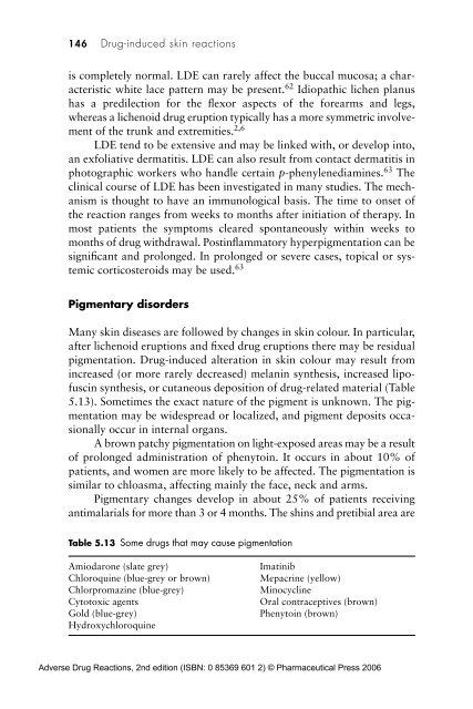
pigmentation. <strong>Drug</strong>-<strong>induced</strong> alteration in <strong>skin</strong> colour may result from<br />
increased (or more rarely decreased) melanin synthesis, increased lipofuscin<br />
synthesis, or cutaneous deposition of drug-related material (Table<br />
5.13). Sometimes the exact nature of the pigment is unknown. The pigmentation<br />
may be widespread or localized, and pigment deposits occasionally<br />
occur in internal organs.<br />
A brown patchy pigmentation on light-exposed areas may be a result<br />
of prolonged administration of phenytoin. It occurs in about 10% of<br />
patients, and women are more likely to be affected. The pigmentation is<br />
similar to chloasma, affecting mainly the face, neck and arms.<br />
Pigmentary changes develop in about 25% of patients receiving<br />
antimalarials for more than 3 or 4 months. The shins and pretibial area are<br />
Table 5.13 Some drugs that may cause pigmentation<br />
Amiodarone (slate grey)<br />
Chloroquine (blue-grey or brown)<br />
Chlorpromazine (blue-grey)<br />
Cytotoxic agents<br />
Gold (blue-grey)<br />
Hydroxychloroquine<br />
Imatinib<br />
Mepacrine (yellow)<br />
Minocycline<br />
Oral contraceptives (brown)<br />
Phenytoin (brown)<br />
Adverse <strong>Drug</strong> Reactions, 2nd edition (ISBN: 0 85369 601 2) © <strong>Pharmaceutical</strong> <strong>Press</strong> 2006