A comparative structural analysis of direct and indirect shoot ...
A comparative structural analysis of direct and indirect shoot ...
A comparative structural analysis of direct and indirect shoot ...

Create successful ePaper yourself
Turn your PDF publications into a flip-book with our unique Google optimized e-Paper software.
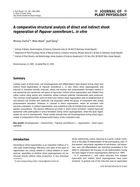
J. Plant Physiol. 157. 281–289 (2000)<br />
© Urban & Fischer Verlag<br />
http://www.urbanfischer.de/journals/jpp<br />
A <strong>comparative</strong> <strong>structural</strong> <strong>analysis</strong> <strong>of</strong> <strong>direct</strong> <strong>and</strong> in<strong>direct</strong> <strong>shoot</strong><br />
regeneration <strong>of</strong> Papaver somniferum L. in vitro<br />
Miroslav Ovečka 1 *, Milan Bobák 2 , Jozef Šamaj 3<br />
1 Institute <strong>of</strong> Botany, Slovak Academy <strong>of</strong> Sciences, Dúbravská cesta 14, SK-842 23 Bratislava, Slovak Republic<br />
2 Department <strong>of</strong> Plant Physiology, Faculty <strong>of</strong> Natural Sciences, Comenius University, Mlynská dolina B-2, SK-84215, Bratislava, Slovak Republic<br />
3 Institute <strong>of</strong> Plant Genetics <strong>and</strong> Biotechnology, Slovak Academy <strong>of</strong> Sciences, Akademická 2, P.O. Box 39 A, SK-950 07 Nitra, Slovak Republic<br />
Received January 11, 2000 · Accepted May 22, 2000<br />
Summary<br />
Cellular origin <strong>of</strong> <strong>shoot</strong> buds, cell morphogenesis <strong>and</strong> differentiation were studied during <strong>direct</strong> <strong>and</strong><br />
in<strong>direct</strong> <strong>shoot</strong> regeneration <strong>of</strong> Papaver somniferum L. in vitro. Direct <strong>shoot</strong> organogenesis was<br />
induced in immature somatic embryos, where cell division <strong>and</strong> protomeristem formation started in<br />
sub-epidermal <strong>and</strong> epidermal cell layers <strong>of</strong> hypocotyl. In<strong>direct</strong> <strong>shoot</strong> regeneration was initiated from<br />
callus culture using auxins <strong>and</strong> cytokinins, where compact globular meristemoids were produced.<br />
The common morphogenetic event <strong>of</strong> <strong>direct</strong> <strong>and</strong> in<strong>direct</strong> <strong>shoot</strong> regeneration was an establishment <strong>of</strong><br />
non-r<strong>and</strong>om cell division <strong>and</strong> restricted cell expansion within the group <strong>of</strong> competent cells during<br />
protomeristem formation. However, in contrast to <strong>direct</strong> regeneration, where all activated cells<br />
became competent, in in<strong>direct</strong> regeneration, only peripheral cells <strong>of</strong> meristemoids acquired morphogenetic<br />
competence. The second difference occurred in <strong>shoot</strong> tunica formation: original hypocotyl<br />
epidermal cells participated in tunica formation during <strong>direct</strong> organogenesis, while this layer regenerated<br />
de novo in meristemoids. These results indicate that cell morphogenesis during <strong>shoot</strong> regeneration<br />
is independent <strong>of</strong> the developmental history <strong>of</strong> the competent cells.<br />
Key words: morphogenesis – morphometry – Papaver somniferum L. – regeneration – <strong>shoot</strong> organogenesis<br />
Introduction<br />
Adventitious <strong>shoot</strong> regeneration is an important method <strong>of</strong> in<br />
vitro plant biotechnology. Efficiency <strong>and</strong> yield <strong>of</strong> this type <strong>of</strong><br />
regeneration are closely related to culture initiation in some<br />
species, regardless <strong>of</strong> whether <strong>shoot</strong> organogenesis can be<br />
induced <strong>direct</strong>ly without callus production. A very important<br />
* E-mail corresponding author: botuove@savba.savba.sk<br />
factor determining culture response to a given culture conditions<br />
is the state <strong>of</strong> differentiation <strong>of</strong> the participating cells. In<br />
this respect, cell polarity, regulation <strong>of</strong> cell division, cell expansion,<br />
<strong>and</strong> cell differentiation are important parameters in the<br />
effort to underst<strong>and</strong> the process <strong>of</strong> cell determination during<br />
the early stages <strong>of</strong> <strong>shoot</strong> organogenesis (Šamaj et al. 1997).<br />
In P. somniferum L., only callus induction from isolated<br />
hypocotyls <strong>and</strong> in<strong>direct</strong> <strong>shoot</strong> organogenesis have been<br />
achieved. A general rule <strong>of</strong> the induction was an application<br />
0176-1617/00/157/03-281 $ 15.00/0
282 Miroslav Ovečka, Milan Bobák, Jozef Šamaj<br />
<strong>of</strong> auxins <strong>and</strong> cytokinins (Nessler <strong>and</strong> Mahlberg 1979, Kamo<br />
et al. 1982, Yoshikawa <strong>and</strong> Furuya 1985, Griffing et al. 1989),<br />
or a transformation <strong>of</strong> the callus tissue originating from hypocotyl<br />
by Agrobacterium rhizogenes (Yoshimatsu <strong>and</strong> Shimomura<br />
1992). Organogenesis in the callus is connected to the<br />
formation <strong>of</strong> meristematic centres–meristemoids with non-r<strong>and</strong>om<br />
distribution <strong>of</strong> starch <strong>and</strong> lipids (Nessler <strong>and</strong> Mahlberg<br />
1979, Šamaj et al. 1990 a, Ovečka et al. 1997). Both starch<br />
<strong>and</strong> lipids have been shown to be related to <strong>shoot</strong> regeneration:<br />
the amount <strong>of</strong> lipids in callus cells is 1.6–2.6 %, while<br />
meristemoid cells contain 13 % in solid culture <strong>and</strong> 34.1% in<br />
liquid culture (Yoshikawa <strong>and</strong> Furuya 1985). Cell specification<br />
in meristemoids takes place during transition phase <strong>of</strong> organogenesis<br />
(meristemoid “maturation”) when the initial accumulation<br />
<strong>and</strong> subsequent utilisation <strong>of</strong> starch <strong>and</strong> lipids change<br />
the nature <strong>of</strong> the cells. As a consequence, the meristemoid<br />
cells express <strong>structural</strong> changes <strong>of</strong> nuclei (Bobák et al.<br />
1990), plastids (Šamaj et al. 1988), <strong>and</strong> vacuolar system<br />
(Šamaj et al. 1990 b). However, the precise data on cellular<br />
origin <strong>of</strong> <strong>shoot</strong> bud primordia are scarce or nonexistent in<br />
early works dealing with opium poppy <strong>shoot</strong> organogenesis.<br />
In our previous study, we documented some cytological <strong>and</strong><br />
morphometrical differences among meristemoid cells during<br />
in<strong>direct</strong> <strong>shoot</strong> organogenesis <strong>of</strong> P. somniferum L. (Ovečka et<br />
al. 1997). Alternatively, bud production was achieved from<br />
cultivated somatic embryos, <strong>and</strong> <strong>direct</strong> <strong>shoot</strong> regeneration <strong>of</strong><br />
opium poppy was documented for the first time (Ovečka et<br />
al. 1997/98). The aim <strong>of</strong> the present work was to study the<br />
similarities <strong>and</strong> differences in morphogenetical steps <strong>of</strong> <strong>shoot</strong><br />
primordia formation during <strong>direct</strong> <strong>and</strong> in<strong>direct</strong> <strong>shoot</strong> regeneration.<br />
We characterised in detail: i. the cellular origin <strong>of</strong><br />
<strong>shoot</strong> primordia <strong>and</strong> <strong>shoot</strong> buds <strong>direct</strong>ly regenerated from<br />
somatic embryos; <strong>and</strong> ii. the cell differentiation during <strong>shoot</strong><br />
primordia formation from meristemoids. In addition to cytological<br />
<strong>and</strong> histological analyses, in<strong>direct</strong> <strong>shoot</strong> regeneration<br />
was studied by morphometrical <strong>analysis</strong>.<br />
Direct <strong>shoot</strong> regeneration was induced from somatic embryos <strong>of</strong> P.<br />
somniferum L. Embryogenic callus culture was initiated from septa <strong>of</strong><br />
poppy capsules using a combination <strong>of</strong> α-naphtaleneacetic acid<br />
(0.537–5.37 µmol/L) <strong>and</strong> kinetin (0.23–0.46 µmol/L). Regeneration <strong>of</strong><br />
somatic embryos took place on the growth regulator-free medium, as<br />
described by Ovečka et al. (1996). Arrested torpedo somatic embryos<br />
unable to develop functional root, spontaneously produced<br />
secondary somatic embryos <strong>and</strong> adventitious <strong>shoot</strong>s during cultivation<br />
on the regeneration medium without growth regulators (Ovečka et<br />
al. 1997/98). Organogenic callus culture was induced from unripe<br />
seeds on solidified MS media (Murashige <strong>and</strong> Skoog 1962), supplemented<br />
with 0.537 µmol/L α-naphtaleneacetic acid <strong>and</strong> 0.46 µmol/L<br />
kinetin. Long-term <strong>shoot</strong> regeneration continued during culture cultivation<br />
on the: (I) induction medium, (II) medium with 0.57 µmol/L indole-3-acetic<br />
acid <strong>and</strong> 2.22 µmol/L 6-benzylaminopurine, <strong>and</strong> (III)<br />
growth regulator-free medium (Ovečka et al. 1997).<br />
Microscopy<br />
Morphological observations were performed <strong>direct</strong>ly on living material<br />
or using scanning electron microscopy (SEM). The samples for SEM<br />
were fixed with 3 % glutaraldehyde (48 h, 0.1mol/L phosphate buffer,<br />
pH 7.2) <strong>and</strong> 2 % OsO 4 (1h, the same buffer <strong>and</strong> pH). After washing in<br />
buffer, the samples were dehydrated in ethanol, critical point dried in<br />
liquid CO 2 , sputter coated with 20 nm layer <strong>of</strong> gold-palladium, <strong>and</strong><br />
observed with a JEOL JXA 840A (JEOL, Japan) microscope. Samples<br />
for transmission electron microscopy (TEM) were fixed in 5 %<br />
glutaraldehyde (5 h, 0.1mol/L phosphate buffer, pH 7) <strong>and</strong> 1% OsO 4<br />
(2 h, 0.1 mol/L phosphate buffer, pH 7), dehydrated in acetone <strong>and</strong><br />
embedded in Durcupan ACM (Fluca, Buchs, Switzerl<strong>and</strong>). Ultrathin<br />
sections were stained according to Reynolds (1963) <strong>and</strong> observed in<br />
ATEM 2000FX (JEOL, Japan) <strong>and</strong> TESLA BS 500 (Tesla, Czech Republic)<br />
electron microscopes. Histological <strong>and</strong> cytological analyses<br />
<strong>of</strong> <strong>shoot</strong> regeneration were performed at the light microscopy level.<br />
Paraffin sections (7–10 µm) were prepared after sample fixation in FAA<br />
(formalin 40 %, acetic acid 5 %, ethyl alkohol 50 %), embedded in<br />
Histoplast S (Serva, Heidelberg, Germany) <strong>and</strong> stained with hematoxylin-eosin,<br />
PAS reaction or Feuglen reaction. Sections (1–2 µm<br />
thick) for the fine cytological observation were prepared from the<br />
samples embedded for TEM. After SEM observation, some dried bulk<br />
samples were immersed in ethanol <strong>and</strong> embedded in Histoplast S<br />
(Serva, Heidelberg, Germany). The sections (7–10 µm thick) were observed<br />
without additional staining under bright-field microscopy<br />
(Ovečka <strong>and</strong> Bobák 1999). All prepared samples were studied using<br />
ZEISS Jenalumar (Carl Zeiss, Jena, Germany) dissection light microscope.<br />
Pictures digitalised using a Kappa CF 8/1 FMCC camera<br />
(Kappa Messtechnik, Germany), <strong>and</strong> a Leica Q500MC image <strong>analysis</strong><br />
system (Leica Cambridge Ltd. Engl<strong>and</strong>, UK) were processed<br />
using Corel Photo Paint 8 (Corel Corporation, Canada).<br />
Material <strong>and</strong> Methods<br />
In vitro cultures<br />
Morphometry<br />
All planimetric measurements <strong>of</strong> meristemoid <strong>and</strong> <strong>shoot</strong> primordia<br />
cells during in<strong>direct</strong> <strong>shoot</strong> regeneration were made using an ASBA<br />
(Wild, Heerbrugg, Switzerl<strong>and</strong>) image analyser. Cell size, cell shape,<br />
<strong>and</strong> nucleus size were measured on the basis <strong>of</strong> cell cross-section<br />
area in 1–2 µm thick sections. The mean values were compared<br />
among the above-mentioned three culture media with different<br />
amounts <strong>of</strong> growth regulators. The cell shape (form factor) was calculated<br />
from the cell area <strong>and</strong> cell projections (a, b, a+b, a–b). The<br />
form factor 5.09 means a circular shape, 6 means a square <strong>and</strong> 7 a<br />
rectangle shape with a side ratio <strong>of</strong> 2 : 1 (Baluška et al. 1990). Morphometrical<br />
values <strong>of</strong> cells in central <strong>and</strong> peripheral zones <strong>of</strong> the regenerated<br />
<strong>shoot</strong> apical meristems were measured as st<strong>and</strong>ards for<br />
the <strong>comparative</strong> characterisation <strong>of</strong> meristemoid cells. The computed<br />
sizes, shapes <strong>of</strong> cells, <strong>and</strong> N/C ratios were used as parameters <strong>of</strong><br />
meristemoid cell activity (cell division <strong>and</strong> cell expansion). Linear regression<br />
analyses, coefficients <strong>of</strong> correlation, <strong>and</strong> N/C ratios were calculated<br />
using Sigma Plot (J<strong>and</strong>el Scientific, USA) statistical s<strong>of</strong>tware.
Shoot regeneration in opium poppy<br />
283<br />
Results<br />
Shoot regeneration was initiated from hypocotyls <strong>of</strong> primary<br />
somatic embryos (Fig. 1 a). The emerging <strong>shoot</strong> primordia<br />
appeared on the hypocotyl surface <strong>of</strong> the somatic embryo<br />
hypocotyl, but they did not disrupt the surface tissues (Fig.<br />
1 b). In addition, no general tissue reorganisation was observed<br />
(Fig. 1b). Due to these features, this kind <strong>of</strong> regeneration<br />
was termed <strong>direct</strong> <strong>shoot</strong> organogenesis in this study.<br />
In<strong>direct</strong> <strong>shoot</strong> regeneration involved tissue dedifferentiation,<br />
callus formation, <strong>and</strong> cell re-differentiation leading to de novo<br />
<strong>shoot</strong> regeneration (Fig. 1 c). Tissue organisation <strong>and</strong> cell<br />
morphogenesis <strong>of</strong> in<strong>direct</strong>ly regenerated <strong>shoot</strong> primordia were<br />
completely different in comparison to underlying callus tissue<br />
(Fig.1d).<br />
Direct <strong>shoot</strong> regeneration<br />
Surface integrity <strong>of</strong> the embryo hypocotyl suggested that adventitious<br />
<strong>shoot</strong>s arose from superficial cell layers (Fig. 1 b).<br />
Initially, sub-epidermal cells were activated <strong>and</strong> divided. The<br />
plane <strong>of</strong> the first cell division seemed to be anticlinal (Fig.<br />
2 a), but subsequent cell divisions were observed in both anti-<br />
<strong>and</strong> periclinal planes (Fig. 2 b). Afterwards, dividing subepidermal<br />
<strong>and</strong> epidermal cells created organogenic nodules,<br />
the first recognisable structures on the hypocotyl surface<br />
(Figs. 2 c, d). The two most important morphogenetic features<br />
<strong>of</strong> these cells were dense non-vacuolated cytoplasm (Figs.<br />
2 a, b, c) <strong>and</strong> reduced cell size (Fig. 2 d).<br />
The first apparent indication <strong>of</strong> cell specification was observed<br />
in the phase <strong>of</strong> apical meristem formation. The cells<br />
within nodule apices adopted tunica-corpus zonation (Figs.<br />
2 e, f). The original epidermal cells participated in differentiation<br />
<strong>of</strong> the tunica layer (Fig. 2 c). This explains why the tunica<br />
<strong>and</strong> protomeristem maintained morphological <strong>and</strong> histological<br />
continuity between the forthcoming buds <strong>and</strong> the remaining<br />
hypocotyl surface (Figs. 1b, 2 c, d, g). Cell specialisation<br />
continued by both histodifferentiation <strong>of</strong> the <strong>shoot</strong> apical meristem<br />
(Figs. 2 e, f) <strong>and</strong> transformation <strong>of</strong> organogenic nodules<br />
into buds (Figs. 2 g, h). Adventitious buds were visible on<br />
the hypocotyl surface (Figs. 2 g, h) <strong>and</strong> regular activity <strong>of</strong> the<br />
apical meristem was detected, including formation <strong>and</strong> growth<br />
<strong>of</strong> leaf primordia (Figs. 2 h, i).<br />
In<strong>direct</strong> <strong>shoot</strong> regeneration<br />
Distinct mode <strong>of</strong> in<strong>direct</strong> induction <strong>of</strong> organogenesis was<br />
based on callus production. Cell activation triggered a morphogenetic<br />
switch in some cells within rarely dividing callus<br />
tissue, resulting in the establishment <strong>of</strong> dividing, meristemlike,<br />
<strong>and</strong> <strong>shoot</strong>-forming tissue (Fig. 3 a). Typical features <strong>of</strong><br />
meristemoids (representing population <strong>of</strong> competent cells)<br />
were small cell size, cytoplasmic density, minimal vacuolation<br />
(Figs. 3 a, b), <strong>and</strong> cell adhesion after cell division (Figs. 1 d,<br />
3 a, b, c). The first cell differentiation events within the multicellular<br />
meristemoids resulted in the formation <strong>of</strong> both peripheral<br />
<strong>and</strong> central cell layers, with only peripheral cells continuing<br />
their divisions (Fig. 3 b). Reserves were stored in central<br />
cells in the form <strong>of</strong> starch (Figs. 3 b, d) <strong>and</strong> lipids (Figs. 3 e, f).<br />
These cells were non-dividing but their cytological parameters<br />
indicated high metabolic activity. Frequent endo- <strong>and</strong><br />
exocytosis in these cells (Fig. 3 e) <strong>and</strong> cell wall ingrowth for-<br />
Figure 1. Shoot buds regenerated in the callus culture <strong>of</strong> P.<br />
somniferum L. a. Somatic embryo (SE) in the culture where<br />
the <strong>shoot</strong> (SH) regeneration from the hypocotyl was<br />
induced. Bar = 1 mm. b. Directly regenerated <strong>shoot</strong>s with<br />
leaf primordia, visible as protuberances on the hypocotyl<br />
surface <strong>of</strong> somatic embryo. Bar = 100 µm. c. Shoot bud<br />
regenerated in the organogenic callus culture. The white<br />
compact tissue represents meristemoids. Bar = 1 mm. d.<br />
Shoot primordium on the surface <strong>of</strong> callus tissue. Bar =<br />
100 µm.
284 Miroslav Ovečka, Milan Bobák, Jozef Šamaj<br />
Figure 2. Direct <strong>shoot</strong> regeneration. a., b. Longitudinal section<br />
<strong>of</strong> the embryo hypocotyl in the stage <strong>of</strong> cell activation.<br />
a. First localised anticlinal cell divisions <strong>of</strong> sub-epidermal<br />
cells (arrows). Bar = 50 µm b. Subsequent anticlinal <strong>and</strong><br />
periclinal cell divisions changed size <strong>and</strong> shape <strong>of</strong> sub-epidermal<br />
activated cells. Bar = 50 µm. c. Localised cell divisions<br />
in organogenic nodule. Frequent cell divisions <strong>of</strong> subepidermal<br />
cells considerably reduced their size. First activated<br />
epidermal cells are detected (arrows). Bar = 50 µm.<br />
d. Organogenic nodules visible on the hypocotyl surface.<br />
Bar = 50 µm. e. Concentration <strong>of</strong> cell proliferation in the<br />
centre <strong>of</strong> organogenic nodule. Bar = 50 µm. f. Protomeristem<br />
with established tunica-corpus zonation in the stage<br />
<strong>of</strong> protomeristem transformation into <strong>shoot</strong> apical meristem.<br />
Bar = 50 µm. g. Cross-section <strong>of</strong> the <strong>shoot</strong> bud primordium<br />
in the vicinity <strong>of</strong> meristematic root pole <strong>of</strong> somatic embryo.<br />
Note the involvement only <strong>of</strong> the epidermal <strong>and</strong> several<br />
sub-epidermal cell layers in adventitious <strong>shoot</strong> production.<br />
Bar = 50 µm. h. Surface view on the <strong>shoot</strong> apical meristem<br />
with well defined three layered tunica (arrow) <strong>and</strong> initiation<br />
<strong>of</strong> leaf primordia outgrowth (arrowheads). Bar = 100 µm. i.<br />
Developing leaves from the flattened apical meristem <strong>of</strong><br />
adventitious <strong>shoot</strong> bud. Bar = 100 µm.<br />
mations in the cells possessing numerous mitochondria (Fig.<br />
3 g) indicated active cell-to-cell transport.<br />
Division <strong>of</strong> the peripheral meristemoid cells was not properly<br />
regulated. Divisions took place in all <strong>direct</strong>ions, resulting<br />
in a r<strong>and</strong>om arrangement <strong>of</strong> meristemoid cells (Fig. 3 c).<br />
However, the selection <strong>of</strong> dividing meristemoid periphery was<br />
very important in cell determination during <strong>shoot</strong> primordia<br />
formation. The change in morphogenesis was connected<br />
with regulation <strong>of</strong> cell division <strong>of</strong> the outermost peripheral<br />
meristemoid cells. The cells exp<strong>and</strong>ed in a radial <strong>direct</strong>ion<br />
<strong>and</strong> divided periclinally (Fig. 4 a). Later, peripherally located<br />
daughter cells formed tunica cell layer undergoing anticlinal<br />
divisions (Fig. 4 b). Organogenesis by protomeristem formation<br />
continued, regularly associated with starch depletion in<br />
the <strong>shoot</strong>-forming meristemoids (Figs. 4 c, d). The <strong>shoot</strong> primordium<br />
was established when cell proliferation in the procambium<br />
region <strong>and</strong> cell division in the peripheral zone <strong>of</strong><br />
the apical meristem were detected (Figs. 4 c, e).<br />
Cell division <strong>and</strong> cell differentiation in procambial region <strong>of</strong><br />
both <strong>direct</strong>ly <strong>and</strong> in<strong>direct</strong>ly regenerated <strong>shoot</strong>s were important<br />
for differentiation <strong>of</strong> vascular tissue <strong>and</strong> laticifer system<br />
typical for P. somniferum L. Laticifers appeared first as laticifer<br />
initials (Fig. 4 f); later they formed an articulated system<br />
during cell expansion (Fig. 4 g). Cell wall perforation also<br />
contributed to laticifer coupling in the lateral <strong>direct</strong>ion to promote<br />
anastomosing <strong>of</strong> laticifer arrays. Early stages <strong>of</strong> perforation<br />
were characterised by thinner <strong>and</strong> elastic cell wall within<br />
the site <strong>of</strong> future perforation, which allowed impressing <strong>and</strong><br />
partial movement <strong>of</strong> cell contents between neighbouring cells<br />
(Figs. 4 h, i). After cell wall perforation had been completed,<br />
typical multinuclear, articulated, anastomosing laticifer system<br />
was observed in both developing <strong>shoot</strong> procambium<br />
(Fig. 4 j) <strong>and</strong> developing leaves (Fig. 4 k) <strong>of</strong> adventitious<br />
<strong>shoot</strong>s regenerated in vitro.<br />
Our histological observations revealed that peripheral meristemoid<br />
cells represent the original site <strong>of</strong> <strong>shoot</strong> primordia<br />
formation. We used morphometric measurements <strong>of</strong> undifferentiated<br />
<strong>and</strong> dividing cells <strong>of</strong> regenerated <strong>shoot</strong> apical meristems<br />
in our effort to properly characterise meristemoid<br />
cells during the course <strong>of</strong> <strong>shoot</strong> primordia formation. All cells<br />
in the central <strong>and</strong> peripheral zones <strong>of</strong> <strong>shoot</strong> apical meristems<br />
expressed high correlation between the volumes <strong>of</strong> the cell<br />
<strong>and</strong> nucleus (N/C ratio). When we compared the measured<br />
N/C ratio values <strong>of</strong> central <strong>and</strong> peripheral cells within the<br />
apical meristem <strong>of</strong> regenerated <strong>shoot</strong>s <strong>and</strong> peripheral meristemoid<br />
cells, we found a high degree <strong>of</strong> similarity (Fig. 5).<br />
Cell size was only one distinct morphometrical parameter. As<br />
the cells <strong>of</strong> peripheral <strong>and</strong> central zones <strong>of</strong> apical meristems,<br />
peripheral meristemoid cells were larger. As the cells in the<br />
rib-zone <strong>of</strong> <strong>shoot</strong> apical meristem (Fig. 6 a), central meristemoid<br />
cells were larger. Mathematical evaluation <strong>of</strong> the cell<br />
shape (form factor) revealed an additional difference: lower<br />
values for peripheral meristemoid cells <strong>and</strong> higher values for<br />
<strong>shoot</strong> apical meristem cells (Fig. 6 b). This means that periph-
Shoot regeneration in opium poppy<br />
285<br />
Figure 3. In<strong>direct</strong> <strong>shoot</strong> regeneration. 1. Cell determination.<br />
a. Production <strong>of</strong> meristemoids within the callus tissue. Bar =<br />
50 µm. b. Tight cell adhesion <strong>and</strong> meristematic nature <strong>of</strong><br />
the meristemoid cells. Note the cytological differences<br />
(cytoplasmic density, thickness <strong>and</strong> stainability <strong>of</strong> the cell<br />
wall) between peripheral dividing cells <strong>and</strong> cells accumulating<br />
starch granules. Bar = 50 µm. c. Numerous globular<br />
meristemoids appearing mostly on the callus surface. Cell<br />
arrangement in the meristemoids was r<strong>and</strong>om, but they had<br />
compact, non-friable nature. Bar = 50 µm. d, e, f, g, – Ultrastructure<br />
<strong>of</strong> the central meristemoid cells. d. Transparent<br />
cytoplasm <strong>and</strong> large deposits <strong>of</strong> starch in the central cells.<br />
Bar = 10 µm. e. Insertion <strong>of</strong> vesicular <strong>and</strong> matrix material<br />
into the cell wall <strong>of</strong> central meristemoid cells by exocytosis.<br />
L-lipid droplet. Bar = 2 µm. f. Lipid droplets which fill up the<br />
cells in the transition layer between centre <strong>and</strong> periphery <strong>of</strong><br />
the meristemoid. Bar = 10 µm. g. Cell wall ingrowths (asterisk)<br />
in the central meristemoid cell. Note the presence <strong>of</strong><br />
numerous mitochondria. Bar = 2 µm.<br />
eral meristemoid cells were more rounded <strong>and</strong> cells <strong>of</strong> <strong>shoot</strong><br />
apical meristem were more oblong. However, unlike these differences<br />
in cell size <strong>and</strong> shape, in both meristemoid periphery<br />
(Fig. 7a) <strong>and</strong> <strong>shoot</strong> apical meristem (Fig. 7b), cell growth<br />
was closely related to the similar negative correlation<br />
between cell size <strong>and</strong> cell shape. Increase in the mean <strong>of</strong> cell<br />
size was found to be followed by general decrease in the<br />
mean <strong>of</strong> form factor (Figs. 7 a, b). In conclusion, most <strong>of</strong> the<br />
cytological <strong>and</strong> morphometrical observations indicate that<br />
only peripheral meristemoid cells can be identified as determined<br />
cells during in<strong>direct</strong> <strong>shoot</strong> organogenesis.<br />
Discussion<br />
This study deals with cytological characterisation <strong>of</strong> de novo<br />
<strong>shoot</strong> regeneration <strong>of</strong> P. somniferum L. in vitro, focusing from<br />
the morphogenetical point <strong>of</strong> view on the differences between<br />
<strong>direct</strong> <strong>and</strong> in<strong>direct</strong> <strong>shoot</strong> primordia formation. Shoot organogenesis<br />
is a suitable experimental system for use in discerning<br />
patterns <strong>of</strong> cell division <strong>and</strong> expansion in order to<br />
compare <strong>direct</strong> <strong>and</strong> in<strong>direct</strong> caulogenetic determination.<br />
Three developmental steps have been identified in distinct<br />
<strong>shoot</strong>-forming cultures: (I) initiation <strong>of</strong> meristematic activity in<br />
callus or explant as an expression <strong>of</strong> the morphogenetic<br />
competence; (II) cell determination during formation <strong>of</strong> meristematic<br />
nodules; <strong>and</strong> (III) differentiation <strong>of</strong> adventitious <strong>shoot</strong><br />
buds (Von Arnold <strong>and</strong> Eriksson 1985, Bobák et al. 1989,<br />
Sharma <strong>and</strong> Bhojwani 1990, Colby et al. 1991, Lakshmanan<br />
et al. 1997). In contrast to well established models <strong>of</strong> <strong>shoot</strong><br />
regeneration, the <strong>structural</strong> details <strong>of</strong> <strong>shoot</strong> primordia initiation<br />
<strong>and</strong> development are missing in studies <strong>of</strong> organogenic<br />
poppy callus culture. In addition, <strong>direct</strong> <strong>shoot</strong> organogenesis<br />
<strong>of</strong> opium poppy has not been published before now. The<br />
question here was what aspects <strong>of</strong> cell division, cell expansion,<br />
<strong>and</strong> cell differentiation are related to cellular origin <strong>of</strong><br />
<strong>shoot</strong> primordia during either <strong>direct</strong> or in<strong>direct</strong> <strong>shoot</strong> regeneration.
286 Miroslav Ovečka, Milan Bobák, Jozef Šamaj<br />
Figure 4. In<strong>direct</strong> <strong>shoot</strong> regeneration. 2. Formation <strong>of</strong> <strong>shoot</strong><br />
primordia. a. Morphogenesis <strong>of</strong> peripheral meristemoid<br />
cells during <strong>shoot</strong> primordia formation. Cells exp<strong>and</strong>ed in<br />
anticlinal <strong>direct</strong>ion (arrowhead) <strong>and</strong> subsequently divided<br />
periclinally (arrows). Bar = 50 µm. b. Establishment <strong>of</strong> the<br />
tunica layer on the meristemoid surface by anticlinal cell<br />
division. Bar = 50 µm. c. Histodifferentiation <strong>of</strong> the <strong>shoot</strong><br />
apical meristem (SAM). Bar = 50 µm. d. Cell proliferation<br />
concentrated in the dome-like protomeristem. Cells located<br />
away <strong>of</strong> the centre <strong>of</strong> cell division are devoid <strong>of</strong> starch <strong>and</strong><br />
start to vacuolate. Bar = 50 µm. e. Developed <strong>shoot</strong> bud<br />
with leaf primordia <strong>and</strong> vascular tissue (VT) in the procambium.<br />
Bar = 100 µm. f. Laticifer initials (arrows) in the procambial<br />
region <strong>of</strong> <strong>shoot</strong> apical meristem. Bar = 50 µm. g.<br />
Initial perforation <strong>of</strong> cross cell wall between two laticifer initials.<br />
Bar = 50 µm. h. Formation <strong>of</strong> articulated, anastomosing<br />
laticifer system. Note the elastic properties <strong>of</strong> longitudinal<br />
cell wall in the site <strong>of</strong> future perforation (arrow). Bar =<br />
50 µm. i. Insertions <strong>of</strong> cell volume between future anastomosing<br />
laticifers in cross-section (arrows). Bar = 50 µm. j.<br />
Articulated, anastomosed laticifer system in <strong>shoot</strong> procambium.<br />
Bar = 50 µm. k. Articulated, anastomosed laticifer<br />
system in developing leaves <strong>of</strong> regenerated <strong>shoot</strong>s. Bar =<br />
50 µm.<br />
Figure 5. Similarity in the correlation curves between cell<br />
size <strong>and</strong> nucleus size <strong>of</strong> peripheral meristemoid cells<br />
(——) <strong>and</strong> cells <strong>of</strong> peripheral <strong>and</strong> central zones <strong>of</strong> the<br />
apical meristems in regenerated <strong>shoot</strong>s (——). 30–50<br />
cells <strong>of</strong> each zone were measured on the medium supplemented<br />
with 0.537 µmol/L <strong>of</strong> α-naphtaleneacetic acid <strong>and</strong><br />
0.46 µmol/L kinetin (I), or with 0.577 µmol/L indole-3-acetic<br />
acid <strong>and</strong> 2.22 µmol/L 6-benzylaminopurine (II), or growth<br />
regulator-free medium (III). Cell size values <strong>and</strong> nucleus<br />
size values are presented as planimetric values <strong>of</strong> cell <strong>and</strong><br />
nucleus area measured from cross sections.<br />
Direct <strong>shoot</strong> formation was initiated in the culture where<br />
somatic embryos had failed to develop correctly, but maintained<br />
continuously juvenile, undifferentiated state <strong>of</strong> their<br />
cells in the root pole which were able to generate new somatic<br />
embryos (Ovečka et al. 1997/98). However, as we show<br />
here, adventitious <strong>shoot</strong>s originated from the differentiated,<br />
non-dividing tissue <strong>of</strong> hypocotyl. The competent cells have to<br />
be re-activated <strong>and</strong> they enter cell division. The culture underwent<br />
spontaneous repetitive regeneration including <strong>shoot</strong><br />
organogenesis <strong>and</strong> somatic embryogenesis (Ovečka et al.<br />
1997/98). Some internal factors determining cell competence<br />
in the course <strong>of</strong> <strong>shoot</strong> bud regeneration have been identified,<br />
such as reduced cellulase activity in tobacco callus culture<br />
(Truelsen <strong>and</strong> Ulvskov 1995), or importance <strong>of</strong> polar auxin<br />
transport, because its inhibition by TIBA (2,3,5-triiodobenzoic<br />
acid) strongly enhanced <strong>shoot</strong> regeneration, but not somatic
Shoot regeneration in opium poppy<br />
287<br />
Figure 6. Mean <strong>of</strong> the cell cross section area (a), <strong>and</strong> mean<br />
<strong>of</strong> the form factor describing cell shape (b), observed during<br />
<strong>shoot</strong> regeneration in the medium supplemented with<br />
0.537 µmol/L <strong>of</strong> α-naphtaleneacetic acid <strong>and</strong> 0.46 µmol/L<br />
kinetin. Peripheral meristemoid cells (A) were compared to<br />
cells <strong>of</strong> central <strong>and</strong> peripheral zones <strong>of</strong> regenerated <strong>shoot</strong><br />
apical meristems (B), <strong>and</strong> central meristemoid cells (C)<br />
were compared to cells in the rib-zone <strong>of</strong> <strong>shoot</strong> apical<br />
meristems (D). Percentage amount <strong>of</strong> cells with isodiametric<br />
(a side ratio app. 1 : 1), <strong>and</strong> oval shape (side ratio up to<br />
2 : 1) is indicated (b). The rest <strong>of</strong> the cells had a more elongated<br />
shape. Results from measurements <strong>of</strong> app. 300 cells<br />
<strong>of</strong> each selected part <strong>of</strong> meristemoids <strong>and</strong> <strong>shoot</strong> apical<br />
meristems. ** – significant at P
288 Miroslav Ovečka, Milan Bobák, Jozef Šamaj<br />
Shoot regeneration in callus culture requires a combination<br />
<strong>of</strong> auxins <strong>and</strong> cytokinins (Christianson <strong>and</strong> Warnick 1983, De<br />
Klerk et al. 1997). The main change in cell division <strong>and</strong> cell<br />
expansion was observed during competence acquisition.<br />
Meristemoids were produced from rarely dividing callus cells.<br />
Cell division was more patterned within meristemoids. Their<br />
cells were small, micro-vacuolated, <strong>and</strong> able to stick together.<br />
This indicates that the fate <strong>of</strong> the callus culture is altered<br />
through cell morphogenesis <strong>of</strong> the activated competent<br />
cells as a result <strong>of</strong> the action <strong>of</strong> growth regulators.<br />
The second switching point in cell morphogenesis <strong>of</strong><br />
poppy callus culture occurred in the multicellular meristemoids<br />
as a defined step <strong>of</strong> cell determination within the <strong>shoot</strong>forming<br />
tissue. A critical event was the localisation <strong>of</strong> cell<br />
division to the meristemoid periphery. The cell determination<br />
has been expressed when modified polar periclinal cell division<br />
occurred in meristemoids. Apical daughter cells generated<br />
dividing peripheral layers with morphogenetic competence<br />
while the distal daughter cells became more differentiated<br />
<strong>and</strong> generated storage cells possessing metabolic<br />
sources. The previously observed differences in cell size <strong>and</strong><br />
shape showed that the peripheral cells were smaller, having<br />
an oblong shape as a result <strong>of</strong> frequent cell division, while<br />
the central cells became larger due to globular expansion<br />
(Ovečka et al. 1997). Starch-containing cells were the largest<br />
ones. They had isodiametric shape, <strong>and</strong> any changes (cell<br />
division, plastid division, vacuolation) took place only after<br />
starch utilisation had been initiated (not shown). Central<br />
meristemoid cells thus can support organogenesis by the<br />
energy. It was clearly shown that some metabolic indications<br />
<strong>of</strong> <strong>shoot</strong> regeneration (protein inclusions, starch, lipids, high<br />
enzyme activities) were found only in meristemoids (Ross et<br />
al. 1973, Nessler <strong>and</strong> Mahlberg 1979, Patel <strong>and</strong> Berlyn 1983).<br />
Meristemoids possessed a high starch content, while high<br />
activities <strong>of</strong> starch-degrading enzymes were observed in<br />
<strong>shoot</strong> buds (Thorpe <strong>and</strong> Murashige 1970, Thorpe <strong>and</strong> Meier<br />
1974, Patel <strong>and</strong> Berlyn 1983). A high N/C ratio <strong>of</strong> peripheral<br />
meristemoid cells <strong>of</strong> opium poppy, comparable with cells in<br />
<strong>shoot</strong> apical meristem (Fig. 5), together with comparable rate<br />
<strong>of</strong> chromatin de-condensation (Ovečka et al. 1997) are also<br />
important parameters <strong>of</strong> cell determination within the meristemoids.<br />
Variability in the size <strong>of</strong> nucleus <strong>and</strong> the correlation<br />
with DNA amount is closely related to <strong>shoot</strong> regeneration<br />
(Flinn et al. 1989, Fournier et al. 1991). A two-phase organogenesis,<br />
with meristemoid formation during the first phase<br />
<strong>and</strong> formation <strong>of</strong> globular structures from the surface during<br />
the second phase, was observed in sundew callus tissue<br />
(Bobák et al. 1993). This data indicates that “mitotic zonation”<br />
established a distinct morphological competence <strong>of</strong> meristemoid<br />
cells in the process <strong>of</strong> <strong>shoot</strong> primordia formation<br />
when the site <strong>of</strong> origin <strong>of</strong> <strong>shoot</strong> buds was located at the<br />
meristemoid periphery.<br />
Concluding our <strong>comparative</strong> <strong>analysis</strong> <strong>of</strong> <strong>direct</strong> <strong>and</strong> in<strong>direct</strong><br />
<strong>shoot</strong> regeneration in P. somniferum L., important changes in<br />
cell morphogenesis, characterised by non-r<strong>and</strong>om cell divisions,<br />
took place during dermal layer <strong>and</strong> protomeristem formation<br />
in <strong>shoot</strong> primordia. We observed a r<strong>and</strong>om orientation<br />
<strong>of</strong> cell divisions in meristemoids, while protomeristem <strong>and</strong> especially<br />
tunica formation were accompanied by regulated<br />
periclinal <strong>and</strong> subsequently anticlinal cell divisions. In <strong>direct</strong>ly<br />
regenerated <strong>shoot</strong>s, the dermal layer was formed by original<br />
epidermal cells through anticlinal cell divisions. This type <strong>of</strong><br />
division maintains epidermal cell polarity. Peripheral meristemoid<br />
cells are polarised with non-homogenous cell surfaces<br />
due to the presence <strong>of</strong> adhered <strong>and</strong> outer non-adhered cell<br />
walls (Ovečka <strong>and</strong> Bobák 1999). This polarity <strong>and</strong> cell position<br />
are important morphogenetic factors in cell determination.<br />
Cell expansion was found to be minimal in cells involved<br />
in formative, morphogenetically important cell division. The<br />
cells exp<strong>and</strong>ed, but not over the cell volume necessary for a<br />
subsequent cell division. In addition, cell size <strong>and</strong> cell shape<br />
have to be negatively correlated. This relationship would indicate<br />
an independence <strong>of</strong> post-mitotic cell growth <strong>and</strong> an extensive<br />
cell vacuolation, originally observed in maize root<br />
apex (Baluška et al. 1990). A <strong>comparative</strong> <strong>analysis</strong> revealed<br />
that cell activation in <strong>shoot</strong> organogenesis <strong>of</strong> P. somniferum<br />
L. depends on the stage <strong>of</strong> cell differentiation before initiation,<br />
while cell morphogenesis <strong>of</strong> the determined cells is similarly<br />
regulated both spatially <strong>and</strong> temporally, during <strong>direct</strong><br />
<strong>and</strong> in<strong>direct</strong> <strong>shoot</strong> regeneration.<br />
Acknowledgements. We gratefully acknowledge D. Jantová for technical<br />
assistance, <strong>and</strong> Dr. M. Čiamporová for the English correction.<br />
This work was supported by Grant Agency VEGA, Grant No.1/6030/99<br />
<strong>and</strong> Grant No. 1/6107/99 from the Slovak Academy <strong>of</strong> Sciences. M.O.<br />
gratefully acknowledges the Alex<strong>and</strong>er von Humboldt-Stiftung (Bonn,<br />
Germany) for donation <strong>of</strong> the Leica Q500MC image <strong>analysis</strong> system.<br />
References<br />
Altamura MM, Capitani F, Falasca G, Gallelli A, Scaramagli S, Bueno<br />
M, Torrigiany P, Bagni N (1995) Morphogenesis in cultured thin<br />
layers <strong>and</strong> pith explants <strong>of</strong> tobacco. I. Effect <strong>of</strong> putrescine on cell<br />
size, xylogenesis <strong>and</strong> meristemoid organization. J Plant Physiol<br />
147: 101–106<br />
Baluška F, Kubica Š, Hauskrecht M (1990) Postmitotic “isodiametric”<br />
cell growth in the maize root apex. Planta 181: 269–274<br />
Bobák M, Blehová A, Erdelský K (1989) Histological <strong>and</strong> cytological<br />
study <strong>of</strong> early phases <strong>of</strong> organogenesis on excised leaves <strong>of</strong> Drosera<br />
spathulata L. cultivated in vitro. Biológia (Bratislava) 44: 785–792<br />
Bobák M, Blehová A, Šamaj J, Ovečka M, Krištín J (1993) Studies <strong>of</strong><br />
organogenesis from the callus culture <strong>of</strong> the sundew (Drosera<br />
spathulata Labill.). J Plant Physiol 142: 251–253<br />
Bobák M, Šamaj J, Erdelský K (1990) Ultra<strong>structural</strong> polymorphysm <strong>of</strong><br />
interkinetic nuclei <strong>and</strong> nucleoli in the cells <strong>of</strong> organogenic callus<br />
cultures <strong>of</strong> Papaver somniferum L. in in vitro conditions. Acta FRN<br />
Univ Comen – Physiol Plant XXV: 3–20<br />
Bronner R, Jeannin G, Hahne G (1994) Early cellular events during organogenesis<br />
<strong>and</strong> somatic embryogenesis induced on immature<br />
zygotic embryos <strong>of</strong> sunflower (Helianthus annuus). Can J Bot 72:<br />
239–248
Shoot regeneration in opium poppy<br />
289<br />
Burritt DJ, Leung DWM (1996) Organogenesis in cultured petiole explants<br />
<strong>of</strong> Begonia × erythrophylla: the timing <strong>and</strong> specificity <strong>of</strong> the<br />
inductive stimuli. J Exp Bot 47: 557–567<br />
Choi YE, Kim HS, Soh WY, Yang DC (1997) Developmental <strong>and</strong> <strong>structural</strong><br />
aspects <strong>of</strong> somatic embryos formed on medium containing<br />
2,3,5-triiodobenzoic acid. Plant Cell Rep 16: 738–744<br />
Christianson ML, Warnick DA (1983) Competence <strong>and</strong> determination<br />
in the process <strong>of</strong> in vitro <strong>shoot</strong> organogenesis. Dev Biol 95: 288–<br />
293<br />
Colby SM, Juncosa AM, Stamp JA, Meredith CP (1991) Developmental<br />
anatomy <strong>of</strong> <strong>direct</strong> <strong>shoot</strong> organogenesis from leaf petioles <strong>of</strong><br />
Vitis vinifera (Vitaceae). Amer J Bot 78: 260–269<br />
De Klerk G-J, Arnholdt-Schmitt B, Lieberei R, Neumann K-H (1997)<br />
Regeneration <strong>of</strong> roots, <strong>shoot</strong>s <strong>and</strong> embryos: physiological, biochemical<br />
<strong>and</strong> molecular aspects. Biol Plant 39: 53–66<br />
Flinn BS, Webb DT, Newcomb W (1989) Morphometric <strong>analysis</strong> <strong>of</strong> reserve<br />
substances <strong>and</strong> ultra<strong>structural</strong> changes during caulogenic<br />
determination <strong>and</strong> loss <strong>of</strong> competence <strong>of</strong> Eastern white pine (Pinus<br />
strobus) cotyledons in vitro. Can J Bot 67: 779–789<br />
Fournier D, Bonnelle Ch, Tourte Y (1991) Ultra<strong>structural</strong> features <strong>of</strong><br />
soybean somatic cells at the beginning <strong>of</strong> an organogenic process:<br />
toward a new concept. Biol Cell 73: 99–105<br />
Gilissen LJW, Van Staveren MJ, Hakkert JC, Smulders MJM, Verhoeven<br />
HA, Creemers-Molenaar J (1994) The competence <strong>of</strong> cells for cell<br />
division <strong>and</strong> regeneration in tobacco explants depends on cellular<br />
location, cell cycle phase <strong>and</strong> ploidy level. Plant Sci 103: 81–91<br />
Griffing LR, Fowke LC, Constabel F (1989) Radioimmunoassay <strong>of</strong><br />
morphinan alkaloids in Papaver somniferum hypokotyls, callus <strong>and</strong><br />
suspension cultures. J Plant Physiol 134, 645–650<br />
Kamo KK, Kimoto W, Hsu AF, Mahlberg PG, Bills DD (1982) Morphine<br />
alkaloids in cultured tissues <strong>and</strong> redifferentiated organs <strong>of</strong> Papaver<br />
somniferum. Phytochemistry 21: 219–222<br />
Konieczny R (1996) Plant regeneration from immature embryo culture<br />
<strong>of</strong> Trifolium michelianum Savi. – histological observations on adventitious<br />
<strong>shoot</strong> induction. Acta Soc Bot Pol 65: 261–266<br />
Lakshmanan P, Ng SK, Loh CS, Goh CJ (1997) Auxin, cytokinin <strong>and</strong><br />
ethylene differentially regulate specific developmental states associated<br />
with <strong>shoot</strong> bud morphogenesis in leaf tissues <strong>of</strong> mangosteen<br />
(Garcinia mangostana L.) cultured in vitro. Plant Cell Physiol<br />
38: 59–64<br />
Marcotrigiano M (1986) Origin <strong>of</strong> adventitious <strong>shoot</strong>s regenerated<br />
from cultured tobacco leaf tissue. Amer J Bot 73: 1541–1547<br />
Murashige T, Skoog F (1962) A revised medium for rapid growth <strong>and</strong><br />
bioassay with tobacco tissue cultures. Physiol Plant 15: 473–497<br />
Nessler CL, Mahlberg PG (1979) Ultrastructure <strong>of</strong> laticifers in redifferentiated<br />
organs on callus from Papaver somniferum. Can J Bot 57:<br />
675–685<br />
Ovečka M, Bobák M (1999) Structural diversity <strong>of</strong> Papaver somniferum<br />
L. cell surfaces in vitro depending on particular steps <strong>of</strong> plant<br />
regeneration <strong>and</strong> morphogenetic program. Acta Physiol Plant 21:<br />
117–126<br />
Ovečka M, Bobák M, Erdelský K, Šamaj J, Blehová A, Krištín J (1996)<br />
Morphology <strong>and</strong> conversion ability <strong>of</strong> somatic embryos in longterm<br />
embryogenic callus culture <strong>of</strong> Papaver somniferum. Biologia<br />
(Bratislava) 51: 417–423<br />
Ovečka M, Bobák M, Šamaj J (1997) Development <strong>of</strong> <strong>shoot</strong> primordia<br />
in tissue culture <strong>of</strong> Papaver somniferum L. Biol Plant 39: 499–506<br />
Ovečka M, Bobák M, Blehová A, Krištín J (1997/98) Papaver somniferum<br />
regeneration by somatic embryogenesis <strong>and</strong> <strong>shoot</strong> organogenesis.<br />
Biol Plant 40: 321–328<br />
Patel KR, Berlyn GP (1983) Cytochemical investigations on multiple<br />
bud formation in tissue cultures <strong>of</strong> Pinus coulteri. Can J Bot 61:<br />
575–585<br />
Reynolds ES (1963) The use <strong>of</strong> the lead citrate at high pH as an electron<br />
opaque in electron microscopy. J Cell Biol 17: 208–213<br />
Ross MK, Thorpe TA, Costerton JW (1973) Ultra<strong>structural</strong> aspects <strong>of</strong><br />
<strong>shoot</strong> initiation in tobacco callus cultures. Amer J Bot 60: 788–795<br />
Sharma KK, Bhojwani SS (1990) Histological aspects <strong>of</strong> in vitro root<br />
<strong>and</strong> <strong>shoot</strong> differentiation from cotyledon explants <strong>of</strong> Brassica juncea<br />
(L.) Czern. Plant Sci 69: 207–214<br />
Šamaj J, Bobák M, Erdelský K (1990a) Histological-anatomical studies<br />
<strong>of</strong> the structure <strong>of</strong> the organogenic callus in Papaver somniferum<br />
L. Biol Plant 32: 14–18<br />
Šamaj J, Bobák M, Erdelský K (1990 b) Vacuolar system in the cells <strong>of</strong><br />
organogenic callus cultures <strong>of</strong> Papaver somniferum L. in vitro.<br />
Acta FRN Univ Comen Physiol Plant XXV: 21–32<br />
Šamaj J, Bobák M, Erdelský K (1988) Plastids in cells <strong>of</strong> Papaver<br />
somniferum L. organogenic calli grown on various media. Biológia<br />
(Bratislava) 43: 577–588, in Slovak<br />
Šamaj J, Bobák M, Ovečka M, Blehová A, Pret’ová A (1997) Structural<br />
features <strong>of</strong> plant morphogenesis in vitro. Veda, Bratislava<br />
Tétu T, Sangwan RS, Sangwan-Norreel BS (1990) Direct somatic embryogenesis<br />
<strong>and</strong> organogenesis in cultured immature zygotic embryos<br />
<strong>of</strong> Pisum sativum L. J Plant Physiol 137: 102–109<br />
Thorpe TA, Murashige T (1970) Some histochemical changes underlying<br />
<strong>shoot</strong> initiation in tobacco callus cultures. Can J Bot 48: 277–<br />
285<br />
Thorpe TA, Meier DD (1974) Starch metabolism in <strong>shoot</strong>-forming tobacco<br />
callus. J Exp Bot 25: 288–294<br />
Truelsen TA, Ulvskov P (1995) The role <strong>of</strong> cellulase in hormonal regulation<br />
<strong>of</strong> <strong>shoot</strong> morphogenesis in tobacco callus. Planta 196: 727–<br />
731<br />
Von Arnold S, Eriksson T (1985) Initial stages in the course <strong>of</strong> adventitious<br />
bud formation on embryos <strong>of</strong> Picea abies. Physiol Plant 64:<br />
41– 47<br />
Yoshikawa T, Furuya T (1985) Morphinan alkaloid production by tissue<br />
differentiated from cultured cells <strong>of</strong> Papaver somniferum. Planta<br />
Med 44: 110–113<br />
Yoshimatsu K, Shimomura K (1992) Transformation <strong>of</strong> opium poppy<br />
(Papaver somniferum L.) with Agrobacterium rhizogenes MAFF<br />
O3-O1724. Plant Cell Rep 11: 132–136
ACTA<br />
PHYSIOLOGIAE<br />
PLANTARUM<br />
Vol. 21. No. 2. 1999:117-126<br />
Structural diversity <strong>of</strong> Papaver somniferum L. cell surfaces in vitro depending<br />
on particular steps <strong>of</strong> plant regeneration <strong>and</strong> morphogenetic program<br />
Miroslav Overka, Milan Bobrk*<br />
Institute <strong>of</strong> Botany, Slovak Academy <strong>of</strong> Sciences, Dfibravskfi cesta 14, SK-84223 Bratislava, Slovak Republic<br />
*Department <strong>of</strong> Plant Physiology, Faculty <strong>of</strong> Natural Sciences, Comenius University, Mlynskfi dolina B-2,<br />
SK-84215, Bratislava, Slovak Republic<br />
Key words: cell surface, cell adhesion, extracellular<br />
matrix, morphogenesis, Papaver somniferum<br />
L., plant regeneration, somatic embryogenesis,<br />
<strong>shoot</strong> organogenesis<br />
Abstract<br />
Culture <strong>of</strong> Papaver somniferum in vitro was used for a characterisation<br />
<strong>of</strong> cell surface structures <strong>and</strong> mode <strong>of</strong> cell adhesion<br />
<strong>and</strong> cell separation during cell differentiation <strong>and</strong> plant regeneration<br />
in somatic embryogenesis <strong>and</strong> <strong>shoot</strong> organogenesis. In<br />
early stages <strong>of</strong> somatic embryogenesis, cell type-specific <strong>and</strong><br />
developmentally regulated change <strong>of</strong> cell morphogenesis was<br />
demonstrated. Cell wall <strong>of</strong> separated embryonic cells were<br />
self-covered with external tubular network, whereas morphogenetic<br />
co-ordination <strong>of</strong> adhered cells <strong>of</strong> somatic proembryos<br />
was supported by fine <strong>and</strong> fibrillar external cell wall continuum<br />
<strong>of</strong> peripheral cells, interconnecting also local sites <strong>of</strong> cell separation.<br />
Such type <strong>of</strong> cell contacts disappeared during histogenesis,<br />
when the protodermis formation took place. Tight cell<br />
adhesion <strong>of</strong> activated cells with polar cell wall thickening, <strong>and</strong><br />
production <strong>of</strong> extent mucilage on the periphery were the crucial<br />
aspects <strong>of</strong> meristemoids. Fine amorphous layer covered developing<br />
<strong>shoot</strong> primordia, but we have not observed such comparable<br />
external fibrillar network. On the contrary intercellular<br />
separation <strong>of</strong> differentiated cells in regenerated organs, <strong>and</strong> accepting<br />
distinct develdpmental system <strong>of</strong> somatic embryogenesis<br />
<strong>and</strong> <strong>shoot</strong> organogenesis, cell adhesion in early stages<br />
<strong>and</strong> ultra<strong>structural</strong> changes associated with tissue disorganisation,<br />
<strong>and</strong> the subsequent reorganisation into either embryos or<br />
<strong>shoot</strong>s appear to be regulatory morphogenetical events <strong>of</strong> plant<br />
regeneration in vitro.<br />
Introduction<br />
Cell <strong>and</strong> tissue cultivation in vitro induces activation<br />
<strong>of</strong> different cellular response mechanisms, mediating<br />
cell adaptation under new environmental<br />
conditions. As a consequence <strong>of</strong> cell adaptation, the<br />
shift <strong>of</strong> existing morphogenetic program by inducing<br />
<strong>of</strong> undifferentiated cell state, <strong>and</strong> (or) reactivation<br />
<strong>of</strong> cells in a new redifferentiation program<br />
could be manipulated by in vitro conditions. Cell<br />
reactivation during induction <strong>of</strong> somatic embryogenesis<br />
is connected with the reprogramming <strong>of</strong><br />
gene expression <strong>and</strong> reorganisation <strong>of</strong> internal cellular<br />
architecture, affecting cell morphology <strong>and</strong><br />
pattern <strong>of</strong> cell division (Cyr et al. 1987, Dijak <strong>and</strong><br />
Simmonds 1988, Dudits et al. 1991, Emons 1994).<br />
Cell morphology <strong>and</strong> cell synchrony during plant<br />
regeneration in vitro are controlled by particular<br />
morphogenesis, where cell-to-cell communications<br />
are responsible for recognition <strong>of</strong> cell Position<br />
<strong>and</strong> behaviour in early stages <strong>of</strong> regeneration. Cell<br />
position with a population <strong>of</strong> meristematic cells appears<br />
to be an important factor in the determination<br />
<strong>of</strong> cell fate (Poethig 1989). Intercellular communications<br />
together with perception <strong>and</strong> transduction<br />
<strong>of</strong> environmental stimuli could be <strong>of</strong> different nature.<br />
From the morphological <strong>and</strong> physiological<br />
117
M. OVECKA & M. BOBf~K<br />
point <strong>of</strong> view redefined plant cell wall (Roberts<br />
1989) appears to be a dynamic <strong>and</strong> active cellular<br />
compartment, mediating cell signalling through the<br />
special wall integral part referred to as an extracellular<br />
matrix (Roberts 1994). It is not surprising that<br />
plant cell <strong>and</strong> tissue surfaces became very attractive<br />
objects in the study <strong>of</strong> cell adhesion <strong>and</strong> separation<br />
during the cell differentiation <strong>and</strong> tissue morpho-<br />
~-enesis (Roberts 1989, 1994, Knox 1992a, b).<br />
In embryogenic culture <strong>of</strong> Papaver somniferum L.<br />
in vitro, we evaluated morphological variability <strong>of</strong><br />
somatic embryos (Ove~ka et al. 1996), <strong>and</strong> the secondary<br />
regeneration ability <strong>of</strong> embryo cells during<br />
subsequent long-term cultivation was described<br />
(Ove~ka et al. 1997/98). The aim <strong>of</strong> this study is the<br />
description <strong>of</strong> the fine structure <strong>of</strong> cell <strong>and</strong> early<br />
embryo surfaces (including cell wall, <strong>and</strong> structures<br />
located extracellularly) during long-term somatic<br />
embryogenesis, compared to the <strong>structural</strong> characteristics<br />
<strong>of</strong> cell <strong>and</strong> tissue surface organisation in<br />
early stages <strong>of</strong> opium poppy <strong>shoot</strong> regeneration in<br />
vitro.<br />
Materials <strong>and</strong> Methods<br />
Somatic embryogenesis <strong>of</strong> Papaver somniferum L.<br />
was initiated <strong>and</strong> maintained as previously described<br />
(Ove~ka et al. 1996). Briefly, embryogenic<br />
callus culture was induced from unripped seeds on<br />
MS induction medium (Murashige <strong>and</strong> Skoog<br />
1962), supplemented with various concentrations<br />
<strong>of</strong> ct-naphtaleneacetic acid <strong>and</strong> kinetin. Somatic<br />
embryos regenerating on hormone-free medium<br />
followed a long-term proliferation with the capacity<br />
<strong>of</strong> secondary somatic embryogenesis.<br />
Long-term organogenic culture <strong>of</strong> Papaver somniferum<br />
L. was induced from unripe seeds on Murashige<br />
<strong>and</strong> Skoogs (1962) medium, supplemented<br />
with c~-naphtaleneacetic acid <strong>and</strong> benzylaminopurine,<br />
or indoleneacetic acid <strong>and</strong> kinetin (Samaj et al.<br />
1990, Ove6ka et al. 1997).<br />
Resin-embedded samples for transmission electron<br />
microscopy (TEM) were fixed in 5 % glutaraldehyde,<br />
buffered with phosphate buffer for 5 h, <strong>and</strong><br />
postfixed in 2 % osmium tetroxide for 2 h, buffered<br />
with the same buffer. After dehydration in acetone,<br />
the samples were embedded in Durcupan ACM<br />
(Fluca). Ultrathin sections stained with uranyl acetate<br />
<strong>and</strong> lead citrate were examined using Tesla BS<br />
500 electron microscope. Semithin resin sections <strong>of</strong><br />
1-1.5 pm thickness prepared for light microscopy<br />
were stained with 1% aqueous toluidine blue <strong>and</strong> 2<br />
% aqueous basic fuchsine. Samples for scanning<br />
electron microscope (SEM) were fixed in 3 % buffered<br />
(phosphate buffer) glutaraldehyde for 48 h <strong>and</strong><br />
2 % buffered osmium tetroxide for 1 h. Samples dehydrated<br />
in ethyl alcohol were critical point dried in<br />
CO2, sputter-coated with gold (20 nm) <strong>and</strong> examined<br />
in scanning electron microscope JXA 840A<br />
(JEOL) at 15 kV. Some critical point dried samples<br />
were immersed in ethyl alcohol, transferred to butyl<br />
alcohol, infiltrated <strong>and</strong> embedded in Histoplast S<br />
(Serva). Sections <strong>of</strong> 8-10 pm thickness were dewaxed<br />
<strong>and</strong> prepared for light microscopy without<br />
additional staining, or stained with the periodic<br />
acid-Shifts reaction (PAS).<br />
Results<br />
Somatic embryogenesis was induced in embryogenic<br />
callus culture by activation <strong>and</strong> determination<br />
<strong>of</strong> competent cells with growth regulators, <strong>and</strong><br />
competent cells expressed their embryogenic potential<br />
on hormone-free medium. Cetlular organisation<br />
<strong>and</strong> cell adhesion were different depending<br />
on particular steps <strong>of</strong> embryonic activation, determination<br />
<strong>and</strong> expression. Population <strong>of</strong> competent<br />
cells were arranged in compact embryogenic clusters,<br />
located predominantly in surface <strong>and</strong> subsurface<br />
cell layers <strong>of</strong> callus. The cells in clusters undergoing<br />
frequent cell divisions maintained<br />
meristemic-like appearance, however r<strong>and</strong>om orientation<br />
<strong>of</strong> cell division plane determined cell rearrangement<br />
within the clumps after cell division<br />
(Fig. 1 a). On the other h<strong>and</strong>, differentiated protodermis<br />
<strong>of</strong> globular, heart-shaped <strong>and</strong> torpedo somatic<br />
embryos comprising files <strong>of</strong> tightly arranged,<br />
convex-shaped cells elongated in the <strong>shoot</strong>-root <strong>direct</strong>ion<br />
was the most remarkable aspect <strong>of</strong> surface<br />
morphology <strong>of</strong> regenerated somatic embryos since<br />
the late globular stage (Fig. 1 b). Embryogenic clusters<br />
included somatic proembryos arising from<br />
competent cells together with later developmental<br />
stages <strong>of</strong> somatic embryos (Fig. lb). When secondary<br />
somatic embryogenesis was induced from the<br />
primary somatic embryos, similar clusters <strong>of</strong> differ-<br />
118
CELL SURFACE STRUCTURE AND MORPHOGENESIS IN VITRO<br />
Fig. 1<br />
119
o<br />
"i<br />
J
CELL SURFACE STRUCTURE AND MORPHOGENESIS IN VITRO<br />
Fig. 3<br />
Fig. 4<br />
121
M. OVECKA & M. BOB,4K<br />
Fig. 1. Scanning electron microscopy <strong>of</strong> critical point dried embryogenic tissue culture.<br />
a. Dividing activated ceils <strong>and</strong> somatic proembryos in clusters on the surface <strong>of</strong> embryogenic callus culture. Bar = 100 lain<br />
b. Asynchronously developing somatic proembryos (p), globular (g), heart-shaped (h) <strong>and</strong> torpedo (t) somatic embryos. Bar = 100<br />
lam<br />
c. Clustered secondary somatic embryos (arrows) originating from primary somatic embryo (E). Bar = 500 ~m<br />
d. External interconnecting strip-like <strong>and</strong> fibrillar "bridges" among individual proembryos within the cluster in early stages <strong>of</strong> regeneration.<br />
Bar = 10 ~am<br />
e. Surface cell wails were covered with continuous discrete layer, which partly disappeared into fibrillar network in the sites <strong>of</strong> cell<br />
separation (arrowheads). Bar = 10 lam<br />
f. Distinct mode <strong>of</strong> cell adhesion <strong>and</strong> cell separation between embryonic competent cells (A) <strong>and</strong> protodermal cells <strong>of</strong> globular somatic<br />
embryo (B). Embryonic cells were self-covered with tubular extension <strong>of</strong> the cell wall, which were absent on the surface <strong>of</strong><br />
the protodermis. Bar = 10 IJm<br />
g. Smooth protodermis <strong>of</strong> heart-shaped somatic embryo. Bar = 10 lam<br />
Fig. 2. Organogenic culture.<br />
a. Globular meristemoid shape supported by anticlinal (thick arrow) <strong>and</strong> periclinat (thin arrow) cell divisions <strong>of</strong> isodiametric cells.<br />
Mucilage layer (asterisk). Bar = 50 lam<br />
b. Transmission electron microscopy <strong>of</strong> peripheral meristemoid ceils. Note the apparently thicker outer "boundary" cell walls <strong>of</strong> the<br />
meristemoid with presence <strong>of</strong> wall-connecting elemental fibrils (arrows). Bar = 5 IJm<br />
c. Initial stages <strong>of</strong> globular meristemoid formation (arrows). Bar = 100 lam<br />
d. Synchrony in cell death <strong>of</strong> the senescing meristemoid. Bar = 100 Inn<br />
e. Two meristemoids surrounded by the dense amorphous mucilage. Bar = 50 lam<br />
f. Extent mucilage accumulation in the region <strong>of</strong> meristemoid senescence. Bar -- 100 lam<br />
g. Giant starch grains as a source <strong>of</strong> metabolic energy for future organogenesis in promeristemoid cells. Bar = 50 lam<br />
Fig. 3. Scanning electron microscopy <strong>of</strong> <strong>shoot</strong> regeneration.<br />
a. Developing <strong>shoot</strong> primordium (asterisk) between callus cells. Note the presence <strong>of</strong> fine layer <strong>of</strong> amorphous material on the cell<br />
surface (arrows). Bar = 100 lain<br />
b. Leaf <strong>of</strong> regenerated <strong>shoot</strong>. Bar = 100 lam<br />
c. Smooth surface <strong>of</strong> leaf epidermal <strong>and</strong> guard stomatal cells. Bar = I0 lam<br />
Fig. 4. Formation <strong>of</strong> intercellular spaces.<br />
a. Intercellular reticular structure in developing leaf tissue. Bar = 1 lam<br />
b. Reticular (R) <strong>and</strong> fibrillar (F) filling <strong>of</strong> intercellular space within the developed leaf parenchyma. Ch-chloroplast, V-central vacuole.<br />
Bar = 1 lam<br />
c, d. Light microscopy <strong>of</strong> serial section <strong>of</strong> critical point dried parenchymatic tissue in the region <strong>of</strong> vascular str<strong>and</strong>s <strong>of</strong> primary somatic<br />
embryo during secondary somatic embryogenesis. Extracellular fibrillar <strong>and</strong> reticular network was present in large intercellular<br />
spaces. The majority <strong>of</strong> PAS reaction-positive material occurred on the surface <strong>of</strong> cell walls (arrows). Bar = 50 lam.<br />
entiating somatic embryos were produced (Fig. lc).<br />
Detailed surface view shows fine structures covering<br />
individual proembryos, connecting them to<br />
each other (Fig. ld). Strip-like <strong>and</strong> fibrillar str<strong>and</strong>s<br />
bridged proembryos over distances among them<br />
continuing around the neighbouring "connected"<br />
proembryos. Strip-like bridges interconnected proembryos<br />
over longer distances <strong>and</strong> mostly fine fibrillar<br />
network covered tight, short distances (Fig.<br />
ld). Similar fibrillar network was found to interconnect<br />
surface proembryo ceils themselves continuing<br />
as a more or less coherent layer on the cell<br />
surface (Fig. le). The fine network could cover the<br />
proembryo itself probably since the first cell divisions<br />
(not shown), but single cells in embryogenic<br />
cluster were covered by long tubular extensions radiating<br />
from the surface <strong>of</strong> each globular cell (Fig.<br />
lf). Late globular somatic embryos where the pro-<br />
todermis have been established together with<br />
heart-shaped <strong>and</strong> torpedo somatic embryos with<br />
differentiated protodermis (Fig. lg) lacked completely<br />
tubular or fibrillar extensions <strong>of</strong> outer tangential<br />
cell walls (Figs. le, f) including fibrillar <strong>and</strong><br />
strip-like connections, which were observed among<br />
the proembryos (Fig. ld).<br />
Formation <strong>of</strong> globular meristemoids as a consequence<br />
<strong>of</strong> frequent mitotic division <strong>of</strong> activated<br />
cells took place during <strong>shoot</strong> organogenesis. Meristemoid<br />
cells achieved <strong>and</strong> propagated their globular<br />
appearance through periclinal, anticlinal <strong>and</strong> mixoriented<br />
cell divisions predominantly in the meristemoid<br />
periphery followed by isotropic cell expansion<br />
(Fig. 2a). Fibrillar network was not observed<br />
on the meristemoid surface (not shown), however<br />
peripheral cells expressed position-dependent<br />
assymetry in cell wall thickness. Thicker outer cell<br />
122
CELL SURFACE STRUCTURE AND MORPHOGENESIS IN VITRO<br />
walls (Fig. 2b) serve as a mechanical meristemoid<br />
separation apart from the surrounding tissue <strong>and</strong><br />
conditions localised outside. Globular proliferation<br />
<strong>of</strong> the meristemoids was clearly visible indicating<br />
division synchrony since early stages <strong>of</strong> development<br />
(Fig. 2c). Globular shape (or shape deformation<br />
depending on the inter-meristemoid pressure)<br />
thicker outer periclinal wall <strong>of</strong> peripheral cells <strong>and</strong><br />
cell synchronisation persisted throughout the meristemoid<br />
development until the meristemoid life<br />
terminated in the case <strong>of</strong> unefficient organogenesis<br />
without formation <strong>of</strong> the <strong>shoot</strong> primordium (Fig.<br />
2d). Meristemoid surfaces <strong>and</strong> relating callus cells<br />
were covered by mucilaginous layer extently<br />
stained with basic fuchsine (Fig. 2e). This extracellularly<br />
located mucilage was present around the<br />
meristemoids during their differentiation (Fig. 2a)<br />
where the first indications <strong>of</strong> mucilage accumulation<br />
on the meristemoid surface appeared in the<br />
stage <strong>of</strong> frequent cell division <strong>of</strong> peripheral cells<br />
(Fig. 2b). Mucilage accumulation was supported by<br />
initiation <strong>of</strong> starch utilisation in the meristemoids<br />
<strong>and</strong> the amount <strong>of</strong> external mucilage increased during<br />
the long-term culture cultivation presumably in<br />
the regions <strong>of</strong> meristemoid senescence (Fig. 2f).<br />
Development <strong>of</strong> the meristemoids was closely related<br />
to sugar metabolism <strong>and</strong> restricted to <strong>shoot</strong>forming<br />
tissue. Extent local starch accumulation<br />
preceded formation <strong>of</strong> meristemoids, when the surrounding<br />
mucilage was absent (Fig. 2c, g). Formation<br />
<strong>of</strong> <strong>shoot</strong> primordia from meristemoids required<br />
utilisation <strong>of</strong> starch reserves (not shown) <strong>and</strong> decreasing<br />
starch amount in some meristemoids (Fig.<br />
2e) or complete starch depletion in collapsed meristemoids<br />
(Fig. 2d, f) correlated with increasing<br />
amount <strong>of</strong> mucilage on their surface (Figs. 2d, e, f).<br />
Developing <strong>shoot</strong> primordium was free <strong>of</strong> fibrillar<br />
matrix on the surface (Fig. 3a), however underlying<br />
<strong>shoot</strong>-forming meristemoids were overlaid by fine<br />
layer <strong>of</strong> amorphous material (Fig. 3a). When <strong>shoot</strong>s<br />
were regenerated the outgrowing leaves contained<br />
discrete epidermis (Fig. 3b) with developed stomata<br />
(Fig. 3c) but lacking both fibrillar <strong>and</strong> amorphous<br />
surface structures (Fig. 3c). Other most conspicuous<br />
extracellular material appeared in intercellular<br />
spaces <strong>of</strong> organised parenchyma tissues <strong>of</strong><br />
developed leaves in the organogenic culture <strong>and</strong> in<br />
the tissues <strong>of</strong> primary somatic embryos. Intercellular<br />
spaces <strong>of</strong> leaf chlorenchyma tissue possessed<br />
more or less dense fibrilIar or reticular network<br />
(Figs. 4a, b). Internal histological tissue organization<br />
<strong>of</strong> primary somatic embryos became malformed<br />
during secondary somatic embryogenesis<br />
forming large intercellular "caves". These intercellular<br />
spaces were filled with a dense reticular network<br />
radiating from the surface <strong>of</strong> the parenchyma<br />
cells (Figs. 4c, d). The type <strong>of</strong> extracellular matrix<br />
interconnecting cells <strong>of</strong> re-differentiated tissues<br />
<strong>and</strong> organs expressed different <strong>structural</strong> characteristics<br />
compared to external cell <strong>and</strong> tissue surfaces<br />
in early stages <strong>of</strong> plant regeneration.<br />
Discussion<br />
Histological <strong>and</strong> morphological study <strong>of</strong> opium<br />
poppy culture in vitro revealed distinct modes <strong>of</strong><br />
cell adhesion, cell-to-cell communication <strong>and</strong> cell<br />
surface structures in different stages <strong>of</strong> plant regeneration.<br />
In somatic embryogenesis, the relationship<br />
between cell shape, cell connection <strong>and</strong> presence or<br />
absence <strong>of</strong> cell wall external fibrillar network in<br />
embryonic tissue <strong>and</strong> differentiating somatic embryos<br />
was clearly demonstrated. Competent embryonic<br />
cells were localized in dividing regions on<br />
the surface <strong>of</strong> the long-term cultivated embryogenic<br />
callus tissue. Small cell size <strong>and</strong> mostly isodiametric<br />
cell shape show their meristematic character.<br />
Their cell walls expressed tubular extensions<br />
on the surface <strong>of</strong> the embryogenic clusters. Somatic<br />
proembryos formed in the embryogenic clusters<br />
were composed <strong>of</strong> similar isodiametric cells but<br />
fibrillar network interconnected all peripheral proembryo<br />
cells until the protodermis was formed. The<br />
presence <strong>of</strong> this interconnecting network strongly<br />
correlated with tighter adherence <strong>of</strong> proembryo<br />
cells. The surface network became a common<br />
structure for peripheral proembryo cells being a supramolecular<br />
communication continuum. On the<br />
other h<strong>and</strong>, embryo-forming cells in the programmed<br />
state when they must be separated<br />
showed limited adherence <strong>and</strong> distinct contacts via<br />
an external wall structures which were rather selfcovering<br />
(Fig. lf). The presence <strong>of</strong> interconnecting<br />
fibrillar network provided an early detection <strong>of</strong> somatic<br />
proembryos within the embryogenic clusters.<br />
Single-cell origin <strong>of</strong> secondary somatic embryos<br />
from hypocotyl <strong>of</strong> primary somatic embryos were<br />
demonstrated in repetitive regenerations (Ove~ka<br />
123
M. OVECKA & M. BOBAK<br />
et al. 1997/98). Surface pattern <strong>of</strong> clustered secondary<br />
somatic proembryos <strong>and</strong> globular embryos<br />
originating from activated hypocotyl cells (Ove~ka<br />
et al. 1997/98) expressed similar developmentally<br />
regulated cell connection <strong>and</strong> surface patterning <strong>of</strong><br />
proembryos <strong>and</strong> globular embryos described here.<br />
The protoderm <strong>of</strong> somatic embryos from the globular<br />
stage with the special cell shape <strong>and</strong> tight cell adhesion<br />
lacked such external wall structures found<br />
common for all regenerated organs covered by epidermis<br />
(<strong>shoot</strong>s <strong>and</strong> leaves in organogenic culture).<br />
Our observations <strong>of</strong> early stages during somatic<br />
embryogenesis are in agreement with spatial <strong>and</strong><br />
temporal cell-to-cell communication responsible<br />
for a co-ordinated embryonic pattern expression<br />
before globular stage previously published. Cells in<br />
the embryogenic tissue during development <strong>of</strong> the<br />
globular embryos were linked with the fibrillar<br />
structure before acquisition <strong>of</strong> tight adhering cell<br />
contacts, but somatic embryos from the globular<br />
stage were free <strong>of</strong> such structure in C<strong>of</strong>fea arabica<br />
(Sondahl et al. 1979), in <strong>direct</strong> somatic embryogenesis<br />
<strong>of</strong> Cichorium from root (Dubois et al. 1990,<br />
1992) <strong>and</strong> leaf tissue (Dubois etal. 1991) <strong>and</strong> in in<strong>direct</strong><br />
somatic embryogenesis <strong>of</strong> Drosera <strong>and</strong> Zea<br />
(Samaj et al. 1995). In Cichorium, the extracellular<br />
matrix <strong>of</strong> tubular structure (Verdus et al. 1993) were<br />
shown <strong>of</strong> a glycoprotein nature functionally interconnecting<br />
the cytoskeleton (Dubois et al. 1992).<br />
The continuum crossing the cell wall <strong>and</strong> the<br />
plasma membrane <strong>and</strong> especially extracellular matrix<br />
within this continuum were precisely characterised<br />
in polarity fixing during Fucus embryogenesis<br />
(see Goodner <strong>and</strong> Qatrano 1993, Brownlee <strong>and</strong><br />
Berger 1995).<br />
Expression <strong>and</strong> co-ordination <strong>of</strong> embryonic development<br />
especially establishment <strong>and</strong> maintenance<br />
<strong>of</strong> cell division pattern could be (or must be) regulated<br />
by embryo-intrinsic mechanisms requiring effective<br />
signalling machinery in the stage <strong>of</strong> somatic<br />
proembryo when cells are still undifferentiated.<br />
Cell signalling must be able to block some signals<br />
during cell determination but widely spread other<br />
signals during subsequent development. Stagedependent<br />
regulation <strong>of</strong>J4e epitope expression recognised<br />
by JIM4 antibody was shown in somatic<br />
embryogenesis <strong>of</strong> carrot (Stacey et aI. 1990). The<br />
epitope expressed in single cultured cells was re-<br />
expressed by cells at the periphery <strong>of</strong> developing<br />
cell clusters in embryogenesis-promoting conditions<br />
<strong>and</strong> subsequently the expression in the protoderm<br />
cells <strong>of</strong> the globular somatic embryo was detected<br />
(Stacey etal. 1990). Developmental <strong>and</strong> spatial<br />
cell regulation through cell surface epitopes is<br />
documented also by emerging data on involvement<br />
<strong>of</strong> some AGPs in cell-cell signalling <strong>and</strong> cellmatrix<br />
interactions during tissue development <strong>and</strong><br />
cell proliferation (Duet al. 1996 for a review). In<br />
embryogenic clumps <strong>of</strong> carrot callus, cell position<br />
<strong>and</strong> cell type-specific distribution <strong>of</strong> AGP epitope<br />
at the plasma membrane <strong>of</strong> the superficial cells <strong>of</strong><br />
the clump were shown (Knox 1990). In Cichorium<br />
two embryogenesis-specific proteins absent in<br />
non-embryogenic line were detected <strong>and</strong> their presence<br />
was correlated with formed external glycoprotein<br />
fibrillar network (Hilbert et al. 1992). From<br />
this point <strong>of</strong> view any <strong>structural</strong> components <strong>of</strong> extracellular<br />
matrix (Sondahl et al. 1979, Goodner<br />
<strong>and</strong> Qatrano 1993, Brownlee <strong>and</strong> Berger 1995,<br />
Dubois et al. 1991, 1992, Samaj et al. 1995, <strong>and</strong><br />
structures reported here) could be evaluated as an<br />
integral part <strong>of</strong> the signalling cascade through extracellular<br />
matrix-plasmomembrane-cytoskeleton<br />
continuum (Reuzeau <strong>and</strong> Point-Lezica 1995) allowing<br />
peripheral proembryo cells to be involved in<br />
co-ordinated development. Temporal occurrence <strong>of</strong><br />
external matrix in developmentally restricted stage<br />
indicates its important role in fixing <strong>of</strong> cell position<br />
<strong>and</strong> in morphogenesis before the protodermis formation.<br />
The differentiated protodermis was free <strong>of</strong><br />
fibrillar <strong>and</strong> tubular network (Fig. I g, Sondahl et al.<br />
1979, Dubois et al. 1992, Samaj et aL 1995) while<br />
other protoderm-specific marker expressions<br />
within the somatic embryo could be detected such<br />
as EP2 expression in carrot encoding a lipid transfer<br />
protein (Sterk et al. 1991).<br />
Arrangement <strong>of</strong> embryogenic cell clusters indicate<br />
association <strong>of</strong> individual proembryos rather than<br />
globular appearance <strong>of</strong> meristemoids in the organogenic<br />
culture where cell layer specialisation precedes<br />
the <strong>shoot</strong> regeneration (Ovetka et al. 1997).<br />
Plane <strong>of</strong> cell division r<strong>and</strong>omly oriented within the<br />
meristemoids was associated with initiated morphogenetic<br />
program <strong>of</strong> cells activated for regeneration,<br />
still undifferentiated, but keeping a tight cell<br />
adhesion within the meristemoid lacking any inter-<br />
124
CELL SURFACE STRUCTURE AND MORPHOGENESIS IN VITRO<br />
celIular spaces. We have not observed <strong>and</strong> it has not<br />
been confirmed elsewhere, the presence <strong>of</strong> external<br />
connecting network on the surface <strong>of</strong> peripheral<br />
meristemoid cells corresponding to the fibrillar network<br />
around somatic proembryos. The mucilage<br />
surrounding the meristemoid surface was involved<br />
in <strong>direct</strong> contact with outer walls <strong>of</strong> peripheral cells<br />
which are regularly thicker compared to internal<br />
cell walls (Fig. 2b, Ove~ka et al. 1997). The mucilage<br />
layer together with thick outer cell wall could<br />
serve as a mechanical separation <strong>of</strong> meristemoids.<br />
There was clearly demonstrated the correlation between<br />
mucilage accumulation <strong>and</strong> starch depletion<br />
under unfavourable conditions which indicate mechanical<br />
<strong>and</strong> metabolic functions <strong>of</strong> external mucilage<br />
within the meristemoid polysaccharide pool.<br />
In <strong>shoot</strong>-promoting conditions accumulation <strong>of</strong><br />
starch is a prerequisite <strong>of</strong> the <strong>shoot</strong> regeneration<br />
(Thorpe <strong>and</strong> Murashige 1968) <strong>and</strong> metabolic reserves<br />
are subsequently utilised during <strong>shoot</strong> primordia<br />
formation without extent mucilage production<br />
(Ove6ka et al. 1997). Plant mucilages are complex<br />
acidic or neutral polysaccharide polymers <strong>of</strong><br />
high molecular weight (Fahn 1979) in some cases<br />
having a micr<strong>of</strong>ibrillar network texture (Mariani et<br />
al. 1988, Sawidis 1991). Material observed within<br />
the intercellular spaces <strong>of</strong> parenchyma tissues in redifferentiated<br />
leaves indicating the polysaccharide<br />
nature after PAS reaction (Figs. 4c, d) occurred as a<br />
fibrillar <strong>and</strong> reticular network mediating intercellular<br />
contacts <strong>of</strong> the differentiated cells. Communication<br />
within intercellular space containing tissue is<br />
developmentally regulated requiring specialised<br />
modifications <strong>of</strong> the wall while consequenced by<br />
the loss <strong>of</strong> cell adhesion (Knox 1992b) <strong>and</strong> though<br />
probably distinctly regulated from communication<br />
<strong>of</strong> tightly adherent proembryo cells. Mucilageous<br />
meristemoid structures as well as reticular filling <strong>of</strong><br />
intercellular spaces are truly extracellular matrix<br />
(Connolly <strong>and</strong> Berlyn 1996), however the function<br />
<strong>of</strong> these materials is probably not comparable with<br />
the function <strong>of</strong> fibfillar network in early somatic<br />
embryogenesis. On the other h<strong>and</strong>, co-ordinated<br />
behaviour <strong>of</strong> the meristemoid cells have been documented.<br />
Meristemoid individuality visible from<br />
early developmental stages to <strong>shoot</strong> primordia establishment<br />
remaining throughout in the synchrony<br />
<strong>of</strong> meristemoid cell death indicates the existence <strong>of</strong><br />
an effective communication system within the mer-<br />
istemoid although the character <strong>and</strong> the basic principle<br />
<strong>of</strong> such communication is still not fully understood.<br />
The morphological description <strong>of</strong> fine plant cell<br />
wall structure shows a greatimportance <strong>of</strong> cell adhesion<br />
in early stages <strong>of</strong> plant regeneration in vitro.<br />
However, more detailed description <strong>of</strong> fine plant<br />
cell wall structure together with molecular characterisation<br />
<strong>of</strong> cell surface epitopes is necessary for<br />
the underst<strong>and</strong>ing <strong>of</strong> the role <strong>of</strong> cell surfaces in the<br />
regulation <strong>of</strong> cell-to-cell communication <strong>and</strong> cell<br />
behaviour in asexual plant propagation.<br />
Acknowledgement<br />
This work was partly supported by Grant Academy<br />
VEGA, Grant No. 1/6107/99 from the Slovak<br />
Academy <strong>of</strong> Sciences.<br />
References<br />
Brownlee C, Berger F. 1995. Extracellular matrix <strong>and</strong><br />
pattern in plant embryos: on the look out for developmental<br />
information. Trends in Genetics 11: 344-348.<br />
Connoly JH, Berlyn G. 1996. The plant extracellular<br />
matrix. Canadian Journal <strong>of</strong> Botany 74: 1545-1546.<br />
Cyr RJ, Bustos MM, Guiltinan M J, Fosket DE. 1987.<br />
Developmental modulation <strong>of</strong> tubulin protein <strong>and</strong><br />
mRNA levels during somatic embryogenesis in cultured<br />
carrot cells. Planta 171: 365-376.<br />
Dijak M, Simmonds DH. 1988. Microtubule organization<br />
during early embryogenesis from mesophyll protoplasts<br />
<strong>of</strong>Medicago sativa L. Plant Science 58:183-191.<br />
Domon J-M, Meyer Y, Faye L, David A, David H.<br />
1994. Extracellular (glyco)proteins in embryogenic <strong>and</strong><br />
non-embryogenic cell lines <strong>of</strong> Carribean pine. Comparison<br />
betweem phenotypes <strong>of</strong> stage one somatic embryos.<br />
Plant Physiology <strong>and</strong> Biochemistry 32:137-147.<br />
Du It, Clarke AE, Bacie A. 1996. Arabinogalactanproteins:<br />
a class <strong>of</strong> extracellular matrix proteoglycans involved<br />
in plant growth <strong>and</strong> development. Trends in Cell<br />
Biology 6:411-414.<br />
Dubois T, Guedira M, Dubois J, Vasseur J. 1990. Direct<br />
somatic embryogenesis in roots <strong>of</strong> Cichorium: Is<br />
callose an early marker? Annals <strong>of</strong> Botany 65: 539-545.<br />
Dubois T, Guedira M, Dubois J, Vasseur J. 1991. Direct<br />
somatic embryogenesis in leaves <strong>of</strong> Cichorium. A<br />
histological <strong>and</strong> SEM study <strong>of</strong> early stages. Protoplasma<br />
162: 120-129.<br />
Dubois T, Dubois J, Guedira M, Diop A, Vasseur J.<br />
1992. SEM characterisation <strong>of</strong> an extracellular matrix<br />
125
M. OVECKA ,~ M. BOBAK<br />
around somatic proembryos in roots <strong>of</strong> Cichorium. Annals<br />
<strong>of</strong> Botany 70:119-124.<br />
Dudits D, Brge L, Gyrgyey J. 1991. Molecular <strong>and</strong> cellular<br />
approaches to the <strong>analysis</strong> <strong>of</strong> plant embryo development<br />
from somatic cells in vitro. Journal <strong>of</strong> Cell Science<br />
99: 475-484.<br />
Emons AMC. 1994. Somatic embryogenesis: cell biological<br />
aspects. Acta Botanica Neerl<strong>and</strong>ica 43: 1-14.<br />
Fahn A. 1979. Secretory tissues in plants. London: Academic<br />
Press.<br />
Goodner B, Quatrano RS. 1993. Fucus embryogenesis:<br />
a model to study the establishment <strong>of</strong> polarity. Plant<br />
Cell 5: 1471-1481.<br />
Hilbert JL, Dubois T, Vasseur J. 1992. Detection <strong>of</strong><br />
embryogenesis-related proteins during somatic embryo<br />
formation in Cichorium. Plant Physiology <strong>and</strong> Biochemistry<br />
30: 733-741.<br />
Knox JP. 1990. Emerging patterns <strong>of</strong> organisation at the<br />
plant cell surface. Journal <strong>of</strong> Cell Science 96: 557-561.<br />
Knox JP. 1992a. Molecular probes for the plant cell surface.<br />
Protoplasma 167: 1-9.<br />
Knox JP. 1992b. Cell adhesion, cell separation <strong>and</strong><br />
plant morphogenesis. Plant Journal 2: 137-141.<br />
Mariani P, Raseio N, Baldan B, Paiero P, Urso T.<br />
1988. Epidermal mucilage cells in leaves <strong>of</strong> Salix species.<br />
Flora 181: 137-145.<br />
Mo LH, Egertsdotter U, yon Arnold S. 1996. Secretion<br />
<strong>of</strong> specific extracellular proteins by somatic embryos<br />
<strong>of</strong> Picea abies is dependent on embryo morphology.<br />
Annals <strong>of</strong> Botany 77:143-152.<br />
Murashige T, Skoog F. 1962. A revised medium for<br />
rapid growth <strong>and</strong> bioassays with tobacco tissue cultures.<br />
Physiologia Plantarum 15: 473-497.<br />
Ove~ka M, Bobfik M, Erdelsk~ K, gamaj J, Blehovfi<br />
A, Kri]tin J.1996. Morphology <strong>and</strong> conversion ability<br />
<strong>of</strong> somatic embryos in long-term embryogenic callus<br />
culture <strong>of</strong> Papaver somniferum. Biologia (Bratislava)<br />
51: 417-423.<br />
Ove6ka M, Bobfik M, Samaj J. 1997. Development <strong>of</strong><br />
<strong>shoot</strong> primordia in tissue culture <strong>of</strong> Papaver somniferum<br />
L. Biologia Plantarum 39: 499-506.<br />
Ove~:ka M, Bobfik M, Blehovfi A, Kri~tin J. 1997/98.<br />
Papaver somniferum regeneration by somatic embryogenesis<br />
<strong>and</strong> <strong>shoot</strong> organogenesis. Biologia Plantarum<br />
40: 321-328.<br />
Poethig RS. 1989. Genetic mosaics <strong>and</strong> cell lineage<br />
<strong>analysis</strong> in plants. Trends Genetics 5: 273-277.<br />
Poulsen GB, Frugis G, Albrechtsen M, Mariotti D.<br />
1996. Synthesis <strong>of</strong> extracellular proteins in embryogenic<br />
<strong>and</strong> non-embryogenic cell cultures <strong>of</strong> alfalfa. Plant Cell<br />
Tissue <strong>and</strong> Organ Culture 44: 257-260.<br />
Reuzeau C, Pont-Lezica RF. 1995. Comparing plant<br />
<strong>and</strong> animal extracellular matrix-cytoskeleton connections<br />
- are they alike? Protoplasma 186: 113-121.<br />
Roberts K.1989. The plant extracellular matrix. Current<br />
Opinion in Cell Biology 1: 1020-1027.<br />
Roberts K. 1994. The plant extracellular matrix: in a<br />
new expansive mood. Current Opinion in Cell Biology<br />
6: 688-694.<br />
Sawidis T. 1991. A histochemical study <strong>of</strong> nectaries <strong>of</strong><br />
Hibiscus rosa-sinensis. Journal <strong>of</strong> Experimental Botany<br />
42: 1477-1487.<br />
Sondahl MR, Salisbury JL, Sharp WR. 1979. SEM<br />
characterization <strong>of</strong> embryogenic tissue <strong>and</strong> globular embryos<br />
during high frequency somatic embryogenesis in<br />
c<strong>of</strong>fee callus cells. Zeitschrift fr Pflanzenphysiologie 94:<br />
t85-188.<br />
Stacey N J, Roberts K, Knox JP. 1990. Patterns <strong>of</strong> expression<br />
<strong>of</strong> the JIM4 arabinogalactan-protein epitope in<br />
cell cultures <strong>and</strong> during somatic embryogenesis in Daucus<br />
carota. Planta 180: 285-292.<br />
Sterk P, Booij H, Schellekens GA, Van Kammen A,<br />
De Vries SC. 1991. Cell-specific expression <strong>of</strong> the carrot<br />
EP2 lipid transfer protein gene. Plant Cell 3: 907-921.<br />
Samaj J, Bobfik M, Erdelsk~ K. 1990. Histologicalanatomical<br />
studies <strong>of</strong> the structure <strong>of</strong> the organogenic<br />
callus in Papaver somniferum L. Biologia Plantarum 32:<br />
14-18.<br />
~amaj J, Bobfik M, Blehovfi A, Kri~tin J, Auxtovfi-<br />
Samajovfi O. 1995. Developmental SEM observations<br />
on an extracellular matrix in embryogenic ca]li <strong>of</strong> Drosera<br />
rotundifolia <strong>and</strong> Zea mays. Protoplasma I86: 45-49.<br />
Thorpe TA, Murashige T. 1968. Starch accumulation<br />
in <strong>shoot</strong>-forming tobacco callus cultures. Science 160:<br />
421-422.<br />
Verdus M-C, Dubois T, Dubois J, Vasseur J. 1993.<br />
Ultra<strong>structural</strong> changes in leaves <strong>of</strong> Cichorium during<br />
somatic embryogenesis. Annals <strong>of</strong> Botany 72: 375-383.<br />
Received May 26, 1998; accepted December 21, 1998<br />
126
Protoplasma (2000) 212:262-267<br />
PROTOPLASMA<br />
9 Springer-Verlag 2000<br />
Printed in Austria<br />
Salt stress induces changes in amounts <strong>and</strong> localization<br />
<strong>of</strong> the mitogen-activated protein kinase SIMK in alfalfa roots<br />
Frantigek Balugka 1,2, Miroslav Ovecka 2, <strong>and</strong> Heribert Hirt 3'*<br />
Institute <strong>of</strong> Botany, University <strong>of</strong> Bonn, Bonn, ; Institute <strong>of</strong> Botany, Slovak Academy <strong>of</strong> Sciences, Bratislava, <strong>and</strong> 3 Institute<br />
<strong>of</strong> Microbiology <strong>and</strong> Genetics, University <strong>of</strong> Vienna, Vienna Biocenter, Vienna<br />
Received October 1, 1999<br />
Accepted December 22, 1999<br />
Dedicated to Pr<strong>of</strong>essor Walter Gustav Url on the occasion <strong>of</strong> his 70th birthday<br />
Summary. SIMK is an alfalfa mitogen-activated protein kinase<br />
(MAPK) that is activated by salt stress <strong>and</strong> shows a nuclear localization<br />
in suspension-cultured cells. We investigated the localization<br />
<strong>of</strong> SIMK in alfalfa (Medicago sati a) roots. Although SIMK was<br />
expressed in most tissues <strong>of</strong> the root apex, cells <strong>of</strong> the quiescent<br />
center <strong>and</strong> statocytes showed much lower SIMK protein amounts.<br />
In cells <strong>of</strong> the elongation zone, SIMK was present in much higher<br />
amounts in epidermal than in cortex cells. In dividing cells <strong>of</strong> the<br />
root tip, SIMK revealed a cell cycle phase-dependent localization,<br />
being predominantly nuclear in interphase but associating with the<br />
cell plate <strong>and</strong> the newly formed cell wall in telophase <strong>and</strong> early G~<br />
phase. In dividing cells, salt stress resulted in an association <strong>of</strong> part<br />
<strong>of</strong> the SIMK with the preprophase b<strong>and</strong>. Generally, salt stress<br />
resulted in much higher amounts <strong>of</strong> SIMK in dividing cells <strong>of</strong><br />
the root apex <strong>and</strong> epidermal cells <strong>of</strong> the elongation zone. These<br />
data demonstrate that amounts <strong>and</strong> subcellular localization <strong>of</strong><br />
SIMK in roots is highly regulated <strong>and</strong> sensitive to environmental<br />
stress.<br />
Keywords: Mitogen-activated protein kinase; Salt stress; Osmotic<br />
stress; Medicago sati a.<br />
Introduction<br />
Hyperosmotic-stress signaling in yeasts, mammals, <strong>and</strong><br />
plants is mediated through highly conserved MAPK<br />
cascades (Brewster et al. 1993, Galcheva-Gargova<br />
et al. 1994, Han et al. 1994, Munnik et al. 1999). MAP<br />
kinase pathways are found in all eukaryotes, including<br />
* Correspondence <strong>and</strong> reprints: Institute <strong>of</strong> Microbiology <strong>and</strong><br />
Genetics, University <strong>of</strong> Vienna, Vienna Biocenter, Dr.-Bohr-Gasse<br />
9, A-1030 Vienna, Austria.<br />
E-mail: hehi@gem.univie.ac.at<br />
plants, <strong>and</strong> are involved in transducing a variety <strong>of</strong><br />
extracellular signals including growth factors, hormones,<br />
as well as biotic <strong>and</strong> abiotic stresses (Jonak<br />
et al. 1999, Waskiewicz <strong>and</strong> Cooper 1995). MAPK cascades<br />
are usually composed <strong>of</strong> three protein kinases<br />
that upon activation undergo sequential phosphorylation<br />
(Robinson <strong>and</strong> Cobb 1997). By phosphorylation<br />
<strong>of</strong> conserved threonine <strong>and</strong> tyrosine residues, a MAPK<br />
becomes activated by a specific MAPK kinase<br />
(MAPKK). A MAPKK kinase (MAPKKK) activates<br />
MAPKK through phosphorylation <strong>of</strong> conserved<br />
threonine <strong>and</strong>/or serine residues. MAPK pathways<br />
may integrate a variety <strong>of</strong> upstream signals through<br />
interaction with other kinases or G proteins (Robinson<br />
<strong>and</strong> Cobb 1997). The latter factors <strong>of</strong>ten <strong>direct</strong>ly<br />
serve as coupling agent between a plasma-membranelocated<br />
receptor protein that senses an extracellular<br />
stimulus <strong>and</strong> a cytoplasmic MAPK module. At the<br />
downstream end <strong>of</strong> the module, activation <strong>of</strong> the cytoplasmic<br />
MAPK module <strong>of</strong>ten induces the translocation<br />
<strong>of</strong> the MAPK into the nucleus, where the kinase<br />
activates certain sets <strong>of</strong> genes through phosphorylation<br />
<strong>of</strong> specific transcription factors (Treisman 1996).<br />
In other cases, a given MAPK may translocate to<br />
other sites in the cytoplasm to phosphorylate specific<br />
enzymes (protein kinases, phosphatases, lipases, etc.)<br />
or cytoskeletal components (Cohen 1997, Robinson<br />
<strong>and</strong> Cobb 1997). By tight regulation <strong>of</strong> MAPK localization<br />
<strong>and</strong> through expression <strong>of</strong> certain signaling<br />
components <strong>and</strong> substrates in particular cells, tissues,<br />
or organs, particular MAPK pathways can mediate
E Balus"ka et al.: Mitogen-activated protein kinase SIMK in alfalfa 263<br />
signaling <strong>of</strong> a multitude <strong>of</strong> extracellular stimuli <strong>and</strong><br />
bring about a large variety <strong>of</strong> specific responses.<br />
We have investigated whether hyperosmotic stress<br />
in plants is also mediated by MAP kinase pathways.<br />
We have found that SIMK (stress-inducible MAP<br />
kinase) (originally named MsK7; Jonak et al. 1993) is<br />
activated by hyperosmotic stress in alfalfa cells<br />
(Munnik et al. 1999). In suspension-cultured cells,<br />
SIMK was found to be a constitutively nuclear protein<br />
<strong>and</strong> was not undergoing changes in its intracellular<br />
location upon salt stress. In this report, we have investigated<br />
the presence <strong>and</strong> localization <strong>of</strong> SIMK in<br />
alfalfa roots. Our results indicate that the tissuespecific<br />
pattern <strong>and</strong> subcellular localization <strong>of</strong> SIMK<br />
is influenced by both cell cycle cues <strong>and</strong> environmental<br />
conditions.<br />
Material <strong>and</strong> methods<br />
Plant material <strong>and</strong> sample preparation<br />
Seeds <strong>of</strong> Medicago sati a L. cv. Europa were placed on moist filter<br />
paper in petri dishes <strong>and</strong> germinated for 3 days in cultivating chambers<br />
in darkness at 25 ~ For salt stress treatment roots <strong>of</strong> 3-dayold<br />
seedlings were incubated in 0 or 200 mM NaC1. 8 mm long root<br />
tips were fixed in 3.7% formaldehyde in stabilizing buffer [SB;<br />
50 mM piperazine-N, N9-bis(2-ethanesulfonic acid), 5 mM MgSO4,<br />
5 mM EGTA, pH 6.9] for 1 h. After rinsing in SB <strong>and</strong> phosphatebuffered<br />
saline (PBS), root tips were dehydrated in a graded ethanol<br />
series diluted with PBS. The root tips were then infiltrated with<br />
Steedman's wax diluted in absolute ethanol in step with the proportion<br />
1 : 1 (ethanol <strong>and</strong> wax, v/v), followed by 2 steps in pure<br />
wax. The infiltrated root tips were allowed to polymerize in pure<br />
wax at room temperature overnight. All chemicals, if not stated<br />
otherwise, were obtained from Sigma Chemical Co. (St. Louis, Mo.,<br />
U.S.A.).<br />
Antibody production <strong>and</strong> specificity<br />
M23 antibody was produced against a synthetic peptide, encoding<br />
t]~e carboxyl-terminal amino acids FNPEYQQ <strong>of</strong> SIMK (Jonak<br />
et al. 1993). The antibody was found to monospecifically detect<br />
SIMK, <strong>and</strong> preincubation <strong>of</strong> the antibody with an excess <strong>of</strong> the<br />
synthetic petide completely blocked immun<strong>of</strong>luorescent labeling <strong>of</strong><br />
SIMK in suspension-cultured cells (Munnik et al. 1999) <strong>and</strong> root<br />
sections (data not shown).<br />
In<strong>direct</strong> immun<strong>of</strong>luorescence microscopy<br />
7 mm thick median longitudinal sections were prepared from the<br />
fixed root tips <strong>and</strong> mounted on slides coated with glycerol-albumen<br />
(Serva, Heidelberg, Federal Republic <strong>of</strong> Germany). Sections<br />
exp<strong>and</strong>ed in a drop <strong>of</strong> distilled water, <strong>and</strong> they were allowed to<br />
adhere to the slides. For easy penetration <strong>of</strong> antibodies, the sections<br />
were first dewaxed in ethanol, rehydrated in an ethanol <strong>and</strong> PBS<br />
series, <strong>and</strong> kept in PBS for 30 min After a 10 rain methanol treatment<br />
at -20 ~ <strong>and</strong> a SB rinse for 30 min at room temperature, the<br />
sections were incubated with affinity-purified SIMK-specific antibody<br />
diluted 1 : 1000 with PBS for 60 rain at room temperature.<br />
After a rinse in SB the sections were incubated with secondary fluorescein<br />
isothiocyanate-conjugated anti-rabbit immunoglobulin G<br />
raised in goat (Sigma) diluted 1 : 100 with PBS for 60 min at room<br />
temperature in darkness. Nuclear DNA was stained with 49,6-<br />
diamidino-2-pheuylindole (DAPI) diluted at 1 g/ml PBS for 10 rain.<br />
After rinsing with PBS, the sections were stained with 0.01%<br />
Toluidine Blue in PBS for 10 rain <strong>and</strong> mounted in antifade mountant<br />
medium containing p-phenylenediamine. Immun<strong>of</strong>luorescence<br />
observation was examined with an Axiovert 405M inverted light<br />
microscope (Zeiss, Oberkochen, Federal Republic <strong>of</strong> Germany) <strong>and</strong><br />
Olympus BX 50 (Olympus, Tokyo, Japan) microscope equipped with<br />
epifluorescence <strong>and</strong> st<strong>and</strong>ard FITC exciter <strong>and</strong> barrier filters (BP<br />
450-490 nm wavelength, LP 520 nm wavelength). Photographs were<br />
taken on Kodak T-Max films rated at 400 ASA.<br />
Results<br />
Cell cycle phase-dependent localization <strong>of</strong> SIMK<br />
SIMK encodes a MAPK that was recently identified<br />
to be activated by salt stress in suspension-cultured<br />
cells, leaves, <strong>and</strong> roots (Munnik et al. 1999). In suspension-cultured<br />
cells, salt stress was not found to<br />
alter the subcellular localization <strong>of</strong> SIMK. However,<br />
suspension-cultured cells are constantly exposed to<br />
mechanical stress (shaking), conditions that were<br />
found to activate SIMK, albeit to a much smaller<br />
extent than salt stress (Munnik et al. 1999). To study<br />
the behavior <strong>and</strong> localization <strong>of</strong> SIMK in a more<br />
natural system, roots <strong>of</strong> 3-day-old alfalfa seedlings<br />
were analyzed before <strong>and</strong> after salt stress. Figure 1<br />
shows immun<strong>of</strong>luorescence pictures taken from longitudinal<br />
sections <strong>of</strong> nonstressed root tips. To correlate<br />
SIMK localization with the cell cycle stages, the sections<br />
were also stained with DAPI <strong>and</strong> compared. By<br />
this <strong>analysis</strong>, proliferating cells <strong>of</strong> the meristematic<br />
region revealed a predominantly nuclear localization<br />
<strong>of</strong> SIMK in interphase cells. In accordance with<br />
the cell cycle phase-dependent expression found in<br />
suspension-cultured cells (Jonak et al. 1993), G2 phase<br />
cells (Fig. 1 A) had higher amounts <strong>of</strong> SIMK protein<br />
than cells in other stages. Interestingly, in mitosis <strong>and</strong><br />
early-Gl-phase cells, S1MK was found to accumulate<br />
in the newly formed nuclei as well as with the assembling<br />
cell plates <strong>and</strong> young cell walls (Fig. 1 A). These<br />
results prompted us to perform a more thorough<br />
<strong>analysis</strong> <strong>of</strong> the localization <strong>of</strong> SIMK during different<br />
mitotic stages. In prophase, metaphase, <strong>and</strong> anaphase,<br />
SIMK was clearly excluded from the condensing chromosomes<br />
<strong>and</strong> the spindle apparatus (Fig. 1 C-J). In<br />
telophase (lower cell in Fig. 1 K, L), SIMK started to<br />
accumulate at the periphery <strong>of</strong> the reforming nuclei<br />
(Fig. 1 K). At a slightly later stage <strong>of</strong> mitosis (upper cell
264 F. Balugka et al.: Mitogen-activated protein kinase SIMK in alfalfa<br />
Fig. 1A-P. Cell cycle phase- <strong>and</strong> salt-stress-dependent localization <strong>of</strong> SIMK. A-L Root tip ceils <strong>of</strong> the proliferation zone that represent<br />
different cell cycle stages. A <strong>and</strong> B SIMK is a nuclear protein in nonstressed interphase ceils. Stars in A indicate cells in the G2 phase <strong>of</strong><br />
the cell cycle. C <strong>and</strong> D During prophase, SIMK has disappeared from the nucleus <strong>and</strong> is now excluded from both condensing chromatids<br />
<strong>and</strong> the spindle apparatus (arrowheads in C). E <strong>and</strong> F Early metaphase (indicated by star). G <strong>and</strong> H Metaphase (indicated by star); SIMK<br />
is still excluded from chromatids <strong>and</strong> the spindle apparatus. I <strong>and</strong> l Anaphase (indicated by star). K <strong>and</strong> L Telophase (lower cell in K <strong>and</strong><br />
L); SIMK is located at the periphery <strong>of</strong> newly forming nuclei (arrows in K). The phragmoplast is shown by an asterisk in K. After cytokinesis<br />
(upper cell in K <strong>and</strong> L), SIMK has re-entered the daugtber nuclei (stars in K) but is also associated with the new cell wall (arrowhead<br />
in K). M-O Cells <strong>of</strong> the proliferation zone from salt-stressed roots (200 mM NaC1, 1 h). M <strong>and</strong> N Salt-stressed interphase cells show<br />
SIMK as a nuclear protein, but preprophase b<strong>and</strong>s (arrows in M) are also labeled. O <strong>and</strong> P In some salt-stressed telophase cells, SIMK is<br />
localized to phragmoplasts but not to the cell plate (asterisk in O). A, C, E, G, I, K, M, <strong>and</strong> O are fluorescence micrographs <strong>of</strong> sections that<br />
were decorated with SIMK-specific antibody. B, D, F, H, J, L, N, <strong>and</strong> P are the corresponding sections that were stained with DAPI. Bar in<br />
P: for A-N, 5 gm; for O <strong>and</strong> R 8 am
E Balus'ka et al.: Mitogen-activated protein kinase SIMK in alfalfa 265<br />
in Fig. 1 K, L), SIMK was seen to have re-entered the<br />
nuclei (Fig. 1 K), but a clear association with the young<br />
cell wall was also apparent (Fig. 1 K). The association<br />
<strong>of</strong> SIMK with young cell walls was consistently found<br />
to extend well into the G1 phase, when cells had clearly<br />
finished cytokinesis, as can be seen by inspecting the<br />
cells in Fig. 1 A, E, <strong>and</strong> I.<br />
Salt-stress-associated changes in intracellular<br />
localization <strong>of</strong> SIMK<br />
A concentration <strong>of</strong> 200 mM NaC1 induces the activation<br />
<strong>of</strong> SIMK in suspension-cultured cells, leaves, <strong>and</strong><br />
roots (Munnik et al. 1999). Root tip cells <strong>of</strong> alfalfa<br />
seedlings that were treated at this salt concentration<br />
for t h showed little changes in the intracellular localization<br />
<strong>of</strong> SIMK. In interphase cells (Fig. 1M, N),<br />
SIMK was found to be still predominantly nuclear.<br />
However, some cells also showed an association <strong>of</strong><br />
SIMK with preprophase b<strong>and</strong>s (Fig. 1M). Whereas<br />
prophase <strong>and</strong> metaphase cells showed a SIMK staining<br />
pattern that was similar to nonstressed cells (data<br />
not shown), some salt-treated root tip cells in telophase<br />
(Fig. 1 O, P) revealed colocalization <strong>of</strong> SIMK<br />
with phragmoplasts (Fig. 1 O). In these cells, SIMK<br />
labeling <strong>of</strong> the newly forming cell walls was distinctly<br />
absent.<br />
Tissue-specific expression <strong>of</strong> SIMK influenced<br />
by salt stress<br />
When root tips were analyzed for SIMK expression at<br />
a lower magnification, it became apparent that SIMK<br />
was abundantly present in most tissues <strong>of</strong> the root<br />
tip (Fig. 2 A). An exception to this rule were the quiescent<br />
center <strong>and</strong> the statocytes at the very root apex<br />
(Fig. 2A) showing much lower amounts <strong>of</strong> SIMK.<br />
Interestingly, root tips <strong>of</strong> salt-stressed seedlings<br />
showed much higher SIMK protein amounts (Fig.<br />
2B). In the transition zone <strong>of</strong> unstressed roots, SIMK<br />
showed considerably higher expression levels in the<br />
epidermis than in the adjacent cortex (Fig. 2C), <strong>and</strong><br />
salt stress exacerbated this difference even further<br />
(Fig. 2 D). When cells <strong>of</strong> the elongation region were<br />
analyzed after salt stress, a large percentage <strong>of</strong> epidermal<br />
cells was observed to have undergone plasmolysis<br />
(Fig. 2E). In these cells, the protoplast<br />
retained most <strong>of</strong> SIMK protein, but some SIMK<br />
protein was also found to be associated with the<br />
plasma membrane <strong>and</strong> the cell wall (Fig. 2 E).<br />
Discussion<br />
In animals, yeasts, <strong>and</strong> plants, specific MAP kinase<br />
pathways are involved in mediating responses to<br />
hyperosmotic stress. The family <strong>of</strong> mammalian MAP<br />
kinases including the SAPK(stress-activated protein<br />
kinase)/JNK(Jun N-terminal kinase)/p38 is activated<br />
by hyperosmotic as well as various other types <strong>of</strong> stress<br />
(Waskiewicz <strong>and</strong> Cooper 1995). In Saccharomyces<br />
cerevisiae, the HOG1 MAP kinase pathway is exclusively<br />
used for mediating hyperosmotic stress (Brewster<br />
et al. 1993). In alfalfa plants, the SIMK pathway is<br />
involved in signaling hyperosmotic stress (Munnik<br />
et al. 1999).<br />
Activation <strong>of</strong> MAP kinases <strong>of</strong>ten involves the<br />
nuclear import <strong>of</strong> the MAP kinase from the cytoplasm<br />
after activation. Studies on the mammalian ERK1/<br />
ERK2 kinases showed that the phosphorylation <strong>of</strong> the<br />
MAP kinase by the upstream MAP kinase kinase was<br />
an essential step to induce the nuclear translocation<br />
(Chen et al. 1992, Lenorm<strong>and</strong> et al. 1993). Recent data<br />
revealed that MAP kinases can shuttle between cytoplasmic<br />
<strong>and</strong> nuclear compartments, <strong>and</strong> that the time<br />
spent in any one compartment can be influenced by a<br />
number <strong>of</strong> parameters that may differ in a systemspecific<br />
way. In mammalian cells, MAPKKs may act as<br />
cytoplasmic anchors for inactive MAPKs (Fukuda<br />
et al. 1997). Phosphorylation <strong>of</strong> MAPK is thought to<br />
induce dissociation from the MAPKK, thereby allowing<br />
nuclear import <strong>of</strong> the MAPK. Other evidence<br />
suggests that phosphorylation-induced dimerization<br />
may also contribute to nuclear import <strong>of</strong> MAPKs<br />
(Khokhlatchev et al. 1998). Investigations <strong>of</strong> the Schizosaccharomyces<br />
pornbe Spcl/Styl stress-signaling<br />
MAP kinase pathway revealed that the nuclear target<br />
<strong>of</strong> the Spcl/Styl kinase, the transcription factor Atfl,<br />
plays an active role in retaining the MAPK in the<br />
nucleus (Gaits et al. 1998). Finally, studies on the<br />
HOG1 kinase in budding yeast added more complexity<br />
by showing that besides regulation <strong>of</strong> entry <strong>and</strong><br />
anchoring <strong>of</strong> the MAPK in the nucleus, nuclear export<br />
<strong>of</strong> the MAPK also contributes to the overall time <strong>of</strong><br />
MAPK nuclear residence (Ferrigno et al. 1998).<br />
To study the intracellular localization <strong>of</strong> SIMK, in<strong>direct</strong><br />
immun<strong>of</strong>luorescence microscopy was performed<br />
on root tip sections <strong>of</strong> seedlings that were either<br />
untreated or stressed with 200 mM NaC1 for different<br />
times. Our <strong>analysis</strong> in nonstressed roots revealed a cell<br />
cycle phase-dependent localization <strong>of</strong> SIMK, showing<br />
constitutive nuclear staining during interphase. The
266 E Balu~ka et al.: Mitogen-activated protein kinase SIMK in alfalfa<br />
Fig. 2A-E. Salt-stress-induced changes in tissue-specific expression <strong>of</strong> SIMK in root apices. A With the exception <strong>of</strong> the quiescent center<br />
<strong>and</strong> statocytes, SIMK is abundantly present in all other tissues in nonstressed root apices. B Salt stress results in enhanced amounts <strong>of</strong><br />
SIMK in all tissues <strong>of</strong> the root apex. C In the transition zone <strong>of</strong> nonstressed alfalfa roots, SIMK shows preferential epidermal expression.<br />
D Salt stress (200 mM NaCI, 1 h) results in enhanced levels <strong>of</strong> SIMK protein in the epidermis. To visualize the SIMK staining pattern in<br />
epidermal cells, micrograph D was exposed for a much shorter time than micrograph C. SIMK labeling <strong>of</strong> cortex cells in D was similar to<br />
that seen in C. E Salt-stress-induced plasmolyzed cells <strong>of</strong> the elongation zone. SIMK is still mostly present in the nuclei but also shows<br />
clear labeling <strong>of</strong> material that is associated with the plasma membrane <strong>and</strong> cell walls. A-E are fluorescence micrographs <strong>of</strong> sections that<br />
were decorated with SIMK-specific antibody. Bar in E: for A <strong>and</strong> B, 20 gm; for C-E, 7 gm<br />
early mitotic stages revealed SIMK to be excluded<br />
from the mitotic spindle <strong>and</strong> chromatids, whereas<br />
during telophase <strong>and</strong> early G1, the most obvious localization<br />
was found with the assembling cell plate <strong>and</strong><br />
with the new cell wall. In fact, we observed close colocalization<br />
<strong>of</strong> SIMK with myosin VIII in alfalfa root<br />
apices (data not shown). Myosin VIII was shown to<br />
accumulate at assembling cell plates <strong>and</strong> young cell<br />
walls <strong>of</strong> cytokinetic root cells <strong>of</strong> diverse plant species<br />
(Reichelt et al. 1999). Since myosin activity was shown<br />
to be regulated via a MAP kinase pathway in animal<br />
cells (Klemke et al. 1997), it could be interesting to test<br />
if plant myosin VIII is a target <strong>of</strong> the SIMK cascade.<br />
SIMK was also found to give more intense labeling<br />
in cells in the G2 phase, supporting our previous results<br />
that SIMK steady-state mRNA levels fluctuate with<br />
the cell cycle, giving maximum levels in the G2 phase<br />
(Jonak et al. 1993). A possible role <strong>of</strong> SIMK during the<br />
cell cycle is also supported by the finding that cells <strong>of</strong><br />
the quiescent center contained much lower amounts<br />
<strong>of</strong> SIMK protein than cells that were actively proliferating.<br />
However, more <strong>direct</strong> evidence is required to<br />
support the idea that SIMK has a function in the cell<br />
cycle.<br />
Salt stress was also found to affect the localization<br />
<strong>of</strong> SIMK. Although interphase cells retained SIMK in
E Balugka et al.: Mitogen-activated protein kinase SIMK in alfalfa 267<br />
the nucleus, some late-G2 cells showed association <strong>of</strong><br />
SIMK with preprophase b<strong>and</strong>s. This could be brought<br />
about by translocation <strong>of</strong> some <strong>of</strong> the nuclear SIMK<br />
protein. Alternatively, because cell fractionation <strong>of</strong><br />
suspension-cultured cells had shown that SIMK is<br />
present in both the nucleus <strong>and</strong> the cytoplasm<br />
(Munnik et al. 1999), SIMK association with preprophase<br />
b<strong>and</strong>s may occur by recruitment <strong>of</strong> the<br />
kinase from a diffuse cytoplasmic pool. Whatever the<br />
mechanism, most investigations have so far revealed<br />
that MAPKs are extremely dynamic molecules, <strong>and</strong><br />
green-fluorescent-protein fusion technology may be<br />
necessary to follow changes in MAPK localization in<br />
real time. Besides affecting the subcellular localization<br />
<strong>of</strong> SIMK in dividing cells, the major effect <strong>of</strong> salt stress<br />
was found in increased SIMK protein levels in both<br />
dividing cells <strong>of</strong> the root apex <strong>and</strong> nondividing cells <strong>of</strong><br />
the transition <strong>and</strong> elongation zone. It remains to be<br />
analyzed whether the increased amounts <strong>of</strong> SIMK in<br />
these cells after salt stress are due tO increased gene<br />
expression, mRNA, or protein stability.<br />
Overall, our data show that both expression as well<br />
as localization <strong>of</strong> SIMK in root tips is regulated in a<br />
highly complex manner. Besides revealing a cell cycle<br />
phase dependency, subcellular localization <strong>of</strong> SIMK<br />
was also sensitive to salt stress. In addition to sal t<br />
stress, SIMK may also be involved in sensing <strong>of</strong> other<br />
environmental factors like temperature <strong>and</strong> mechanical<br />
stress (unpubl. data). Future experimental work<br />
will be necessary to reveal the physiological significance<br />
<strong>of</strong> environmental-stress-induced changes in<br />
amounts <strong>and</strong> intracellular localization <strong>of</strong> SIMK.<br />
Although our present knowledge is scarce, the SIMK<br />
pathway appears to be well suited to study the intracellular<br />
dynamics <strong>of</strong> stress signaling in plants.<br />
Acknowledgments<br />
The work was supported grants from the Austrian Science Foundation<br />
(P12188-GEN <strong>and</strong> P11729-GEN) <strong>and</strong> from the TMR program<br />
<strong>of</strong> the European Union. This work was also supported by Grant<br />
Agency VEGA, grant nr. 3009 from the Slovak Academy <strong>of</strong> Sciences.<br />
Financial support to AGRAVIS by the Deutsche Agentur fiir<br />
Raumfahrtangelegenheiten (Bonn, Federal Republic <strong>of</strong> Germany)<br />
<strong>and</strong> the Ministerium fiir Wissenschaft und Forschung (D~sseldorf,<br />
Federal Republic <strong>of</strong> Germany) is gratefully acknowledged.<br />
References<br />
Chen RH, Sarnecki C, Blenis J (1992) Nuclear localisation <strong>and</strong><br />
regulation <strong>of</strong> ERK- <strong>and</strong> RSK-endcoded protein kinases. Mol Cell<br />
Biol 12:915-927<br />
Cheng M, Boulton TG, Cobb MH (1996) ERK3 is a constitutively<br />
nuclear protein kinase. J Biol Chem 271:8951-8958<br />
Cohen P (1997) The search for physiological substrates <strong>of</strong> MAP <strong>and</strong><br />
SAP kinases in mammalian cells. Trends Cell Biol 7:353-361<br />
Ferrigno P, Posas E Koepp D, Saito H, Silver PA (1998) Regulated<br />
nucleo/cytoplasmic exchange <strong>of</strong> HOG1 MAPK requires the<br />
importin [3 homologs NMD5 <strong>and</strong> XPO1. EMBO J 17:5606-5614<br />
Fukuda M, Gotoh Y, Nishida E (1997) Interaction <strong>of</strong> MAP kinase<br />
with MAP kinase kinase, its possible role in the control <strong>of</strong> nucleocytoplasmic<br />
transport <strong>of</strong> MAP kinase. EMBO J 16:1901-1908<br />
Gaits F, Degols G, Shiozaki K, Russell P (1998) Phospohorylation<br />
<strong>and</strong> association with the transcription factor Atfl regulate localisation<br />
<strong>of</strong> Spcl/Styl stress-activated kinase in fission yeast. Genes<br />
Dev 12:1464-1473<br />
Galcheva-Gargova Z, Derijard B, Wu I, Davis R (1994) An<br />
osmosensing signal transduction pathway in mammalian cells.<br />
Science 265:806-808<br />
Han J, Lee J-D, Bibbs L, Ulevitch RJ (1994) A MAP kinase targeted<br />
by endotoxin <strong>and</strong> hyperosmolarity in mammalian cells. Science<br />
265:808-811<br />
Jonak C, Ligterink W, Hirt H (1999) MAP kinases in plant signal<br />
transduction. Cell Mol Life Sci 55:204-213<br />
- Pgty A, B6gre L, Hirt H, Heberle-Bors E (1993) The plant<br />
homolog <strong>of</strong> MAP kinase is expressed in a cell cycle-dependent<br />
<strong>and</strong> organ specific manner. Plant J 3:611-617<br />
Khokhlatchev AV, Canagarajah B, Wilsbacher J, Robinson M, Atkinson<br />
M, Goldsmith E, Cobb MH (1998) Pbosphorylation <strong>of</strong> the<br />
MAP kinase ERK2 promotes its homodimerization <strong>and</strong> nuclear<br />
localisation. Cell 93:605-615<br />
Klemke RL, Cai S, Giannini AL, Gallagher PJ, de Lanerolle P,<br />
Cheresh DA (1997) Regulation <strong>of</strong> cell motility by mitogenactivated<br />
protein kinase. J Cell Biol 137:481-492<br />
Lenorm<strong>and</strong> R Sardet C, Pages G, L'Allemain G, Brunet A, Pouyssegur<br />
J (1993) Growth factors induce nuclear translocation <strong>of</strong> MAP<br />
kinases (p42mapk <strong>and</strong> p44mapk) but not <strong>of</strong> their activator MAP<br />
kinase kinase (p45mapkk) in fibroblasts. J Cell Biol 122: 1079-<br />
1088<br />
Marshall CJ (1995) Specificity <strong>of</strong> receptor tyrosine kinase signaling:<br />
transient versus sustained extracellular signal-regulated kinase<br />
activation. Cell 80:179-185<br />
Munnik T, Ligterink W, Meskiene I, Calderini O, Beyerly J, Musgrave<br />
A, Hirt H (1999) Distinct osmosensing protein kinase pathways<br />
are involved in signaling moderate <strong>and</strong> severe hyperosmotic<br />
stress. Plant J 20:381-388<br />
Murashige T, Skoog F (1962) A revised medium for rapid growth<br />
<strong>and</strong> bioassays with tobacco tissue culture. Physiol Plant 15:<br />
473-479<br />
Reichelt S, Knight AE, Hodge TR Balu~ka F, Samaj J, Volkmann D,<br />
Kendrick-Jones J (1999) Characterization <strong>of</strong> the unconventional<br />
myosin VIII in plant cells <strong>and</strong> its localization at the postcytokinetic<br />
cell wall. Plant J 19:555-569<br />
Robinson MJ, Cobb MH (1997) Mitogen-activated protein kinase<br />
pathways. Curr Opin Cell Biol 9:180-186<br />
Treisman R (1996) Regulation <strong>of</strong> transcription by MAP kinase cascades.<br />
Curr Opin Cell Biol 8:205-215<br />
Waskiewicz AJ, Cooper J (1995) Mitogen <strong>and</strong> stress response pathways,<br />
MAP kinase cascades <strong>and</strong> phosphatase regulation in<br />
mammals <strong>and</strong> yeast. Curr Opin Cell Biol 7:798-802<br />
Brewster JL, deValoir T, Dwyer ND, Winter E, Gustin MC (1993)<br />
An osmosensing signal transduction pathway in yeast. Science<br />
259:1760-1763
The EMBO Journal Vol. 21 No. 13 pp. 3296±3306, 2002<br />
Involvement <strong>of</strong> the mitogen-activated protein kinase<br />
SIMK in regulation <strong>of</strong> root hair tip growth<br />
Jozef SÏ amaj 1 ,2 , Miroslav Ovecka 1 ,3,4 ,<br />
Andrej Hlavacka 3 , Fatma Lecourieux 1 ,<br />
Irute Meskiene 1 , Irene Lichtscheidl 5 ,<br />
Peter Lenart 1 ,JaÂn Salaj 2 , Dieter Volkmann 3 ,<br />
La szlo BoÈ gre 6 , FrantisÏek BalusÏka 3 ,4 <strong>and</strong><br />
Heribert Hirt 1 ,7<br />
1 Institute <strong>of</strong> Microbiology <strong>and</strong> Genetics, Vienna Biocenter, University<br />
<strong>of</strong> Vienna, Dr Bohrgasse 9, A-1030 Vienna, 5 Institut <strong>of</strong> Ecology,<br />
University <strong>of</strong> Vienna, Althanstrasse 14, A-1091 Vienna, Austria,<br />
3 Institute <strong>of</strong> Botany, Plant Cell Biology Department, University <strong>of</strong><br />
Bonn, Kirschallee 1, D-53115 Bonn, Germany, 2 Institute <strong>of</strong> Plant<br />
Genetics <strong>and</strong> Biotechnology, Slovak Academy <strong>of</strong> Sciences,<br />
Akademicka 2, PO Box 39A, SK-950 07 Nitra, 4 Institute <strong>of</strong> Botany,<br />
Slovak Academy <strong>of</strong> Sciences, DuÂbravska cesta 14, SK-842 23<br />
Bratislava, Slovak Republic <strong>and</strong> 6 School <strong>of</strong> Biological Sciences,<br />
Royal Holloway, University <strong>of</strong> London, Egham TW20 0EX, UK<br />
7 Corresponding author<br />
e-mail: hehi@univie.ac.at<br />
Mitogen-activated protein kinases (MAPKs) are<br />
involved in stress signaling to the actin cytoskeleton in<br />
yeast <strong>and</strong> animals. We have analyzed the function <strong>of</strong><br />
the stress-activated alfalfa MAP kinase SIMK in root<br />
hairs. In epidermal cells, SIMK is predominantly<br />
nuclear. During root hair formation, SIMK was activated<br />
<strong>and</strong> redistributed from the nucleus into growing<br />
tips <strong>of</strong> root hairs possessing dense F-actin meshworks.<br />
Actin depolymerization by latrunculin B resulted in<br />
SIMK relocation to the nucleus. Conversely, upon<br />
actin stabilization with jasplakinolide, SIMK co-localized<br />
with thick actin cables in the cytoplasm.<br />
Importantly, latrunculin B <strong>and</strong> jasplakinolide were<br />
both found to activate SIMK in a root-derived cell<br />
culture. Loss <strong>of</strong> tip-focused SIMK <strong>and</strong> actin was<br />
induced by the MAPK kinase inhibitor UO 126 <strong>and</strong><br />
resulted in aberrant root hairs. UO 126 inhibited targeted<br />
vesicle traf®cking <strong>and</strong> polarized growth <strong>of</strong> root<br />
hairs. In contrast, overexpression <strong>of</strong> gain-<strong>of</strong>-function<br />
SIMK induced rapid tip growth <strong>of</strong> root hairs <strong>and</strong><br />
could bypass growth inhibition by UO 126. These data<br />
indicate that SIMK plays a crucial role in root hair tip<br />
growth.<br />
Keywords: actin cytoskeleton/MAP kinase/root hairs/<br />
signaling/tip growth<br />
Introduction<br />
Mitogen-activated protein kinases (MAPKs), a speci®c<br />
class <strong>of</strong> serine/threonine protein kinases, are involved in<br />
controlling many cellular functions in all eukaryotes. A<br />
general feature <strong>of</strong> MAPK cascades is their composition <strong>of</strong><br />
three functionally linked protein kinases. An MAPK is<br />
phosphorylated <strong>and</strong> thereby activated by a MAPK kinase<br />
(MAPKK), which itself becomes activated by another<br />
serine/threonine protein kinase, a MAPKK kinase<br />
(MAPKKK). Targets <strong>of</strong> MAPKs can be various transcription<br />
factors, protein kinases or cytoskeletal proteins<br />
(Whitmarsh <strong>and</strong> Davis, 1998).<br />
Signaling through MAPK cascades is involved in cell<br />
differentiation, division <strong>and</strong> stress responses (Robinson<br />
<strong>and</strong> Cobb, 1997). In plants, a number <strong>of</strong> studies have<br />
demonstrated that MAPKs are signaling biotic <strong>and</strong> abiotic<br />
stresses, including cold <strong>and</strong> drought (Jonak et al., 1996),<br />
wounding (Seo et al., 1995; BoÈgre et al., 1997; Zhang <strong>and</strong><br />
Klessig, 1998) <strong>and</strong> plant±pathogen interactions (Ligterink<br />
et al., 1997; Cardinale et al., 2000; NuÈhse et al., 2000).<br />
Moreover, plant MAPKs are also involved in cell division<br />
<strong>and</strong> hormone action (Ligterink <strong>and</strong> Hirt, 2001).<br />
Studies in animal <strong>and</strong> yeast cells revealed distinct roles<br />
<strong>of</strong> various MAPKs such as dynamic organization <strong>of</strong> the<br />
actin cytoskeleton <strong>and</strong> polarization <strong>of</strong> cell growth<br />
(Mazzoni et al., 1993; Zarzov et al., 1996; Rousseau<br />
et al., 1997; SchaÈfer et al., 1998; Delley <strong>and</strong> Hall, 1999).<br />
Prominent examples <strong>of</strong> MAPKs controlling the actin<br />
cytoskeleton are the p38 kinase <strong>of</strong> animal cells <strong>and</strong> MPK1<br />
<strong>of</strong> budding yeast. Both p38 <strong>and</strong> MPK1 are critical for<br />
polarization <strong>of</strong> the actin cytoskeleton (Mazzoni et al.,<br />
1993; Zarzov et al., 1996; Rousseau et al., 1997). The p38<br />
kinase is also responsible for cell migration <strong>of</strong> animal cells<br />
(Rousseau et al., 1997) <strong>and</strong> MPK1 is involved in yeast cell<br />
growth (Mazzoni et al., 1993; Zarzov et al., 1996).<br />
Recently, the stress-induced MAPK (SIMK) (Munnik<br />
et al., 1999) <strong>and</strong> its upstream activator SIMKK (Kiegerl<br />
et al., 2000) have been characterized <strong>and</strong> shown to be<br />
inducible by osmotic stress <strong>and</strong> various fungal elicitors<br />
(Cardinale et al., 2000). Here, we studied the function <strong>of</strong><br />
SIMK during root hair formation. In trichoblasts, SIMK<br />
was located to peripheral spots predicting root hair<br />
outgrowth. In growing root hairs, SIMK was found at<br />
root hair tips together with F-actin meshworks. After<br />
treatment <strong>of</strong> root hairs with actin drugs <strong>and</strong> the MAPKK<br />
inhibitor UO 126 (Favata et al., 1998), changes in the actin<br />
cytoskeleton were correlated with changes in the subcellular<br />
localization <strong>and</strong> activity <strong>of</strong> SIMK. Moreover,<br />
overexpression <strong>of</strong> gain-<strong>of</strong>-function SIMK in transgenic<br />
plants resulted in increased root hair formation <strong>and</strong><br />
growth. Our data suggest that SIMK plays an important<br />
role in root hair tip growth linking polar growth to MAPK<br />
signaling <strong>and</strong> the actin cytoskeleton.<br />
Results<br />
Tip-focused SIMK localization in growing<br />
root hairs<br />
In situ hybridization with a SIMK antisense probe revealed<br />
that SIMK was strongly expressed in alfalfa root hairs<br />
(data not shown). The polyclonal M23 antibody was<br />
derived against the heptapeptide FNPEYQQ, correspond-<br />
3296 ã European Molecular Biology Organization
Stress MAP kinase SIMK in plant tip growth<br />
Fig. 1. Immunoblot <strong>and</strong> immuno¯uorescence detection <strong>of</strong> total <strong>and</strong> active SIMK. (A) Root extracts were prepared <strong>and</strong> immunoblotted with actin antibody<br />
(lane 1) or with SIMK antibody M23 (lane 2). (B) Salt treatment <strong>of</strong> roots for 10 min activated SIMK as revealed by immunoblotting crude root<br />
extracts with phospho-speci®c SIMK antibody N103 (lane 2) <strong>and</strong> SIMK-speci®c antibody M23 (lane 3). Active SIMK is hardly detected in control<br />
roots with N103 (lane 1). (C) Immuno¯uorescence microscopy <strong>of</strong> SIMK in elongating root cells <strong>of</strong> M.sativa L. using the Steedman's wax embedding<br />
technique. Note that SIMK is localized predominantly to nuclei (indicated by arrowheads), but depleted from nucleoli (indicated by stars). (D) DIC<br />
image <strong>of</strong> (C). (E) Immunodepletion control <strong>of</strong> epidermal root cells (shown in F) with M23 after pre-incubation with FNPEYQQ heptapeptide.<br />
(F) Corresponding DIC image for (E). (G) Trichoblast before root hair initiation showing cell periphery-associated spot-like SIMK labeling at the<br />
outer tangential cell wall (arrows). (H) Trichoblast at the bulging stage: SIMK labeling appears at the outermost domain <strong>of</strong> the developing bulge<br />
(arrows). (I) Growing root hair showing SIMK labeling focused to the tip (arrows) <strong>and</strong> in spot-like structures along the root hair tube. SIMK is depleted<br />
from the nucleus <strong>and</strong> nucleoli (arrowhead <strong>and</strong> star, respectively). (J) Root epidermal cells showing very low levels <strong>of</strong> active SIMK labeled with<br />
N103 antibody. (K) Corresponding DIC image for (J). Nuclei <strong>and</strong> nucleoli in (J) <strong>and</strong> (K) are indicated by arrowheads <strong>and</strong> stars. (L) Tip <strong>of</strong> a growing<br />
root hair showing accumulation <strong>of</strong> active SIMK in spot-like structures at the root hair tip (arrows). (M) Immunodepletion control <strong>of</strong> root hair with<br />
N103 after pre-incubation with CTDFMTpEYpVVTRWC peptide. Bar = 15 mm for (C±F), 10 mm for (G±K) <strong>and</strong> 5 mm for (L) <strong>and</strong> (M).<br />
ing to the C-terminus <strong>of</strong> SIMK (Cardinale et al., 2000),<br />
<strong>and</strong> speci®cally recognizes SIMK but not other related<br />
MAPKs (Munnik et al., 1999; Cardinale et al., 2000).<br />
Immunoblot <strong>analysis</strong> <strong>of</strong> root extracts revealed that M23<br />
recognized a single b<strong>and</strong> <strong>of</strong> 46 kDa that corresponds to<br />
SIMK (Figure 1A, lane 2). A monoclonal actin antibody<br />
used in this study reacts speci®cally with a single b<strong>and</strong> <strong>of</strong><br />
45 kDa in crude root cell extracts (Figure 2A, lane 1). A<br />
phospho-speci®c polyclonal antibody N103 was raised in<br />
rabbit against CTDFMTpEYpVVTRWC peptide <strong>of</strong><br />
3297
J.SÏ amaj et al.<br />
Fig. 2. Co-immunolocalization <strong>of</strong> tubulin <strong>and</strong> SIMK (A±C) or actin <strong>and</strong> SIMK (D±O) in root hairs using the freeze-shattering technique.<br />
(A) Microtubules are organized in longitudinal <strong>and</strong> net-axially arranged arrays in non-growing parts <strong>of</strong> the root hair tube <strong>and</strong> are much less abundant<br />
in subapical <strong>and</strong> apical zones <strong>of</strong> growing root hair apices. (B) SIMK accumulates in root hair apices <strong>and</strong> in distinct spots. (C) Merged image indicating<br />
no signi®cant co-localization (yellow color) <strong>of</strong> microtubules <strong>and</strong> SIMK at root hair tips <strong>and</strong> within root hair tubes. Arrows indicate root hair tip.<br />
(D±F) Control growing root hairs. (G±I) Growing root hairs treated with 10 mM latrunculin B (LB) for 30 min. (J±L) Growing root hairs treated with<br />
5 mM jasplakinolide (JK) for 60 min. (M±O) Growing root hairs treated with 50 mM brefeldin A (BFA) for 60 min. (D) Dense actin meshworks are<br />
present at root tips, <strong>and</strong> F-actin organizes in the form <strong>of</strong> longitudinal bundles further away from the root hair tip. (E) SIMK accumulation in root hair<br />
apices <strong>and</strong> in distinct spots further away from the hair tip. (F) Co-localization (yellow color) <strong>of</strong> actin <strong>and</strong> SIMK at root hair tips (indicated by arrowheads).<br />
Nuclei are indicated by arrows in (E) <strong>and</strong> (F). (G) LB disrupts F-actin in growing root hairs <strong>and</strong> depletes actin from root hair tips (arrowheads).<br />
(H) SIMK relocates from tips (arrowheads) to nuclei (arrows) upon LB treatment. (I) DAPI staining <strong>of</strong> (H). Nuclei are indicated by arrows. (J) JK<br />
induces F-actin stabilization <strong>and</strong> the appearance <strong>of</strong> thick actin cables protruding to root hair tips. (K) SIMK is located to thick cables <strong>and</strong> to roundshaped<br />
cytoplasmic spots after JK treatment. (L) Extensive co-localization (yellow) <strong>of</strong> SIMK with thick F-actin cables in JK-treated hairs. (M) BFA<br />
causes the disappearance <strong>of</strong> the F-actin meshwork from the tip (arrowhead) while F-actin ®laments deeper in the cytoplasm remain intact. (N) SIMK<br />
is relocated from the tip <strong>and</strong> concentrates in patches within the cytoplasm in BFA-treated hairs. (O) SIMK patches are associated with actin ®laments<br />
(arrows) in BFA-treated hairs. Bar = 25 mm for (D±F), 30 mm for (G±I) <strong>and</strong> 15 mm for (A), (B) <strong>and</strong> (J±O).<br />
SIMK. The N103 antibody was puri®ed on protein A <strong>and</strong><br />
immunoaf®nity columns. Because SIMK is activated by<br />
salt stress (Munnik et al., 1999), protein extracts prepared<br />
from salt-treated roots were immunoblotted with N103<br />
antibody. In untreated roots, very little active SIMK was<br />
detected by N103 (Figure 1B, lane 1). Upon salt stress,<br />
N103 speci®cally recognized a 46 kDa b<strong>and</strong> (Figure 1B,<br />
lane 2) corresponding to SIMK as detected by the speci®c<br />
SIMK antibody M23 (Figure 1B, lane 3). In protoplasts cotransformed<br />
with SIMK <strong>and</strong> its activator SIMKK (Kiegerl<br />
3298
Stress MAP kinase SIMK in plant tip growth<br />
et al., 2000), N103 speci®cally recognized activated SIMK<br />
(data not shown). These data show that N103 antibody is<br />
suitable for studying activated SIMK.<br />
In root tips, SIMK is predominantly nuclear in stele <strong>and</strong><br />
cortex tissues, but is more abundant in epidermal cells<br />
(BalusÏka et al., 2000b). Cytological <strong>analysis</strong> <strong>of</strong> longitudinal<br />
sections from the root transition <strong>and</strong> elongation<br />
zones revealed that SIMK was mostly present in nuclei <strong>of</strong><br />
epidermal cells (arrows in Figure 1C <strong>and</strong> D). SIMK<br />
showed a spot-like nuclear staining, but was not detected<br />
in nucleoli (stars in Figure 1C). An immunodepletion<br />
control in which the SIMK antibody was pre-incubated<br />
with the FNPEYQQ heptapeptide con®rmed the speci®city<br />
<strong>of</strong> labeling in roots (Figure 1E <strong>and</strong> F).<br />
When root hair formation was analyzed, SIMK was<br />
found to accumulate in outgrowing bulges <strong>and</strong> at root hair<br />
tips (Figures 1G±I <strong>and</strong> 2E). SIMK accumulated in distinct<br />
spots at the cell periphery facing the outer tangential cell<br />
wall <strong>of</strong> trichoblast (Figure 1G) marking the site <strong>of</strong> bulge<br />
outgrowth. During bulge formation, SIMK showed strong<br />
association with cell periphery-associated spots <strong>of</strong> the<br />
bulge (Figure 1H). In growing root hairs, SIMK was found<br />
to accumulate within root hair tips <strong>and</strong> in spot-like<br />
structures in the root hair tube (Figure 1I). This pattern<br />
<strong>of</strong> SIMK distribution along root hairs was con®rmed by<br />
semi-quantitative measurements <strong>of</strong> ¯uorescence intensity<br />
(data not shown). Using phospho-speci®c N103 antibody,<br />
activation <strong>of</strong> SIMK was observed during root hair<br />
formation. In root epidermal cells, only very weak labeling<br />
was found (Figure 1J <strong>and</strong> K). In growing root hairs, active<br />
SIMK accumulated in root hair tips in the form <strong>of</strong> distinct<br />
spots (Figure 1L). An immunodepletion control in which<br />
the N103 antibody was pre-incubated with the<br />
CTDFMTpEYpVVTRWC peptide revealed no labeling<br />
in roots <strong>and</strong> con®rmed the speci®city <strong>of</strong> N103 antibody<br />
(Figure 1M). These data show that accumulation <strong>of</strong> active<br />
SIMK in root hair tips correlates with root hair formation.<br />
SIMK co-localizes with F-actin meshworks in<br />
root hair tips <strong>and</strong> with F-actin cables after<br />
jasplakinolide treatment<br />
Actin ®laments, but not microtubules, are abundant at tips<br />
<strong>of</strong> growing root hairs (Bibikova et al., 1999; Braun et al.,<br />
1999; Miller et al., 1999) <strong>and</strong> are involved in root hair<br />
initiation <strong>and</strong> polar growth (BalusÏka et al., 2000a).<br />
Because SIMK can associate with mitotic but not cortical<br />
microtubules <strong>of</strong> dividing cells under certain circumstances<br />
(BalusÏka et al., 2000b), we also investigated whether<br />
SIMK co-localized with microtubules in growing root<br />
hairs. We did not ®nd co-localization <strong>of</strong> SIMK with<br />
microtubules in root hair apices <strong>and</strong> in root hair tubes<br />
(Figure 2A±C). In order to study a possible SIMK<br />
association with actin ®laments, we performed co-localization<br />
studies <strong>of</strong> actin <strong>and</strong> SIMK in growing root hairs<br />
(Figure 2D±F) <strong>and</strong> root hairs treated with actin drugs<br />
(Figure 2G±L). SIMK co-localized with the dense F-actin<br />
meshwork at root hair tips (Figure 2F, arrowheads) <strong>and</strong> in<br />
spots along longitudinal cables <strong>of</strong> actin ®laments in<br />
subapical <strong>and</strong> deeper portions <strong>of</strong> growing root hairs.<br />
These data suggest that SIMK is associated with both the<br />
actin cytoskeleton <strong>and</strong> vesicular compartments in root<br />
hairs.<br />
To gain further insight into the importance <strong>of</strong> F-actin<br />
<strong>and</strong> apical SIMK localization, roots were treated with<br />
latrunculin B (LB), an inhibitor <strong>of</strong> actin polymerization<br />
(Gibbon et al., 1999; BalusÏka et al., 2000a), or with<br />
jasplakinolide, an F-actin-stabilizing drug (Bubb et al.,<br />
2000). We performed time course experiments with these<br />
drugs followed by co-localization <strong>of</strong> actin <strong>and</strong> SIMK<br />
within bulges <strong>and</strong> root hairs. After treating roots for 30 min<br />
with LB, effective depolymerization <strong>of</strong> F-actin was<br />
observed in bulging trichoblasts. Under these conditions,<br />
SIMK disappeared from periphery-associated spots <strong>of</strong> root<br />
hair bulges <strong>and</strong> relocated to the nucleus (data not shown).<br />
In growing root hairs treated with LB, ¯uorescent spots<br />
presumably representing G-actin or patches <strong>of</strong> fragmented<br />
F-actin were distributed evenly over the entire length <strong>of</strong><br />
the root hairs (Figure 2G). In root hair apices, LB<br />
treatment resulted in disintegration <strong>and</strong> depletion <strong>of</strong><br />
dense apical F-actin meshworks (Figure 2G). The most<br />
intense SIMK labeling <strong>of</strong> LB-treated root hairs was found<br />
invariably within nuclei <strong>and</strong> nucleoli (Figure 2H),<br />
as revealed by nuclear 4¢,6-diamidino-2-phenylindole<br />
(DAPI) staining (Figure 2I).<br />
To study the association <strong>of</strong> SIMK with F-actin in more<br />
detail, root hairs were treated with jasplakinolide.<br />
Jasplakinolide-induced stabilization <strong>of</strong> F-actin was accompanied<br />
by the appearance <strong>of</strong> thick F-actin cables protruding<br />
into the extreme root hair tips (Figure 2J). In<br />
jasplakinolide-treated root hairs, SIMK co-localized<br />
extensively with these thick F-actin cables (Figure 2K<br />
<strong>and</strong> L). Besides this, SIMK was also located to individual<br />
spots within the cytoplasm (Figure 2K <strong>and</strong> L). These data<br />
suggest that drugs affecting the organization <strong>and</strong> dynamics<br />
<strong>of</strong> the F-actin cytoskeleton (the G-actin/F-actin ratio) have<br />
a <strong>direct</strong> impact on the intracellular localization <strong>of</strong> SIMK.<br />
SIMK is associated with vesicular traf®c in<br />
root hairs<br />
Besides co-localization with tip-focused F-actin meshworks,<br />
SIMK was found in distinct spots along the entire<br />
length <strong>of</strong> the root hair (Figures 1 <strong>and</strong> 2). To investigate<br />
further the vesicle-associated localization <strong>of</strong> SIMK, we<br />
used brefeldin A (BFA) as an ef®cient inhibitor <strong>of</strong><br />
vesicular traf®cking in plant cells (Satiat-Jeunemaitre<br />
et al., 1996). Upon BFA treatment, the apical dense<br />
meshworks <strong>of</strong> F-actin in growing root hairs disappeared<br />
(Figure 2M) <strong>and</strong> SIMK was located throughout the root<br />
hair cytoplasm in spotty <strong>and</strong> patchy structures <strong>of</strong> variable<br />
sizes (Figure 2N). These BFA-induced patches were<br />
distributed along F-actin (Figure 2O), indicating that the<br />
actin cytoskeleton might be important for their formation.<br />
The tip-focused gradient <strong>of</strong> SIMK <strong>and</strong> F-actin was lost in<br />
root hairs treated with BFA (Figure 2N <strong>and</strong> O). These<br />
results suggest that vesicular traf®cking is involved in the<br />
control <strong>of</strong> SIMK distribution in root hair tips.<br />
Jasplakinolide <strong>and</strong> latrunculin B induce activation<br />
<strong>of</strong> SIMK in cultured root cells<br />
In order to investigate a possible role for the actin<br />
cytoskeleton on SIMK activity, we tested two actin drugs<br />
for their effects on SIMK activity in root-derived suspension<br />
cultured cells. Immunokinase <strong>analysis</strong> revealed that<br />
both jasplakinolide (5 mM) <strong>and</strong> LB (10 mM) activate SIMK<br />
(Figure 3, uper panel). After treating cells with LB, SIMK<br />
3299
J.SÏ amaj et al.<br />
Fig. 3. Immunokinase <strong>analysis</strong> <strong>of</strong> the jasplakinolide (JK)- <strong>and</strong> the latrunculin<br />
B (LB)-induced activation <strong>of</strong> SIMK in root-derived suspension-cultured<br />
cells. Alfalfa cells were treated with 5 mM JK or 10 mM<br />
LB for the indicated times. Extracts from treated cells, containing<br />
100 mg <strong>of</strong> total protein, were immunoprecipitated with 5 mg <strong>of</strong><br />
protein A-puri®ed SIMK antibody. Kinase reactions were performed<br />
with 1 mg/ml MBP as a substrate, 0.1 mM ATP <strong>and</strong> 2 mCi <strong>of</strong><br />
[g- 32 P]ATP. Phosphorylation <strong>of</strong> MBP was analyzed by autoradiography<br />
after SDS±PAGE. Corresponding controls with DMSO [DMSO was<br />
used at the same concentrations as for dilution <strong>of</strong> jasplakinolide<br />
(0.25%) <strong>and</strong> latrunculin B (0.1%), respectively] showing no kinase<br />
activity at 0, 10, 30 <strong>and</strong> 60 min are presented in the lower panels. An<br />
immunoblot showing constant levels <strong>of</strong> SIMK protein during treatments<br />
with actin drugs is presented in the lowest panel.<br />
was activated within 10 min, <strong>and</strong> activity decreased at later<br />
time points. In contrast, jasplakinolide treatment resulted<br />
in activation <strong>of</strong> SIMK after 10 min, but maximal activation<br />
was observed at 60 min. Control treatment <strong>of</strong> cells with 0.1<br />
<strong>and</strong> 0.25% dimethylsulfoxide (DMSO), the concentrations<br />
used for applying the actin drugs, showed no SIMK<br />
activation. Importantly, SIMK protein levels were constant<br />
during treatment with both LB <strong>and</strong> jasplakinolide<br />
(Figure 3, lower panel). These data show that the state <strong>of</strong><br />
the actin cytoskeleton affects the activity <strong>of</strong> SIMK.<br />
Overexpression <strong>of</strong> active SIMK affects root hair<br />
formation <strong>and</strong> growth<br />
In order to see whether SIMK is necessary for root hair<br />
growth, both gain-<strong>of</strong>-function <strong>and</strong> loss-<strong>of</strong>-function constructs<br />
<strong>of</strong> SIMK (SIMK-GOF <strong>and</strong> SIMK-LOF) were<br />
produced <strong>and</strong> stably expressed in transgenic tobacco. In<br />
analogy to the Drosophila rolled MAPK-GOF mutant<br />
(Brunner et al., 1994), the D348N exchange was introduced<br />
within the a-L16 helix in the C-terminal part <strong>of</strong> the<br />
protein. This region is involved in binding to upstream<br />
kinases <strong>and</strong> phosphatases. Transient expression assays in<br />
protoplasts showed that SIMK-GOF protein was more<br />
active than wild-type SIMK (data not shown). Biochemical<br />
<strong>analysis</strong> on stably transformed tobacco plants by M23<br />
immunokinase assays <strong>and</strong> immunoblotting revealed that<br />
four SIMK-GOF lines showed clearly increased MAPK<br />
activity in comparison with control plants, while protein<br />
levels were similar in both control <strong>and</strong> SIMK-GOF plants<br />
(Figure 4A, upper panel). Since M23 equally recognizes<br />
SIMK <strong>and</strong> its tobacco homolog SIPK/Ntf4 (Wilson et al.,<br />
1998), M23 immunokinase assays determine both endogenous<br />
SIPK/Ntf4 <strong>and</strong> ectopically expressed SIMK-GOF<br />
in tobacco plants.<br />
In a similar approach, SIMK-LOF transgenic tobacco<br />
lines were produced. The K69M point mutation within the<br />
b-3 sheet <strong>of</strong> subdomain II creates an inactive loss-<strong>of</strong>function<br />
MAPK enzyme by disrupting the ATP-binding<br />
site (Robinson et al., 1996). Both in transient protoplast<br />
assays (data not shown) <strong>and</strong> in transgenic SIMK-LOF<br />
tobacco lines (Figure 4A, lower panel), no signi®cant<br />
changes in MAPK activity <strong>and</strong> protein levels were<br />
observed.<br />
Inspection <strong>of</strong> transgenic tobacco SIMK-GOF plants<br />
revealed that the root hair formation zone was extremely<br />
shortened <strong>and</strong> root hairs were much longer (Figure 4B)<br />
in comparison with control non-transformed roots<br />
(Figure 4C). In contrast, roots <strong>of</strong> SIMK-LOF mutant<br />
plants showed no visible root hair phenotype (Figure 4D).<br />
Taken together, these results indicate that overexpression<br />
<strong>of</strong> active SIMK stimulates root hair growth <strong>and</strong> formation.<br />
MAPKK inhibitor UO 126 inhibits root hair growth<br />
Since overexpression <strong>of</strong> inactive SIMK-LOF was ineffective<br />
in inhibiting root hair formation <strong>and</strong> growth, we<br />
used a MAPKK inhibitor to down-regulate MAPK activity<br />
in root hairs. For this purpose, we treated roots with UO<br />
126, an inhibitor <strong>of</strong> MAPK activation (Favata et al., 1998).<br />
After a 1 h treatment, the ®rst morphological changes in<br />
emerging <strong>and</strong> growing root hairs became evident <strong>and</strong><br />
correlated with inhibition <strong>of</strong> root hair tip growth. After 2 h<br />
<strong>of</strong> UO 126 treatment, ballooning <strong>of</strong> emerging root hairs<br />
(Figure 5A) <strong>and</strong> swelling <strong>of</strong> apical <strong>and</strong> basal parts <strong>of</strong><br />
growing root hairs (Figure 5D) were observed. No such<br />
effect was found using UO 124, an inactive analog <strong>of</strong> UO<br />
126 (Figure 5B <strong>and</strong> E), or DMSO at the same concentration<br />
as a solvent for UO 126 <strong>and</strong> UO 124, respectively<br />
(Figure 5C <strong>and</strong> F). These data suggest that MAPK activity<br />
is necessary for tip growth <strong>of</strong> root hairs.<br />
UO 126 affects actin organization <strong>and</strong> SIMK<br />
distribution within root hairs<br />
To test whether MAPK activity is <strong>direct</strong>ly involved in the<br />
regulation <strong>of</strong> actin organization, roots were treated with<br />
UO 126. After UO 126 treatment, vacuolation <strong>of</strong> root hair<br />
tips (Figure 6A <strong>and</strong> B) was observed. At the same time, we<br />
noticed the partial destruction <strong>of</strong> F-actin cables located<br />
deeper within hairs <strong>and</strong> actin re-arrangement around<br />
newly formed vacuoles (Figure 6A). Under these conditions,<br />
SIMK was distributed evenly in cytoplasm <strong>and</strong><br />
nuclei <strong>of</strong> treated hairs without any preferable accumulation<br />
in subcellular compartments (Figure 6B). In contrast,<br />
treatment with the non-active analog UO 124 caused no<br />
signi®cant changes in tip-focused actin <strong>and</strong> SIMK localization<br />
(Figure 6C±E). These experiments indicate that<br />
inhibition <strong>of</strong> MAPK activity causes remodeling <strong>of</strong> the<br />
actin cytoskeleton in emerging root hairs.<br />
UO 126 inhibits root hair growth by changing<br />
polar vesicular traf®cking <strong>and</strong> root hair<br />
cytoarchitecture<br />
To study the effect <strong>of</strong> UO 126-induced tip growth<br />
inhibition in root hairs in real time, we used videoenhanced<br />
microscopy. In control alfalfa root hairs, the<br />
apex (5±7 mm) was usually ®lled with small vesicles which<br />
were densely packed (Figure 7A) <strong>and</strong> showed r<strong>and</strong>om<br />
motions with a radius <strong>of</strong> 0.5±1 mm. Occasionally, several<br />
vesicles moved in a row over longer distances (3±8 mm) in<br />
the axial <strong>direct</strong>ion <strong>and</strong> also in various other <strong>direct</strong>ions<br />
3300
Stress MAP kinase SIMK in plant tip growth<br />
Fig. 4. Effect <strong>of</strong> overexpression <strong>of</strong> SIMK-GOF <strong>and</strong> SIMK-LOF on kinase activity <strong>and</strong> root hair formation. (A) MAPK activity <strong>and</strong> protein levels in<br />
control (c) tobacco SR1 plants <strong>and</strong> transformed SIMK-GOF (1±4) <strong>and</strong> SIMK-LOF (1 <strong>and</strong> 2) SR1 lines using M23 recognizing both SIMK <strong>and</strong> its<br />
tobacco homolog SIPK/Ntf4. (B) Root <strong>of</strong> a SIMK-GOF tobacco plant showing a very short root hair formation zone (indicated by a bracket) <strong>and</strong><br />
much longer root hairs (indicated by arrow) as compared with (C <strong>and</strong> D). (C <strong>and</strong> D) Roots <strong>of</strong> non-transformed <strong>and</strong> SIMK-LOF plants, respectively,<br />
showing normal root hair formation zones (brackets) <strong>and</strong> regular length <strong>of</strong> root hairs (arrows). Bar = 400 mm for (B±D).<br />
within the apical dome. Whereas large organelles were<br />
slow, vesicles moved with velocities between 4 <strong>and</strong><br />
6 mm/s. At the plasma membrane, secretory vesicles are<br />
supposed to fuse <strong>and</strong> deliver their contents to the cell wall,<br />
whereas endocytotic vesicles should appear at the ¯anks <strong>of</strong><br />
the apical dome. These processes cannot be observed,<br />
however, due to the small size <strong>and</strong> fast movement <strong>of</strong> the<br />
vesicles.<br />
The ®rst signs <strong>of</strong> an effect <strong>of</strong> inhibition <strong>of</strong> MAPK<br />
activity appeared after 6±8 min <strong>of</strong> treatment with 10 mM<br />
UO 126 (Figure 7B). At this time, tip growth slowed down<br />
(from 4±6 to 1.5±2 mm/min) <strong>and</strong> ®nally stopped within<br />
15 min. At 6±8 min, the movement <strong>of</strong> the vesicles became<br />
hectic <strong>and</strong> disorganized in the tip region <strong>and</strong> the amplitude<br />
<strong>of</strong> r<strong>and</strong>om displacements increased (1±2 mm), whereas<br />
targeted movements over longer distances disappeared<br />
(Figure 7C). Importantly, putative endocytotic vesicles at<br />
the plasma membrane became visible in cells treated with<br />
UO 126 because they stayed at apical dome ¯anks for 6±8 s<br />
before they disappeared (Figure 7C, H <strong>and</strong> L). In the tube,<br />
vesicles slowed down <strong>and</strong>, after 15 min, became immobile<br />
<strong>and</strong> static. Tubular small vacuoles within the tip<br />
(Figure 7D) in¯ated to large roundish vacuoles that<br />
oscillated in r<strong>and</strong>om motion (Figure 7E). Their tonoplasts<br />
were lined by vesicles <strong>and</strong> these vacuoles <strong>of</strong>ten fused<br />
(Figure 7E <strong>and</strong> F). During this process, the shape <strong>of</strong> the<br />
apical dome became swollen over the whole length where<br />
apical secretory vesicles showed disorganized r<strong>and</strong>om<br />
motions <strong>and</strong> the cell wall appeared thicker (Figure 7D±F).<br />
Similar effects <strong>of</strong> UO126 on root hair cytoarchitecture <strong>and</strong><br />
vesicular traf®cking were detected in root hairs <strong>of</strong> control<br />
tobacco plants where UO 126 caused growth arrest within<br />
5 min (Figure 7G±I). These results suggest that UO 126<br />
inhibits root hair tip growth by modifying cytoarchitecture<br />
<strong>and</strong> polar vesicular targeting <strong>and</strong> traf®cking. Importantly,<br />
the root hairs <strong>of</strong> tobacco SIMK-GOF transgenic plants<br />
continued to grow in the presence <strong>of</strong> UO 126 (growth<br />
was inhibited by 10±15% only) <strong>and</strong> maintained root<br />
hair cytoarchitecture <strong>and</strong> normal vesicular traf®cking<br />
(Figure 7J <strong>and</strong> K). In comparison with control plants,<br />
growth <strong>of</strong> SIMK-GOF root hairs was inhibited only at a<br />
10-fold higher concentration <strong>of</strong> UO 126 (Figure 7L),<br />
showing minor changes in cytoarchitecture <strong>and</strong> no large<br />
<strong>and</strong> round vacuoles appearing in their apices (Figure 7L).<br />
These results show that overexpression <strong>of</strong> active SIMK<br />
can override inhibition <strong>of</strong> root hair growth by UO 126.<br />
3301
J.SÏ amaj et al.<br />
Fig. 5. Morphological changes on emerging (A) <strong>and</strong> growing root hairs (D) induced by 2 h treatment with 10 mM UO 126, a MAPKK inhibitor.<br />
(A) UO 126 causes swelling <strong>of</strong> emerging root hairs (arrows). (B <strong>and</strong> C) Emerging root hairs are not affected by 2 h treatment with 10 mM UO 124 or<br />
0.1% DMSO, respectively. (D) UO 126 induces vacuolation <strong>and</strong> swelling <strong>of</strong> apices <strong>of</strong> growing root hairs (arrows). (E <strong>and</strong> F) UO 124 (10 mM) <strong>and</strong><br />
(0.1%) DMSO have no morphological effect on growing root hairs. Bar = 30 mm for (A), (D) <strong>and</strong> (F), <strong>and</strong> 40 mm for (B), (C) <strong>and</strong> (E).<br />
Discussion<br />
The stress-activated MAP kinase SIMK is strongly<br />
expressed in root hairs. The selective enrichment <strong>of</strong><br />
active SIMK in tips <strong>of</strong> emerging root hairs coincides<br />
with dynamic F-actin meshworks (Braun et al., 1999;<br />
BalusÏka et al., 2000a; BalusÏka <strong>and</strong> Volkmann, 2002).<br />
Depolymerization <strong>and</strong> stabilization <strong>of</strong> F-actin activates<br />
SIMK, indicating that MAPK activity is <strong>direct</strong>ly affected<br />
by altering F-actin dynamics. Inhibition <strong>of</strong> MAPKK<br />
activity caused changes in the subcellular distribution <strong>of</strong><br />
actin <strong>and</strong> SIMK, resulting in tip growth inhibition <strong>and</strong><br />
aberrant root hair morphology. Conversely, overexpression<br />
<strong>of</strong> active SIMK resulted in enhanced growth <strong>of</strong> root<br />
hairs. These data indicate that SIMK functions in root hair<br />
growth.<br />
Active SIMK is present in root hairs<br />
In situ hybridization <strong>and</strong> immunolocalization revealed that<br />
both SIMK transcript <strong>and</strong> protein are present within root<br />
hairs. Previously, we have shown that SIMK is localized<br />
predominantly to nuclei in meristematic cells <strong>of</strong> root<br />
apices (BalusÏka et al., 2000b). SIMK is also found in<br />
nuclei <strong>of</strong> elongating epidermal root cells (Figure 1).<br />
During bulge formation <strong>and</strong> root hair formation, SIMK is<br />
relocated polarly from nuclei towards bulging domains <strong>of</strong><br />
trichoblasts <strong>and</strong> tips <strong>of</strong> growing root hairs, SIMK is also<br />
located at root hair tips in an active form.<br />
MPK1 activation in yeast occurs by a weakening <strong>of</strong> the<br />
cell wall associated with stretch-stressed plasma membranes<br />
(Kamada et al., 1995), with concominant actin repolarization<br />
(Delley <strong>and</strong> Hall, 1999). A similar mechanism<br />
can also be envisaged for plant tip growth within<br />
Fig. 6. Immunolocalization <strong>of</strong> actin (green, A <strong>and</strong> C) <strong>and</strong> SIMK (red,<br />
B <strong>and</strong> D) in root hairs treated with 10 mM UO 126, a MAPKK inhibitor<br />
(A <strong>and</strong> B), or with 10 mM UO 124, an inactive analog <strong>of</strong> UO 126<br />
(C±E) for 60 min. (A) Note the vacuolation at the tips (arrows) <strong>and</strong><br />
F-actin depletion from root hairs. The remaining actin labeling is<br />
evenly distributed. (B) SIMK is distributed evenly in root hair cytoplasm<br />
<strong>and</strong> nuclei, except holes represented by vacuoles (arrows).<br />
(C) The tip actin meshwork (arrows) <strong>and</strong> ®lamentous actin are preserved<br />
in hairs treated with UO 124. (D) SIMK is tip focused in hairs<br />
treated with UO 124 (arrows). (E) Co-localization <strong>of</strong> SIMK with actin<br />
meshworks at the tip (arrows, yellow color). Thick F-actin bundles<br />
within trichoblasts are indicated by arrowheads. Bar = 30 mm for (A)<br />
<strong>and</strong> (B), 18 mm for (C) <strong>and</strong> (D) <strong>and</strong> 23 mm for (E±G).<br />
outgrowing bulges <strong>and</strong> root hair tips, both representing<br />
weak cell periphery domains (BalusÏka et al., 2000a). In<br />
order to relieve the high stretch stress imposed on the<br />
plasma membrane, both abundant exocytosis (Kell <strong>and</strong><br />
Glaser, 1993; Fricker et al., 2000) <strong>and</strong> dense F-actin<br />
meshworks (Ko <strong>and</strong> McCulloch, 2000) are essential,<br />
3302
Stress MAP kinase SIMK in plant tip growth<br />
Fig. 7. Video-enhanced microscopy <strong>of</strong> growing root hairs treated with UO 126. (A) An untreated growing alfalfa root hair with a vesicle-rich apical<br />
dome. (B±F) Alfalfa root hair treated with 10 mM UO 126. Note that UO 126 markedly changes the cytoarchitecture <strong>and</strong> shape <strong>of</strong> the root hair apex<br />
causing inhibition <strong>of</strong> root hair tip growth. (G±I) Tobacco growing root hair treated with 10 mM UO 126. This concentration <strong>of</strong> UO 126 caused growth<br />
arrest within 5 min <strong>and</strong> similar changes in the cytoarchitecture <strong>and</strong> shape <strong>of</strong> the root hair apex to those in alfalfa (A±F). (J±L) Tobacco SIMK-GOF<br />
root hair treated with 10 mM UO 126 (J <strong>and</strong> K) or 100 mM UO 126 (L) for the indicated time points. Note that 10 mM UO126 did not cause growth<br />
arrest <strong>and</strong> changes in root hair cytoarchitecture <strong>and</strong> shape within 60 min, while 100 mM UO 126 inhibits root hair growth within 60 min, causing<br />
minor changes in the cytoarchitecture. Bar = 7 mm for (A±F) <strong>and</strong> 5 mm for (G±L).<br />
culminating in the onset <strong>of</strong> root hair tip growth (Braun<br />
et al., 1999; Miller et al., 1999; BalusÏka et al., 2000a).<br />
SIMK localization <strong>and</strong> activity are associated with<br />
actin organization<br />
Dense F-actin meshworks at root hair tips <strong>of</strong> different plant<br />
species were observed in root hairs by immunolabeling<br />
with actin antibodies or in vivo using green ¯uorescent<br />
protein (GFP) fused to the F-actin-binding domain <strong>of</strong> talin<br />
(Braun et al., 1999; BalusÏka et al., 2000a; BalusÏka <strong>and</strong><br />
Volkmann, 2002). An intact actin cytoskeleton is necessary<br />
for root hair tip growth because root hairs treated with<br />
the F-actin disruptors LB or cytochalasin D are inhibited in<br />
their growth <strong>and</strong> show morphological abnormalities<br />
(Miller et al., 1999; BalusÏka et al., 2000a; Ovecka et al.,<br />
2000). F-actin recruitment towards root hair bulges <strong>and</strong><br />
growing tips roughly coincides with local accumulations<br />
<strong>of</strong> actin-depolymerizing factor (ADF) (Jiang et al., 1997),<br />
pro®lin (Braun et al., 1999; BalusÏka et al., 2000a) <strong>and</strong> Rop<br />
GTPases (Molendijk et al., 2001). SIMK was found to<br />
localize within tips <strong>of</strong> growing root hairs. Tip-focused<br />
localization <strong>of</strong> SIMK disappeared after treatment with LB<br />
<strong>and</strong> resulted in nuclear accumulation <strong>of</strong> SIMK. The<br />
association <strong>of</strong> SIMK with ®lamentous actin could be<br />
enhanced by jasplakinolide, an inducer <strong>of</strong> actin polymerization.<br />
These ®ndings indicate that an intact actin<br />
cytoskeleton is necessary for the proper localization <strong>of</strong><br />
SIMK within root hair tips. Interestingly, both actin drugs<br />
activate SIMK in suspension-cultured root cells, suggesting<br />
that the state <strong>and</strong> dynamics <strong>of</strong> the actin cytoskeleton<br />
<strong>direct</strong>ly in¯uence SIMK activity. Actin reorganization is<br />
likely to be mediated by actin-binding proteins (e.g.<br />
pro®lin, ADF, villin, ARP, ®mbrin <strong>and</strong> calponin), <strong>and</strong><br />
SIMK might phosphorylate <strong>and</strong> regulate one <strong>of</strong> these<br />
actin-binding proteins. Both ADF <strong>and</strong> pro®lin are actinbinding<br />
proteins responsible for the dynamics <strong>of</strong> F-actin<br />
meshworks (for a review on plant cells see Staiger, 2000).<br />
Presently, it is not clear which actin cytoskeletal proteins<br />
are targeted by SIMK in vivo. In animal cells, p38<br />
regulates the dynamic organization <strong>of</strong> the actin cytoskeleton<br />
via phosphorylation <strong>of</strong> the small heat shock protein<br />
HSP27 (SchaÈfer et al., 1998). HSP27 acts as an F-actincapping<br />
protein inhibiting polymerization <strong>of</strong> G-actin in its<br />
phosphorylated state (Benndorf et al., 1994). Interestingly,<br />
3303
J.SÏ amaj et al.<br />
p38 is homologous to the yeast HOG1 MAP kinase that colocalizes<br />
with actin patches in osmotically stressed yeast<br />
cells (Ferrigno et al., 1998). Recently, it was shown that<br />
extracellular regulated kinase interacts with the actin <strong>and</strong><br />
calponin homology domains <strong>of</strong> actin-binding proteins<br />
(Leinweber et al., 1999).<br />
Our results show that SIMK is activated in response to<br />
F-actin destabilization, suggesting that SIMK might have a<br />
<strong>direct</strong> role in transmitting signals from the actin cytoskeleton<br />
to the tip growth machinery. Disturbances <strong>of</strong> actin<br />
dynamics, induced with either LB or jasplakinolide,<br />
activated SIMK. Interestingly, Gachet et al. (2001)<br />
reported that LB activates stress-activated MAP kinase<br />
(SAPK) <strong>of</strong> ®ssion yeast. This F-actin-dependent SAPK<br />
cascade is part <strong>of</strong> a new mitotic checkpoint ensuring<br />
proper spindle orientation. Similarly, activation <strong>of</strong> the<br />
yeast MAP kinase MPK1 via LB-mediated depolymerization<br />
<strong>of</strong> F-actin triggers the morphogenesis checkpoint in<br />
budding yeast cells (Harrison et al., 2001). One possibility<br />
is that depolymerization <strong>of</strong> F-actin imposes mechanical<br />
stress on the cellular architecture which activates MAPK<br />
cascades in order to regain the balance <strong>of</strong> intracellular<br />
forces (Chicurel et al., 1998). This is supported by our<br />
in vivo observation when inhibition <strong>of</strong> MAPK activity<br />
leads to changes in cytoarchitecture <strong>and</strong> actin-dependent<br />
vesicular motilities within root hair tips. However,<br />
®lamentous actin can also inhibit the activity <strong>of</strong> certain<br />
kinases, as has been shown recently for the c-Abl tyrosine<br />
kinase in mammalian cells (Woodring et al., 2001). This is<br />
probably not the case for SIMK in plant cells because<br />
stabilization <strong>of</strong> F-actin with jasplakinolide activates<br />
SIMK. Possibly, SIMK monitors the balance <strong>of</strong> forces,<br />
<strong>and</strong> activation <strong>of</strong> SIMK could be necessary for the<br />
recovery <strong>of</strong> a balanced cytoarchitecture (Chicurel et al.,<br />
1998). It would be interesting to test this idea in yeast<br />
where MAPKs monitor actin-dependent mitosis <strong>and</strong><br />
morphogenesis checkpoints (Gachet et al., 2001;<br />
Harrison et al., 2001).<br />
UO 126 inhibits root hair growth <strong>and</strong> disrupts<br />
polar actin <strong>and</strong> SIMK distribution<br />
UO 126 inhibited root hair growth <strong>and</strong> affected the<br />
subcellular organization <strong>of</strong> both the actin cytoskeleton <strong>and</strong><br />
SIMK in root hairs. As revealed by video-enhanced<br />
microscopy, the disturbance <strong>of</strong> actin-dependent polar<br />
vesicle traf®cking by UO 126 is most likely responsible<br />
for the swelling <strong>and</strong> diffuse growth <strong>of</strong> root hairs.<br />
Gain-<strong>of</strong>-function SIMK induces longer root hairs<br />
The way in which the D334N mutation acts in the<br />
Drosophila MAPK rolled mutant is not well understood,<br />
but it results in a gain-<strong>of</strong>-function phenotype in the<br />
signaling pathway <strong>of</strong> Drosophila eye development<br />
(Brunner et al., 1994). Exchanging the homologous<br />
amino acid in SIMK, we observed higher MAPK activity<br />
in transformed protoplasts <strong>and</strong> plants. A possible explanation<br />
comes from analyzing a similar mutation in a yeast<br />
FUS3 allele where it was concluded that FUS3 was more<br />
active because the MAPK phosphatases were less able to<br />
inactivate the FUS3 mutant kinase (Hall et al., 1996).<br />
Overexpression <strong>of</strong> SIMK-GOF in tobacco plants showed a<br />
phenotype <strong>of</strong> long root hairs correlated with sustained<br />
activity <strong>of</strong> SIMK. On the other h<strong>and</strong>, overexpression <strong>of</strong><br />
SIMK-LOF in tobacco showed no visible root hair<br />
phenotype. This result could be explained either by<br />
functional redundancy <strong>of</strong> several MAPKs in root hairs<br />
or, alternatively, by the inability <strong>of</strong> SIMK-LOF to compete<br />
suf®ciently with the endogenous tobacco MAPK homolog<br />
(SIPK/Ntf4) to inhibit root hair growth. In support <strong>of</strong><br />
functional redundancy, knockout mutants <strong>of</strong> AtMPK6, the<br />
Arabidopsis homolog <strong>of</strong> SIMK, show no phenotype<br />
(K.Shinozaki, personal communication).<br />
SIMK <strong>and</strong> vesicular traf®c in root hairs<br />
Root hairs are tubular extensions <strong>of</strong> trichoblasts growing<br />
exclusively at their tips by means <strong>of</strong> highly polarized exo<strong>and</strong><br />
endocytosis (Shaw et al., 2000). Exocytotic vesicles<br />
deliver cell wall polysaccharides such us pectins <strong>and</strong><br />
xyloglucan (Sherrier <strong>and</strong> V<strong>and</strong>enBosch, 1994), <strong>and</strong> molecules<br />
such as lectins <strong>and</strong> arabinogalactan proteins were<br />
detected in cell walls <strong>of</strong> root hair tips (Diaz et al., 1989;<br />
S Ï amaj et al., 1999). On the other h<strong>and</strong>, nothing is known<br />
about endocytosis in root hairs which should be responsible<br />
for plasma membrane <strong>and</strong> cell wall recycling. By<br />
immunolocalization, active SIMK was found to accumulate<br />
in spots within root hair tips which are known to be<br />
®lled with exo- <strong>and</strong> endocytotic vesicles (Sherrier <strong>and</strong><br />
V<strong>and</strong>enBosch, 1994; this study). We show here that the<br />
MAPKK inhibitor UO 126 inhibits targeted vesicular<br />
traf®c, resulting in growth arrest <strong>of</strong> root hairs. Since<br />
overexpression <strong>of</strong> SIMK-GOF in tobacco could overcome<br />
growth arrest by UO 126, it is likely that SIMK has an<br />
important function in growth control <strong>of</strong> root hairs. Because<br />
<strong>of</strong> resolution limits <strong>of</strong> video-enhanced microscopy <strong>and</strong> the<br />
dynamic behavior <strong>of</strong> abundant exocytotic vesicles, it is not<br />
possible to conclude that all exocytotic vesicles fuse to the<br />
tip plasma membrane. The vesicles could also undergo a<br />
`kiss <strong>and</strong> run' mode <strong>of</strong> movement in delivering cell wall<br />
material to the tip. Diffuse growth <strong>and</strong> cell wall thickening<br />
at tips <strong>of</strong> UO 126-inhibited root hairs suggest that<br />
exocytosis is not blocked. On the other h<strong>and</strong>, after growth<br />
arrest with UO 126, the putative endocytotic vesicles<br />
appearing on tip ¯anks persist at the plasma membrane for<br />
a much longer time. These observations suggest that<br />
MAPK activity may be required for endocytosis rather<br />
than for exocytosis in growing root hairs, but clari®cation<br />
<strong>of</strong> this issue awaits further advances in plant endocytosis<br />
research, <strong>and</strong> future work will have to determine the exact<br />
molecular mechanism <strong>of</strong> the interdependent regulation <strong>of</strong><br />
actin <strong>and</strong> SIMK <strong>and</strong> their roles in polar vesicular<br />
traf®cking.<br />
Materials <strong>and</strong> methods<br />
Plant material<br />
Seeds <strong>of</strong> Medicago sativa L. cv. Europa were placed on moist ®lter paper<br />
in Petri dishes <strong>and</strong> germinated for 3 days in cultivating chambers in<br />
darkness at 25°C. Three-day seedlings with straight primary roots<br />
40±50 mm long were selected.<br />
For in vivo observation, M.sativa <strong>and</strong> Nicotiana tabacum L. cv. SR1<br />
(control SIMK-LOF <strong>and</strong> SIMK-GOF) seeds were sterilized <strong>and</strong><br />
germinated on half-strength MS medium or on wet ®lter paper for 2 or<br />
3 days. Seedlings were mounted between slide <strong>and</strong> coverslip that<br />
contained three layers <strong>of</strong> para®lm as spacers. Within such chambers, the<br />
primary roots could develop for at least 24 h. This method ensured that the<br />
roots could adapt to the liquid growth medium, <strong>and</strong> avoided mechanical<br />
stress during mounting <strong>of</strong> roots on the microscope. Chambers were placed<br />
upright into a Petri dish ®lled with Fahraeus growth medium (Fahraeus,<br />
3304
Stress MAP kinase SIMK in plant tip growth<br />
1957). After placing the chambers under the microscope, root hairs <strong>of</strong><br />
short to medium length were selected <strong>and</strong> their growth was measured for<br />
at least 10 min. When elongation was normal, i.e. in the range <strong>of</strong><br />
0.4±0.6 mm/min, the growth medium was removed with ®lter paper <strong>and</strong><br />
replaced by Fahraeus growth medium containing UO 126.<br />
Suspension cultures were prepared from callus derived from alfalfa<br />
roots (M.sativa L.) <strong>and</strong> maintained as described (Baier et al., 1999). Log<br />
phase cells were used 3 days after a weekly 1:3 dilution in fresh medium.<br />
Vector constructs<br />
The SIMK-GOF mutation was created by changing D348 into N348, <strong>and</strong><br />
the SIMK-LOF mutation was created by changing K84 into M84. Site<strong>direct</strong>ed<br />
mutagenesis was performed in pALTER vector (Clontech). For<br />
recloning to plant expression vectors, PCR was performed with the<br />
following primers: SIMK 5¢-NcoI 5¢-AAAACCATGGAAGGAGGA-<br />
GGAGC-3¢ <strong>and</strong> SIMK 3¢-NotI 5¢-AAAGGATCCTCAGCGGCC-<br />
GCCCTGCTGGTACTCAGGGTTAAATGC-3¢.<br />
PCR products were recloned to pBIN-HygTX vector (provided by<br />
C.Gatz, IGF Berlin) with the TMV omega leader as SmaI±XbaI inserts.<br />
Plant transformation<br />
Leaf discs <strong>of</strong> the SR1 tobacco line were transformed with Agrobacterium<br />
containing binary vector with SIMK-GOF or SIMK-LOF. Nine <strong>and</strong> four<br />
primary transformants, respectively, were regenerated from SR1 tobacco<br />
transformed with SIMK-GOF <strong>and</strong> SIMK-LOF.<br />
Treatments with inhibitors<br />
For dilution <strong>of</strong> drugs, distilled water or modi®ed Fahraeus medium were<br />
used. Root apices were treated with 100 mM BFA (Sigma). LB (10 mM;<br />
Calbiochem) in 0.1% DMSO <strong>and</strong> 5 mM jasplakinolide (Molecular Probes)<br />
in 0.25% DMSO were used as actin inhibitors. For UO 126 <strong>and</strong> UO 124<br />
inhibitors (Calbiochem), concentrations ranging from 10 nM up to<br />
100 mM were used.<br />
Antibodies <strong>and</strong> immunoblotting<br />
Antibodies against SIMK (M23, recognizing the FNPEYQQ heptapeptide<br />
<strong>of</strong> SIMK) (Cardinale et al., 2000), phosphorylated SIMK (N103,<br />
recognizing the double-phosphorylated CTDFMTpEYpVVTRWC peptide<br />
<strong>of</strong> SIMK) <strong>and</strong> actin (clone C4, Amersham) were tested on crude root<br />
extracts as described by BoÈgre et al. (1997). Immunoreactive b<strong>and</strong>s were<br />
visualized using enhanced chemiluminescence (ECL kit, Amersham)<br />
according to the manufacturer's instructions. Membranes were exposed to<br />
Biomax X-ray ®lm (Kodak) for 30 s.<br />
In vitro kinase activity assays<br />
Cell extracts containing 100 mg <strong>of</strong> total protein were immunoprecipitated<br />
overnight with 5 mg <strong>of</strong> protein A-puri®ed SIMK antibody. The<br />
immunoprecipitated kinase was washed three times with wash buffer<br />
(50 mM Tris±HCl pH 7.4, 250 mM NaCl, 5 mM EGTA, 0.1% Tween-20,<br />
5 mM NaF) <strong>and</strong> once with kinase buffer (20 mM HEPES pH 7.4, 15 mM<br />
MgCl 2 , 5 mM EGTA, 1 mM dithiothreitol). Kinase reactions were<br />
performed as described (BoÈgre et al., 1997). Brie¯y, the immunocomplexes<br />
were incubated for 30 min at room temperature in 15 ml <strong>of</strong> kinase<br />
buffer containing 1 mg/ml MBP, 0.1 mM ATP <strong>and</strong> 2 mCi <strong>of</strong> [g- 32 P]ATP.<br />
The reaction was stopped by adding SDS±PAGE loading buffer, <strong>and</strong> the<br />
phosphorylation <strong>of</strong> MBP was analyzed by autoradiography after<br />
SDS±PAGE.<br />
Immunolocalization <strong>and</strong> microscopy<br />
Immunolocalizations were performed using both freeze-shattering (Braun<br />
et al., 1999) <strong>and</strong> Steedman's wax embeding methods (BalusÏka et al.,<br />
2000a) except that half-strength stabilization buffer was used for sample<br />
®xation. Immuno¯uorescence images were collected using a Leica<br />
confocal microscope TCS4D (Leica, Heidelberg, Germany) or an<br />
Axioplan 2 microscope (Zeiss, Oberkochen, Germany).<br />
Video microscopy <strong>of</strong> living root hairs<br />
Video microscopy was performed as described by Foissner et al. (1996).<br />
Brie¯y, the bright®eld image from an Univar microscope (Reichert-<br />
Leica, Austria) equipped with a 403 planapo objective was collected<br />
with a high-resolution video camera (Chalnicon C 1000.1, Hamamatsu,<br />
Germany), processed by a digital image processor (DVS 3000,<br />
Hamamatsu Germany) <strong>and</strong> recorded on digital video tape (JVC).<br />
Images <strong>and</strong> video scenes were imported into a personal computer<br />
(Sony Vaio).<br />
Acknowledgements<br />
We thank Markus Braun for his advice <strong>and</strong> help with freeze shattering.<br />
This work was supported by an EU Marie Curie individual fellowship to<br />
J.SÏ. <strong>and</strong> the EU Sokrates Program to A.H. Financial support to F.B. <strong>and</strong><br />
D.V. by the Deutsches Zentrum fuÈr Luft- und Raumfahrt (Bonn,<br />
Germany) is highly appreciated. J.S Ï ., J.S. <strong>and</strong> F.B. receive partial support<br />
from the Slovak Academy <strong>of</strong> Sciences, Grant Agency Vega (grants no. 2/<br />
6016/99 <strong>and</strong> 2031), Bratislava, Slovakia. We thank the Alex<strong>and</strong>er von<br />
Humboldt foundation (Bonn, Germany) for donations <strong>of</strong> equipment. The<br />
work was also supported by grants to H.H. from the Austrian Science<br />
Foundation.<br />
References<br />
Baier,R., Schiene,K., Kohring,B., Flaschel,E. <strong>and</strong> Niehaus,K. (1999)<br />
Alfalfa <strong>and</strong> tobacco cells react differently to chitin oligosaccharides<br />
<strong>and</strong> Sinorhizobium meliloti nodulation factors. Planta, 210, 157±164.<br />
BalusÏka,F. <strong>and</strong> Volkmann,D. (2002) Actin-driven polar growth <strong>of</strong> plant<br />
cells. Trends Cell Biol., 12, 14.<br />
BalusÏka,F., Salaj,J., Mathur,J., Braun,M., Jasper,F., SÏamaj,J., Chua,<br />
N.-H., Barlow,P.W. <strong>and</strong> Volkmann,D. (2000a) Root hair formation:<br />
F-actin-dependent tip growth is initiated by local assembly <strong>of</strong> pro®linsupported<br />
F-actin meshworks accumulated within expansin-enriched<br />
bulges. Dev. Biol., 227, 618±632.<br />
BalusÏka,F., Ovecka,M. <strong>and</strong> Hirt,H. (2000b) Salt stress- <strong>and</strong> cell cycle<br />
phase-dependent changes in expression <strong>and</strong> subcellular localisation <strong>of</strong><br />
the alfalfa mitogen-activated protein kinase SIMK. Protoplasma, 212,<br />
262±267.<br />
Benndorf,R., Hayess,K., Ryazantsev,S., Wieske,M., Behlke,J. <strong>and</strong><br />
Lutsch,G. (1994) Phosphorylation <strong>and</strong> supramolecular organization<br />
<strong>of</strong> murine small heat shock protein HSP25 abolish its actin<br />
polymerization-inhibiting activity. J. Biol. Chem., 269, 20780±20784.<br />
Bibikova,T.N., Blanca¯or,E.B. <strong>and</strong> Gilroy,S. (1999) Microtubules<br />
regulate tip growth <strong>and</strong> orientation in root hairs <strong>of</strong> Arabidopsis<br />
thaliana. Plant J., 17, 657±665.<br />
BoÈgre,L., Ligterink,W., Meskiene,I., Barker,P.J., Heberle-Bors,E.,<br />
Huskisson,N.S. <strong>and</strong> Hirt,H. (1997) Wounding induces the rapid <strong>and</strong><br />
transient activation <strong>of</strong> a speci®c MAP kinase pathway. Plant Cell, 9,<br />
75±83.<br />
Braun,M., BalusÏka,F., von Witsch,M. <strong>and</strong> Menzel,D. (1999)<br />
Redistribution <strong>of</strong> actin, pro®lin <strong>and</strong> phosphatidylinositol-4,5-<br />
bisphosphate (PIP 2 ) in growing <strong>and</strong> maturing root hairs. Planta,<br />
209, 435±443.<br />
Brunner,D., Oellers,N., Szabad,J., Biggs,W.H., Zipursky,L.S. <strong>and</strong><br />
Hafen,E. (1994) A gain <strong>of</strong> function mutation in Drosophila MAP<br />
kinase activates multiple receptor tyrosine kinase signaling pathways.<br />
Cell, 76, 875±888.<br />
Bubb,M.R., Spector,I., Beyer,B.B. <strong>and</strong> Fosen,K.M. (2000) Effects <strong>of</strong><br />
jasplakinolide on the kinetics <strong>of</strong> actin polymerization. An explanation<br />
for certain in vivo observations. J. Biol. Chem., 275, 5163±5170.<br />
Cardinale,F., Jonak,C., Ligterink,W., Niehaus,K., Boller,T. <strong>and</strong> Hirt,H.<br />
(2000) Differential activation <strong>of</strong> four speci®c MAPK pathways by<br />
distinct elicitors. J. Biol. Chem., 275, 36734±36740.<br />
Chicurel,M.E., Chen,C.S. <strong>and</strong> Ingber,D.E. (1998) Cellular control lies in<br />
the balance <strong>of</strong> forces. Curr. Opin. Cell Biol., 10, 232±239.<br />
Delley,P.-A. <strong>and</strong> Hall,M.N. (1999) Cell wall stress depolarizes cell<br />
growth via hyperactivation <strong>of</strong> RHO1. J. Cell Biol., 147, 163±174.<br />
Diaz,C., Melchers,L., Hooykaas,P., Lugtenberg,B. <strong>and</strong> Kijne,J. (1989)<br />
Root lectin as a determinant <strong>of</strong> host-plant speci®city in the<br />
Rhizobium±legume symbiosis. Nature, 338, 579±581.<br />
Fahraeus,G. (1957) The infection <strong>of</strong> clover root hairs by nodule bacteria<br />
studied by a simple glass slide technique. J. Gen. Microbiol., 16,<br />
374±381.<br />
Favata,M.F. et al. (1998) Identi®cation <strong>of</strong> a novel inhibitor <strong>of</strong> mitogenactivated<br />
protein kinase kinase. J. Biol. Chem., 273, 18623±18632.<br />
Ferrigno,P., Posas,F., Koepp,D., Saito,H. <strong>and</strong> Silver,P.A. (1998)<br />
Regulated nucleo/cytoplasmic exchange <strong>of</strong> HOG1 MAPK requires<br />
the importin b homologs NMD5 <strong>and</strong> XPO1. EMBO J., 17, 5606±5614.<br />
Foissner,I., Lichtscheidl,I.K. <strong>and</strong> Wasteneys,G.O. (1996) Actin-based<br />
vesicle dynamics <strong>and</strong> exocytosis during wound wall formation in<br />
Characean internodal cells. Cell Motil. Cytoskel., 35, 35±48.<br />
Fricker,W., Jarvis,M.C. <strong>and</strong> Brett,C.T. (2000) Turgor pressure,<br />
membrane tension <strong>and</strong> the control <strong>of</strong> exocytosis in higher plants.<br />
Plant Cell Environ., 23, 999±1003.<br />
3305
J.SÏ amaj et al.<br />
Gachet,Y., Tournier,S., Millar,J.B.A. <strong>and</strong> Hyams,J.S. (2001) A MAP<br />
kinase-dependent actin checkpoint ensures proper spindle orientation<br />
in ®ssion yeast. Nature, 412, 352±355.<br />
Gibbon,B.C., Kovar,D.R. <strong>and</strong> Staiger,C.J. (1999) Latrunculin B has<br />
different effects on pollen germination <strong>and</strong> tube growth. Plant Cell,<br />
11, 2349±2363.<br />
Hall,P.J., Cherkasova,V., Elion,E., Gustin,M.C. <strong>and</strong> Winter,E. (1996)<br />
The osmoregulatory pathway represses mating pathway activity in<br />
Saccharomyces cerevisiae: isolation <strong>of</strong> a FUS3 mutant that is<br />
insensitive to the repression mechanism. Mol. Cell. Biol., 16,<br />
6715±6723.<br />
Harrison,J.C., Bardes,E.S.G., Ohya,Y. <strong>and</strong> Lew,D.J. (2001) A role for<br />
the Pkc1p/Mpk1p kinase cascade in the morphogenesis checkpoint.<br />
Nat. Cell Biol., 3, 1±5.<br />
Jiang,C.-J., Weeds,A.G. <strong>and</strong> Hussey,P.J. (1997) The maize actindepolymerizing<br />
factor, ZmADF3, redistributes to the growing tip <strong>of</strong><br />
elongating root hairs <strong>and</strong> can be induced to translocate into the<br />
nucleus with actin. Plant J., 12, 1035±1043.<br />
Jonak,C., Kiegerl,S., Ligterink,W., Barker,P.J., Huskisson,N.S. <strong>and</strong><br />
Hirt,H. (1996) Stress signaling in plants: a mitogen-activated protein<br />
kinase pathway is activated by cold <strong>and</strong> drought. Proc. Natl Acad. Sci.<br />
USA, 93, 11274±11279.<br />
Kamada,Y., Jung,U.S., Piotrowski,J. <strong>and</strong> Levin,D.E. (1995) The protein<br />
kinase C-activated MAP kinase pathway <strong>of</strong> Saccharomyces cerevisiae<br />
mediates a novel aspect <strong>of</strong> the heath shock response. Genes Dev., 9,<br />
1559±1571.<br />
Kell,A. <strong>and</strong> Glaser,R.W. (1993) On the mechanical <strong>and</strong> dynamic<br />
properties <strong>of</strong> plant cell membranes: their role in growth, <strong>direct</strong> gene<br />
transfer <strong>and</strong> protoplast fusion. J. Theor. Biol., 160, 41±62.<br />
Kiegerl,S. et al. (2000) SIMKK, a mitogen-activated protein kinase<br />
(MAPK) kinase, is a speci®c activator <strong>of</strong> the salt stress-induced<br />
MAPK, SIMK. Plant Cell, 12, 2247±2258.<br />
Ko,K.S. <strong>and</strong> McCulloch,C.A.G. (2000) Partners in protection:<br />
interdependence <strong>of</strong> cytoskeleton <strong>and</strong> plasma membrane in<br />
adaptations to applied forces. J. Membr. Biol., 174, 85±95.<br />
Leinweber,B.D., Leavis,P.C., Grabarek,Z., Wang,C.L.A. <strong>and</strong><br />
Morgan,K.G. (1999) Extracellular regulated kinase (ERK)<br />
interaction with actin <strong>and</strong> the calponin homology (CH) domain <strong>of</strong><br />
actin-binding proteins. Biochem. J., 344, 117±123.<br />
Ligterink,W. <strong>and</strong> Hirt,H. (2001) Mitogen-activated protein (MAP)<br />
kinase pathways in plants: versatile signaling tools. Int. Rev. Cytol.,<br />
201, 209±215.<br />
Ligterink,W., Kroj,T., zur Nieden,U., Hirt,H. <strong>and</strong> Scheel,D. (1997)<br />
Receptor-mediated activation <strong>of</strong> a MAP kinase in pathogen defense <strong>of</strong><br />
plants. Science, 276, 2054±2057.<br />
Mazzoni,C., Zarzov,P., Rambourg,A. <strong>and</strong> Mann,C. (1993) The SLT2<br />
(MPK1) MAP kinase homolog is involved in polarized cell growth in<br />
Saccharomyces cerevisiae. J. Cell Biol., 123, 1821±1833.<br />
Miller,D.D., de Ruijter,N.C.A., Bisseling,T. <strong>and</strong> Emons,A.M.C. (1999)<br />
The role <strong>of</strong> actin in root hair morphogenesis: studies with lipochitooligosaccharide<br />
as a growth stimulator <strong>and</strong> cytochalasin as an actin<br />
perturbing drug. Plant J., 17, 141±154.<br />
Molendijk,A.J., Bisch<strong>of</strong>f,F., Rajendrakumar,C.S.V., Friml,J., Braun,M.,<br />
Gilroy,S. <strong>and</strong> Palme,K. (2001) Arabidopsis thaliana Rop GTPases are<br />
localized to tips <strong>of</strong> root hairs <strong>and</strong> control polar growth. EMBO J., 20,<br />
2779±2788.<br />
Munnik,T., Ligterink,W., Meskiene,I., Calderini,O., Beyerly,J.,<br />
Musgrave,A. <strong>and</strong> Hirt,H. (1999) Distinct osmosensing protein kinase<br />
pathways are involved in signaling moderate <strong>and</strong> severe hyperosmotic<br />
stress. Plant J., 20, 381±388.<br />
NuÈhse,T., Peck,S.C., Hirt,H. <strong>and</strong> Boller,T. (2000) Microbial elicitors<br />
induce activation <strong>and</strong> dual phosphorylation <strong>of</strong> the Arabidopsis MAP<br />
kinase AtMPK6. J. Biol. Chem., 275, 7521±7526.<br />
Ovecka,M., BalusÏka,F., NadubinskaÂ,M. <strong>and</strong> Volkmann,D. (2000)<br />
Actomyosin <strong>and</strong> exocytosis inhibitors alter root hair morphology in<br />
Poa annua L. Biologia, 55, 105±114.<br />
Robinson,M.J. <strong>and</strong> Cobb,M.H. (1997) Mitogen-activated protein kinase<br />
pathways. Curr. Opin. Cell Biol., 9, 180±186.<br />
Robinson,M.J., Harkins,P.C., Zhang,J., Baer,R., Haycock,J.W.,<br />
Cobb,M.H. <strong>and</strong> Goldsmith,E.J. (1996) Mutation <strong>of</strong> position 52<br />
in ERK2 creates a nonproductive binding mode for adenosine<br />
5¢-triphosphate. Biochemistry, 35, 5641±5646.<br />
Rousseau,S., Houle,F., L<strong>and</strong>ry,J. <strong>and</strong> Huot,J. (1997) p38 MAP kinase<br />
activation by vascular endothelial growth factor mediates actin<br />
reorganization <strong>and</strong> cell migration in human endothelial cells.<br />
Oncogene, 15, 2169±2177.<br />
SÏamaj,J., Braun,M., BalusÏka,F., Ensikat,H.J., Tsumuraya,Y. <strong>and</strong><br />
Volkmann,D. (1999) Speci®c localization <strong>of</strong> arabinogalactan±protein<br />
epitopes at the surface <strong>of</strong> maize root hairs. Plant Cell Phys., 40,<br />
874±883.<br />
Satiat-Jeunemaitre,B., Cole,L., Bourett,T., Howard,R. <strong>and</strong> Hawes,C.<br />
(1996) Brefeldin A effects in plant <strong>and</strong> fungal cells: something new<br />
about vesicle traf®cking? J. Microsc., 181, 162±177.<br />
SchaÈfer,C., Ross,S.E., Bragado,M.J., Groblewski,G.E., Ernst,S.A. <strong>and</strong><br />
Williams,J.A. (1998) A role for the p38 mitogen-activated protein<br />
kinase/Hsp27 pathway in cholecystokinin-induced changes in the actin<br />
cytoskeleton in rat pancreatic acini. J. Biol. Chem., 273, 24173±24180.<br />
Seo,S., Okamoto,M., Seto,H., Ishizuka,K., Sano,H. <strong>and</strong> Ohashi,Y. (1995)<br />
Tobacco MAP kinase: a possible mediator in wound signal<br />
transduction pathways. Science, 270, 1988±1992.<br />
Shaw,S.L., Dumais,J. <strong>and</strong> Long,S.R. (2000) Cell surface expansion in<br />
polarly growing root hairs <strong>of</strong> Medicago truncatula. Plant Physiol.,<br />
124, 959±969.<br />
Sherrier,D.J. <strong>and</strong> V<strong>and</strong>enBosch,K.A. (1994) Secretion <strong>of</strong> cell wall<br />
polysaccharides in Vicia root hairs. Plant J., 5, 185±195.<br />
Staiger,C.J. (2000) Signaling to the actin cytoskeleton in plants. Annu.<br />
Rev. Plant Physiol. Plant Mol. Biol., 51, 257±288.<br />
Whitmarsh,A.J. <strong>and</strong> Davis,R.J. (1998) Structural organization <strong>of</strong> MAPkinase<br />
signaling modules by scaffold proteins in yeast <strong>and</strong> mammals.<br />
Trends Biochem. Sci., 23, 481±485.<br />
Wilson,C., Pfosser,M., Jonak,C., Hirt,H., Heberle-Bors,E. <strong>and</strong><br />
Vicente,O. (1998) Evidence for the activation <strong>of</strong> a MAP kinase<br />
upon phosphate-induced cell cycle re-entry in tobacco cells. Physiol.<br />
Plant., 102, 532±538.<br />
Woodring,P.J., Hunter,T. <strong>and</strong> Wang,J.Y.J. (2001) Inhibition <strong>of</strong> c-Abl<br />
tyrosine kinase activity by ®lamentous actin. J. Biol. Chem., 276,<br />
27104±27110.<br />
Zarzov,P., Mazzoni,C. <strong>and</strong> Mann,C. (1996) The SLT2 (MPK1) MAP<br />
kinase is activated during periods <strong>of</strong> polarized cell growth in yeast.<br />
EMBO J., 15, 83±91.<br />
Zhang,S. <strong>and</strong> Klessig,D.F. (1998) The tobacco wounding-activated<br />
mitogen-activated protein kinase is encoded by SIPK. Proc. Natl<br />
Acad. Sci. USA, 95, 7225±7230.<br />
Received January 11, 2002; revised March 26, 2002;<br />
accepted May 14, 2002<br />
3306
Cell Biology International 27 (2003) 257–259<br />
Short communication<br />
Involvement <strong>of</strong> MAP kinase SIMK <strong>and</strong> actin cytoskeleton in the<br />
regulation <strong>of</strong> root hair tip growth<br />
Jozef Samaj a,e , Miroslav Ovecka a,b,c* , Andrej Hlavacka b , Fatma Lecourieux a ,<br />
Irute Meskiene a , Irene Lichtscheidl d , Peter Lenart a ,Ján Salaj e , Dieter Volkmann b ,<br />
Laszlo Bögre f , Frantisek Baluska b,c , Heribert Hirt a<br />
a Institute <strong>of</strong> Microbiology <strong>and</strong> Genetics, Vienna Biocenter, Dr. Bohrgasse 9, 1030 Vienna, Austria<br />
b Institute <strong>of</strong> Botany, University <strong>of</strong> Bonn, Kirschallee 1, 53115 Bonn, Germany<br />
c Institute <strong>of</strong> Botany, Slovak Academy <strong>of</strong> Sciences, Dúbravská cesta 14, SK – 842 23 Bratislava, Slovak Republic<br />
d Institute <strong>of</strong> Ecology, University <strong>of</strong> Vienna, Althanstrasse 14, 1091 Vienna, Austria<br />
e Institute <strong>of</strong> Plant Genetics <strong>and</strong> Biotechnology, Slovak Academy <strong>of</strong> Sciences, Akademická 2, P. O. Box 39 A, SK – 950 07 Nitra, Slovak Republic<br />
f School <strong>of</strong> Biological Sciences, Royal Holloway, University <strong>of</strong> London, Egham, TW20 0EX London, UK<br />
Accepted 10 October 2002<br />
Cell<br />
Biology<br />
International<br />
www.elsevier.com/locate/jnlabr/ycbir<br />
1. Introduction<br />
Mitogen-activated protein kinases (MAPKs), a<br />
specific class <strong>of</strong> serine/threonine protein kinases, are<br />
involved in controlling many cellular functions in all<br />
eukaryotes. Signaling through MAPK cascades is involved<br />
in cell division, differentiation, <strong>and</strong> stress sensing.<br />
Recently, the stress induced MAPK (SIMK) (Munnik<br />
et al., 1999) <strong>and</strong> its upstream activator SIMKK (Kiegerl<br />
et al., 2000) have been characterized in Medicago sativa<br />
L. <strong>and</strong> shown to be inducible by osmotic stress <strong>and</strong><br />
various fungal elicitors (Cardinale et al., 2000). In<br />
different plant species dense F-actin meshworks at the<br />
tip <strong>of</strong> root hairs were observed by immunolabeling<br />
with actin antibodies or in vivo using GFP fused to<br />
the F-actin binding domain <strong>of</strong> talin (Baluška <strong>and</strong><br />
Volkmann, 2002; Baluška et al., 2000a; Braun et al.,<br />
1999). As MAPKs are involved in stress signaling to the<br />
actin cytoskeleton in yeast <strong>and</strong> animals, we have analyzed<br />
the function <strong>of</strong> the stress-activated alfalfa MAP<br />
kinase SIMK in root hairs.<br />
2. Results <strong>and</strong> discussion<br />
In situ hybridization with an SIMK anti-sense probe<br />
revealed that SIMK was strongly expressed in alfalfa<br />
root hairs (Fig. 1). Previously, we have shown that<br />
* Corresponding author. Tel.: +421-2-59426-102;<br />
fax: +421-2-5477-1948.<br />
E-mail address: botuove@savba.sk (M. Ovecka).<br />
Fig. 1. In situ hybridization on alfalfa root sections with SIMK<br />
anti-sense (A) <strong>and</strong> sense (B, negative control) probes. (A) Superficial<br />
section through root showing that SIMK mRNA (appears as a specific<br />
purple-blue colour) is concentrated to root hairs (indicated by arrows)<br />
emerging along root body surface when section was hybridized with<br />
anti-sense probe. Less intense labeling could also be detected in root<br />
epidermis. (B) Similar superficial section showing no specific labeling<br />
<strong>of</strong> root section hybridized with the sense probe. Root hairs are<br />
indicated by arrows. Bar=70 µm.<br />
SIMK protein is predominantly localized to nuclei in<br />
meristematic cells <strong>of</strong> root apices (Baluška et al., 2000b).<br />
SIMK is also found in nuclei <strong>of</strong> elongating epidermal<br />
root cells. However, during bulge <strong>and</strong> root hair formation<br />
SIMK is not only polarly relocated from nuclei<br />
towards bulging domains <strong>of</strong> trichoblasts <strong>and</strong> tips <strong>of</strong><br />
growing root hairs, but it is also activated <strong>and</strong> located at<br />
root hair tips in an active form. In trichoblasts, SIMK<br />
was located to peripheral spots predicting root hair<br />
outgrowth. In growing root hairs, SIMK was found to<br />
accumulate within root hair tips <strong>and</strong> in spot-like structures<br />
in the root hair tube. The selective enrichment <strong>of</strong><br />
active SIMK in tips <strong>of</strong> emerging root hairs coincides<br />
1065-6995/03/$ - see front matter 2003 Elsevier Science Ltd. All rights reserved.<br />
doi:10.1016/S1065-6995(02)00344-X
258<br />
J. Samaj et al. / Cell Biology International 27 (2003) 257–259<br />
A<br />
B<br />
cortical<br />
cytoplasm<br />
(A)<br />
nucleus<br />
38 µm (±5 µm)<br />
from the tip(B)<br />
basal cytoplasmic<br />
region<br />
10 µm (C)<br />
sub-apex<br />
10 µm<br />
(D)<br />
apex<br />
7 µm<br />
(E)<br />
C<br />
D<br />
maen fluorescence intensity (AU±SEM)<br />
160<br />
140<br />
120<br />
100<br />
80<br />
60<br />
40<br />
20<br />
0<br />
A B C D E<br />
compartments <strong>of</strong> root hairs<br />
Fig. 2. A typical example <strong>of</strong> pr<strong>of</strong>ile distribution <strong>of</strong> SIMK labeling in growing root hairs. (A) Immunodetection <strong>of</strong> SIMK in apical portion <strong>of</strong> root<br />
hair. Majority <strong>of</strong> SIMK labeling is visible in apical <strong>and</strong> sub-apical zones <strong>of</strong> root hair. Outlined nucleus <strong>and</strong> line <strong>of</strong> pr<strong>of</strong>ile measurement are indicated.<br />
(B) Cytological zonation <strong>of</strong> growing root hair with indicated scale. (C) The results <strong>of</strong> relative fluorescence intensities <strong>of</strong> individual pixels along the<br />
hair horizontal axis (represented by line in A) displayed as the SIMK-level pr<strong>of</strong>ile diagram with indicated cytological zones <strong>of</strong> root hair. (D) The<br />
results <strong>of</strong> mean relative fluorescence intensities <strong>of</strong> individual root hair zones displayed as grey histogram. n10, Growing root hairs.<br />
with dynamic F-actin meshworks, essential in the onset<br />
<strong>of</strong> root hair tip growth (Baluška <strong>and</strong> Volkmann, 2002;<br />
Baluška et al., 2000a; Braun et al., 1999; Miller et al.,<br />
1999). The pattern <strong>of</strong> SIMK distribution in root hairs<br />
confirmed that it was tip-focused <strong>and</strong> this cytoarchitectural<br />
gradient was documented by semi-quantitative<br />
fluorescence intensity measurements (Fig. 2).<br />
Tip-focused localization <strong>of</strong> SIMK disappeared after<br />
treatment with the latrunculin B <strong>and</strong> resulted in the<br />
relocation <strong>of</strong> SIMK to the nucleus. Conversely, upon
J. Samaj et al. / Cell Biology International 27 (2003) 257–259 259<br />
actin stabilization with jasplakinolide, an inducer <strong>of</strong><br />
actin polymerization, SIMK co-localized with thick<br />
actin cables in the cytoplasm. Importantly, latrunculin B<br />
<strong>and</strong> jasplakinolide were both found to activate SIMK in<br />
a root-derived cell culture (Samaj et al., 2002). Loss <strong>of</strong><br />
tip-focused SIMK <strong>and</strong> actin was induced by the MAPK<br />
kinase inhibitor UO 126 <strong>and</strong> resulted in aberrant root<br />
hairs. UO 126 inhibits polarized root hair growth by<br />
changing polar vesicle trafficking <strong>and</strong> root hair cytoarchitecture.<br />
After UO 126 treatment, arrest <strong>of</strong> hair<br />
growth <strong>and</strong> vacuolation <strong>of</strong> root hair tips were observed<br />
by video enhanced microscopy. Tubular small vacuoles<br />
within the tip inflated to large roundish vacuoles that<br />
oscillated in r<strong>and</strong>om motion, <strong>and</strong> the shape <strong>of</strong> the apical<br />
dome became swollen. SIMK was distributed evenly in<br />
cytoplasm <strong>and</strong> nuclei <strong>of</strong> UO 126 treated hairs without<br />
any preferable accumulation in subcellular compartments<br />
(Samaj et al., 2002). Thus, UO 126 inhibits root<br />
hair tip growth by modifying the cytoarchitecture <strong>and</strong><br />
actin-dependent polar vesicle targeting <strong>and</strong> trafficking.<br />
In contrast to SIMK inhibition, overexpression <strong>of</strong> gain<strong>of</strong>-function<br />
SIMK induced rapid tip-growth <strong>of</strong> root<br />
hairs <strong>and</strong> could bypass growth inhibition by UO 126.<br />
3. Conclusions<br />
An intact actin cytoskeleton <strong>and</strong> vesicle trafficking<br />
are necessary for the proper localization <strong>of</strong> SIMK within<br />
root hair tips. Depolymerization <strong>and</strong> stabilization <strong>of</strong><br />
F-actin activate SIMK, indicating that the MAPK is not<br />
only associated with the actin cytoskeleton but its<br />
activity is also <strong>direct</strong>ly affected by altering the dynamics<br />
<strong>of</strong> F-actin. Inhibition <strong>of</strong> MAPK activity caused remodeling<br />
<strong>and</strong> changes in the subcellular distribution <strong>of</strong><br />
actin <strong>and</strong> SIMK, resulting in tip-growth inhibition <strong>and</strong><br />
aberrant root hair morphology. On the other h<strong>and</strong>,<br />
overexpression <strong>of</strong> gain-<strong>of</strong>-function SIMK in transgenic<br />
plants resulted in increased root hair formation <strong>and</strong><br />
growth. Taken together, our data suggest that SIMK<br />
plays an important role in root hair tip growth linking<br />
MAPK signaling to actin cytoskeleton.<br />
References<br />
Baluška F, Volkmann D. Actin-driven polar growth <strong>of</strong> plant cells.<br />
Trends Cell Biol 2002;12:14.<br />
Baluška F, Salaj J, Mathur J, Braun M, Jasper F, Šamaj J et al. Root<br />
hair formation: F-actin-dependent tip growth is initiated by local<br />
assembly <strong>of</strong> pr<strong>of</strong>ilin-supported F-actin meshworks accumulated<br />
within expansin-enriched bulges. Dev Biol 2000;227:618–32.<br />
Baluška F, Ovecka M, Hirt H. Salt stress- <strong>and</strong> cell cycle phasedependent<br />
changes in expression <strong>and</strong> subcellular localisation <strong>of</strong> the<br />
alfalfa mitogen-activated protein kinase SIMK. Protoplasma 2000;<br />
212:262–7.<br />
Braun M, Baluška F, Von Witsch M, Menzel D. Redistribution <strong>of</strong><br />
actin, pr<strong>of</strong>ilin <strong>and</strong> phosphatidylinositol-4,5-bisphosphate (PIP 2 )in<br />
growing <strong>and</strong> maturing root hairs. Planta 1999;209:435–43.<br />
Cardinale F, Jonak C, Ligterink W, Niehaus K, Boller T, Hirt H.<br />
Differential activation <strong>of</strong> four specific MAPK pathways by distinct<br />
elicitors. J Biol Chem 2000;275:36734–40.<br />
Kiegerl S, Cardinale F, Siligan C, Gross A, Baudouin E, Liwosz A<br />
et al. SIMKK, a mitogen-activated protein kinase (MAPK)<br />
kinase, is a specific activator <strong>of</strong> the salt stress-induced MAPK,<br />
SIMK. Plant Cell 2000;12:2247–58.<br />
Miller DD, De Ruijter NCA, Bisseling T, Emons AMC. The role<br />
<strong>of</strong> actin in root hair morphogenesis: studies with lipochitooligosaccharide<br />
as a growth stimulator <strong>and</strong> cytochalasin as an actin<br />
perturbing drug. Plant J 1999;17:141–54.<br />
Munnik T, Ligterink W, Meskiene I, Calderini O, Beyerly J, Musgrave<br />
A et al. Distinct osmosensing protein kinase pathways are involved<br />
in signaling moderate <strong>and</strong> severe hyperosmotic stress. Plant J 1999;<br />
20:381–8.<br />
Samaj J, Ovecka M, Hlavacka A, Lecourieux F, Meskiene I,<br />
Lichtscheidl I et al. Involvement <strong>of</strong> the mitogen-activated protein<br />
kinase SIMK in regulation <strong>of</strong> root hair tip-growth. EMBO J 2002;<br />
21:3296–306.
Protoplasma (2010) 243:51–62<br />
DOI 10.1007/s00709-009-0055-6<br />
ORIGINAL ARTICLE<br />
Plasmolysis <strong>and</strong> cell wall deposition in wheat root hairs<br />
under osmotic stress<br />
Michael Volgger & Ingeborg Lang & Miroslav Ovečka &<br />
Irene Lichtscheidl<br />
Received: 13 February 2009 /Accepted: 25 May 2009 /Published online: 17 June 2009<br />
# Springer-Verlag 2009<br />
Abstract We analysed cell wall formation in rapidly growing<br />
root hairs <strong>of</strong> Triticum aestivum under reduced turgor pressure<br />
by application <strong>of</strong> iso- <strong>and</strong> hypertonic mannitol solutions. Our<br />
experimental series revealed an osmotic value <strong>of</strong> wheat root<br />
hairs <strong>of</strong> 150 mOsm. In higher concentrations (200–<br />
650 mOsm), exocytosis <strong>of</strong> wall material <strong>and</strong> its deposition,<br />
as well as callose synthesis, still occurred, but the elongation<br />
<strong>of</strong> root hairs was stopped. Even after strong plasmolysis when<br />
the protoplast retreated from the cell wall, deposits <strong>of</strong> wall<br />
components were observed. Labelling with DiOC 6 (3) <strong>and</strong><br />
FM1-43 revealed numerous Hechtian str<strong>and</strong>s that spanned the<br />
plasmolytic space. Interestingly, the Hechtian str<strong>and</strong>s also led<br />
towards the very tip <strong>of</strong> the root hair suggesting strong<br />
anchoring sites that are readily incorporated into the new cell<br />
wall. Long-term treatments <strong>of</strong> over 24 h in mannitol solutions<br />
(150–450 mOsm) resulted in reduced growth <strong>and</strong><br />
concentration-dependent shortening <strong>of</strong> root hairs. However,<br />
the formation <strong>of</strong> new root hairs does occur in all concentrations<br />
used. This reflects the extraordinary potential <strong>of</strong> wheat<br />
root cells to adapt to environmental stress situations.<br />
Dedicated to Pr<strong>of</strong>essor Cornelius Lütz on the occasion <strong>of</strong> his 65th<br />
birthday<br />
M. Volgger : I. Lang (*) : I. Lichtscheidl<br />
Cell Imaging <strong>and</strong> Ultrastructure Research,<br />
Faculty <strong>of</strong> Life Sciences, The University <strong>of</strong> Vienna,<br />
Althanstrasse 14, 1090 Vienna, Austria<br />
e-mail: ingeborg.lang@univie.ac.at<br />
M. Ovečka<br />
Agrobiotechnology Institute, Campus de Arrosadia,<br />
Mutilva Baja 31192 Navarra, Spain<br />
M. Ovečka<br />
Institute <strong>of</strong> Botany, Slovak Academy <strong>of</strong> Sciences,<br />
Dubravska cesta 14, 84523 Bratislava, Slovakia<br />
Keywords Cell wall . Mannitol osmotic stress .<br />
Plasmolysis/Hechtian str<strong>and</strong>s . Root hairs . Tip growth .<br />
Triticum aestivum<br />
Abbreviations<br />
CESA6 Cellulose synthase 6 protein<br />
DCB 2,6-Dichlorobenonitrile<br />
MAPK Mitogen activated protein kinase<br />
YFP Yellow fluorescent protein<br />
Introduction<br />
Root hairs are tubular elongations <strong>of</strong> the rhizodermis (root<br />
epidermis) <strong>and</strong> serve as contact between plant <strong>and</strong> soil to<br />
improve water <strong>and</strong> mineral uptake. Their uni<strong>direct</strong>ional<br />
growth is facilitated by the deposition <strong>of</strong> cell wall material<br />
at a very limited area which allows for targeted observation<br />
<strong>of</strong> the process <strong>and</strong> has made root hairs a model for <strong>analysis</strong><br />
<strong>of</strong> cell wall formation (Hepler et al. 2001; Baluška et al.<br />
2003; Šamaj et al. 2004). At the same time, their important<br />
role for plant nutrition makes root hairs <strong>and</strong> their response<br />
to soil conditions especially interesting for crop yield.<br />
Cellular organisation <strong>of</strong> root hairs <strong>and</strong> cell wall formation<br />
A growing root hair has a tubular shape <strong>and</strong> can be divided into<br />
three zones (Volkmann 1984): the tip zone with a clear apical<br />
zone <strong>and</strong> subapical zone, the vacuolation zone <strong>and</strong> the basal<br />
zone (Fig. 1). (1) The tip zone consists <strong>of</strong> a polar<br />
accumulation <strong>of</strong> cytoplasm <strong>and</strong> small vesicles (Galway et al.<br />
1997; Galway,2000). Cell elongation occurs at the domeshaped<br />
front <strong>of</strong> this apical zone (“tip growth”) (Hepler et al.<br />
2001; Sieberer et al. 2005). Golgi vesicles that contain
52 M. Volgger et al.<br />
Fig. 1 Zones <strong>of</strong> a growing<br />
root hair: basal zone close to the<br />
rhizodermis (a), vacuolation<br />
zone (b), tip zone with<br />
clear zone at the very front (c).<br />
Bar 10 μm<br />
pectins, hemicelluloses <strong>and</strong> polygalacturonic acid (McNeil<br />
et al. 1984; Varner<strong>and</strong>Lin1989) fuse with the existing<br />
plasma membrane <strong>and</strong> deposit their content towards the<br />
outside <strong>of</strong> the growing cell by exocytosis. At the same time,<br />
excess membrane material is recycled by endocytotic<br />
processes (Ovečka et al. 2005). In addition, some callose<br />
<strong>and</strong> cellulose depositions were observed after staining with<br />
aniline blue (callose) <strong>and</strong> calco fluor white (cellulose) (Strobl<br />
<strong>and</strong> Lichtscheidl 2004). The subapex contains larger cell<br />
organelles <strong>and</strong> compartments like endoplasmatic reticulum<br />
(ER), mitochondria <strong>and</strong> Golgi stacks. The cell walls on the<br />
flanks <strong>of</strong> the root hairs are thicker <strong>and</strong> less plastic than at the<br />
dome. At the sides, cellulose micr<strong>of</strong>ibrils form primary <strong>and</strong><br />
then secondary cellulose textures that support <strong>and</strong> restrain the<br />
growing root hair (Schröter <strong>and</strong> Sievers 1971; Schnepf et al.<br />
1986). (2) In the vacuolation zone, the central vacuole fills<br />
most <strong>of</strong> the cell <strong>and</strong>, except for some cytoplasmic str<strong>and</strong>s, the<br />
cytoplasm is pushed towards the side walls. Here, organelles<br />
show a rotational streaming. It is postulated that turgor<br />
pressure <strong>of</strong> the vacuole is the driving force for cell expansion<br />
in general <strong>and</strong> tip growth in particular. (3) The basal zone <strong>of</strong><br />
the root hair lies close to the rhizodermis. The vacuole<br />
completely fills also this part <strong>of</strong> the root hair, <strong>and</strong> the thin<br />
layer <strong>of</strong> cytoplasm is pushed towards the cell wall.<br />
Cell wall formation under reduced turgor<br />
Turgor pressure <strong>of</strong> the vacuole can be reduced under osmotic<br />
stress conditions, as induced by freezing, drought or soil<br />
salinity. Are tip growth <strong>and</strong> cell wall formation still possible in<br />
a turgorless state? Here, this situation is simulated by<br />
plasmolysis (de Vries 1877; Oparka1994). The hypertonic<br />
(plasmolytic) solution osmotically extracts water from the<br />
vacuole up to a point <strong>of</strong> complete turgor loss <strong>and</strong>, depending<br />
on the concentration <strong>of</strong> the solution, can even cause the<br />
detachment <strong>of</strong> the living protoplast from the cell wall<br />
(Schnepf et al. 1986; Lang-Pauluzzi 2000). Thin, threadlike<br />
connections have been observed between the protoplast <strong>and</strong><br />
the cell wall <strong>of</strong> plasmolysed cells. These connections are<br />
named after Hecht (1912) <strong>and</strong> are referred to as Hechtian<br />
str<strong>and</strong>s (Sitte 1963; Schnepf et al. 1986; Oparkaetal.1996;<br />
Bachewich <strong>and</strong> Heath 1997; Lang-Pauluzzi <strong>and</strong> Gunning<br />
2000;Langetal.2004;DeBoltetal.2007). At the cell wall <strong>of</strong><br />
plasmolysed plant cells, Hechtian str<strong>and</strong>s pass into a cortical<br />
network, the Hechtian reticulation (Pont-Lezica et al. 1993)<br />
that reminds <strong>of</strong> cortical endoplasmatic reticulum. Studies by<br />
Oparka et al. (1993, 1996) on plasmolysed onion epidermal<br />
cells demonstrated that, although the cortical ER remained<br />
strongly attached to the cell wall, it is nonetheless bounded by<br />
the plasma membrane. Together, Hechtian str<strong>and</strong>s <strong>and</strong><br />
Hechtian reticulum conserve the surface area <strong>of</strong> the plasma<br />
membrane in plasmolysis/deplasmolysis cycles (Oparka et al.<br />
1994). Hechtian str<strong>and</strong>s may form the basis for freezing<br />
tolerance in cold-hardened plants: as the temperature <strong>of</strong> the<br />
cell is lowered, ice grows outside the cell by the withdrawal<br />
<strong>of</strong> water from inside. Cold-hardened cells have been shown<br />
to form more Hechtian str<strong>and</strong>s than non-hardened cells<br />
(Johnson-Flanagan <strong>and</strong> Singh 1986; Singh et al. 1987; Buer<br />
et al. 2000). The authors conclude that membrane conservation<br />
by Hechtian str<strong>and</strong>s is a means to successfully retrieve<br />
membrane material during expansion in deplasmolysis.<br />
In the present study, the impact <strong>of</strong> plasmolytic solutions<br />
on cell wall formation was tested in living root hairs <strong>of</strong> the<br />
crop plant Triticum aestivum. The adaptation to osmotic<br />
stress situations <strong>and</strong> the ability to form new root hairs under<br />
these conditions were analysed by long-term culture in<br />
plasmolytic solutions. We provide clear evidences for<br />
continuation <strong>of</strong> the cell wall formation process in root hairs<br />
under turgorless conditions, although no further tip growth<br />
is possible. Together with formation <strong>of</strong> new root hairs in<br />
higher osmolarity, it shed more light to the adaptation<br />
abilities <strong>of</strong> tip-growing root hairs.<br />
Materials <strong>and</strong> methods<br />
Plant material<br />
Living roots <strong>and</strong> root hairs <strong>of</strong> T. aestivum (Poaceae) were<br />
prepared according to the protocol <strong>of</strong> Ovečka et al. (2005).
Plasmolysis <strong>and</strong> cell wall deposition in root hairs 53<br />
In brief, seeds were synchronised in distilled water for<br />
2 days at 4°C <strong>and</strong> then germinated in Petri dishes on wet<br />
filter paper for 1 day at 24°C under continuous light<br />
conditions. Three- to 4-day-old seedlings were transferred<br />
into micro-chambers between a cover slip <strong>and</strong> a glass slide.<br />
Six layers <strong>of</strong> Parafilm “M” (Pechiney Plastic Packaging,<br />
Chicago, IL, USA) acted as a spacer between slide <strong>and</strong><br />
cover slip. In vertical orientation, the seedlings grew in<br />
sterile glass cuvettes for another 24–30 h under continuous<br />
light. As culture medium, we used a phosphate buffer, pH<br />
6.2, 25 mOsm. The medium just reached the open lower<br />
edge <strong>of</strong> the micro-chambers allowing for free exchange <strong>of</strong><br />
the medium between chambers <strong>and</strong> the cuvette. During the<br />
cultivation period, the seedlings were exposed to constant<br />
light at 22°C. To prevent damage or unintentional changes<br />
<strong>of</strong> growth, it was very important to protect the very<br />
sensitive root hairs against mechanical stress. The custommade<br />
growth chambers enabled us to take the roots to the<br />
microscope stage <strong>and</strong> analyse them without further transfer<br />
or manipulation. Prior to every experiment, the regular <strong>and</strong><br />
constant growth <strong>of</strong> the selected cells was controlled. To<br />
assure st<strong>and</strong>ard conditions, we observed root hairs during<br />
the most stable phase <strong>of</strong> growth at a length <strong>of</strong> 100–300 μm.<br />
For adaptation studies <strong>of</strong> whole roots, the seedlings were<br />
prepared as described above <strong>and</strong> grown under the respective<br />
osmotic stress conditions for up to 30 h. Data were<br />
taken at 24 h after transfer. Lengths <strong>of</strong> whole roots were<br />
measured before <strong>and</strong> after transfer to the osmotic solution.<br />
During the time <strong>of</strong> observation, wheat seedlings formed one<br />
primary root <strong>and</strong> two lateral roots. Total root length was<br />
calculated by the addition <strong>of</strong> the lengths <strong>of</strong> the primary root<br />
plus lateral roots. Out <strong>of</strong> three seedlings per concentration,<br />
a mean value was determined reflecting the increment.<br />
Editing <strong>of</strong> data <strong>and</strong> graphic design was done in Micros<strong>of</strong>t<br />
Access <strong>and</strong> Excel (Micros<strong>of</strong>t Office 2003). Statistics were<br />
performed on SPSS[R] 10.0. Correlation between length <strong>of</strong><br />
root hairs <strong>and</strong> osmotic value <strong>of</strong> growth media was tested by<br />
Spearman rank correlation for st<strong>and</strong>ard significance level <strong>of</strong><br />
[alpha]=0.01%.<br />
Osmotic solutions<br />
Osmotic stress <strong>of</strong> the plants was induced by exposure to<br />
iso- <strong>and</strong> hypertonic solutions <strong>of</strong> glucose (Merck, Germany)<br />
<strong>and</strong> D-mannitol (Merck, Germany) in concentrations <strong>of</strong> 100<br />
to 650 mOsm. Under the microscope, the culture medium<br />
<strong>of</strong> the micro-chamber was carefully removed with a strip <strong>of</strong><br />
filter paper <strong>and</strong> replaced by the osmotic solution with a<br />
micropipette (chamber perfusion). For long-term experiments,<br />
the buffer in the cuvette was replaced by the osmotic<br />
solution. After germination <strong>and</strong> transfer to the microchambers,<br />
the seedlings were grown in the osmotic medium<br />
for up to 30 h. Osmotic values <strong>of</strong> solutions were measured in a<br />
Micro-Osmometer (Model 3MO plus, Advanced Instruments,<br />
Massachusetts, USA).<br />
The osmotic value <strong>of</strong> root cells <strong>and</strong> root hairs was<br />
determined microscopically <strong>and</strong> coincides with the state <strong>of</strong><br />
incipient plasmolysis (or limiting plasmolysis; Oparka 1994)<br />
equalling the respective concentration <strong>of</strong> gradient osmotic<br />
solutions.<br />
Aut<strong>of</strong>luorescence <strong>and</strong> fluorescent markers<br />
The deposition <strong>of</strong> new cell wall material, mainly callose,<br />
was visualised by its blue aut<strong>of</strong>luorescence after UV<br />
excitation. Selective staining <strong>of</strong> callose was performed by<br />
chamber perfusion with 1% aniline blue at a concentration<br />
in buffer or osmotic solution, respectively. After UV<br />
excitation, aniline blue gave a yellow fluorescence.<br />
The membrane potential dye DiOC 6 (3) (3,3′-dihexyloxacarbocyanine<br />
iodide) (Molecular Probes, USA) was used<br />
to stain the Hechtian str<strong>and</strong>s, Hechtian reticulum <strong>and</strong> ER <strong>of</strong><br />
plasmolysed root hairs. DiOC 6 (3) was applied by perfusion<br />
<strong>of</strong> the micro-chamber for 5 min, just prior to observation.<br />
We used a concentration <strong>of</strong> 5 μg/ml dissolved in the<br />
respective osmotic solution <strong>and</strong> rinsed <strong>of</strong>f excess dye with<br />
the same osmotic solution. DiOC 6 (3) has been successfully<br />
used to stain Hechtian str<strong>and</strong>s in onion <strong>and</strong> Tradescantia<br />
cells (Oparka et al. 1994; Lang-Pauluzzi <strong>and</strong> Gunning<br />
2000; Lang et al. 2004).<br />
Alternatively, roots were exposed to a membrane selective<br />
non-permeable styryl dye, FM1-43 (N-(3-triethylammoniumpropyl)-4-(4-[dibutylamino]styryl)pyridinium<br />
dibromide)<br />
(Molecular Probes). The dye has been widely used for<br />
observing plasma membrane recycling (Betz et al. 1996;<br />
Emans et al. 2002; Bolte et al. 2004; Ovečka et al. 2005). It<br />
was applied by perfusion for 4 min right before observation.<br />
We used a final concentration <strong>of</strong> 8 μM FM1-43 in buffer or<br />
osmotic solution. With an excitation wavelength <strong>of</strong> 488 nm,<br />
the emission maximum <strong>of</strong> FM1-43 lies at 600 nm.<br />
Microscopy<br />
A Labophot 2 (Nikon) with epi-fluorescence illumination<br />
was used for conventional light microscopy. Aut<strong>of</strong>luorescence<br />
<strong>of</strong> cell wall components <strong>and</strong> aniline blue labelling<br />
was detected after UV excitation in combination with a UV-<br />
2A/DM400 (Nikon) emission filter set.<br />
Furthermore, living root hairs were analysed by confocal<br />
laser scanning microscopy (Leica DMIRE2) with an objective<br />
×63, NA 1.32 (Leica). For DiOC 6 (3) <strong>and</strong> FM1-43 labelling,<br />
the 488 nm excitation wavelength from an argon laser was<br />
selected. Step sizes for z-series varied from 0.25 to 0.8 μm.<br />
Stacks <strong>of</strong> images <strong>and</strong> maximum projections were generated<br />
with the s<strong>of</strong>tware associated to the laser scanning microscope<br />
(Leica Confocal S<strong>of</strong>tware). Images were taken in the
54 M. Volgger et al.<br />
fluorescence <strong>and</strong> transmission mode simultaneously to clarify<br />
the location <strong>of</strong> membranes within plasmolysed root hairs.<br />
Overviews <strong>of</strong> seedlings <strong>and</strong> whole roots were taken with a<br />
Coolpix 990 (Nikon) attached to a stereo microscope (Nikon).<br />
Results<br />
Root hairs are formed by bulging from root epidermal cells.<br />
They grow up to a length <strong>of</strong> 500 to 1,000 μm (Fig. 2a) in<br />
about 13 h. During bulge formation, growth is slow with<br />
0.1 to 0.2 μm/min. At a length <strong>of</strong> 5 to 35 μm, growth<br />
speeds up continuously to 1 μm/min. This velocity is<br />
maintained until the middle <strong>of</strong> the total growth phase (about<br />
7 h). Thereafter, growth continuously slows down till the<br />
end <strong>of</strong> the growth phase (Fig. 2 b, c); figures were taken in<br />
10 min intervals, the light source was switched <strong>of</strong>f between<br />
the takes. The resulting growth curve was confirmed by at<br />
least 100 r<strong>and</strong>om samples <strong>of</strong> root hairs that were not<br />
stressed by long-term microscopic observation.<br />
When root hairs cease elongation, the polar distribution<br />
is lost, <strong>and</strong> the vacuole progresses into the tip zone. The<br />
organelles distribute regularly over the whole length <strong>of</strong> the<br />
tube, <strong>and</strong> also large organelles reach up into the clear zone.<br />
The velocity <strong>of</strong> organelles, however, is maintained.<br />
Isotonic medium causes growth stop<br />
Up to a concentration <strong>of</strong> 150 mOsm mannitol, neither<br />
changes in growth nor cytoarchitecture <strong>of</strong> root hairs could<br />
Fig. 2 a–c Root hair elongation<br />
under normal growth conditions<br />
(n
Plasmolysis <strong>and</strong> cell wall deposition in root hairs 55<br />
be detected (Fig. 3a). At this concentration <strong>of</strong> 150 mOsm,<br />
we observed two different reactions: some root hairs are not<br />
affected by the osmotic medium <strong>and</strong> maintain normal tip<br />
growth, whereas others stop elongation after transfer into<br />
the osmotic medium. In both cases, plasmolysis does not<br />
occur. We therefore conclude that the osmotic value <strong>of</strong> T.<br />
aestivum root hairs is around 150 mOsm; cells in this<br />
situation are in a turgorless state.<br />
Deposition <strong>of</strong> new cell wall in the tip continues <strong>and</strong> the<br />
polar organisation <strong>of</strong> the cytoplasm remains. After only<br />
short treatment with isotonic medium, cell elongation can<br />
be resumed after transfer back to original growth medium.<br />
When we kept the cells, however, for longer periods in<br />
isotonic buffer, growth could not be renewed despite the<br />
continuous asymmetric cellular organisation.<br />
Continuous deposition <strong>of</strong> wall material during plasmolysis<br />
In mildly hypertonic solutions <strong>of</strong> 200–350 mOsm, water<br />
loss from the vacuole into the hypertonic medium occurs,<br />
<strong>and</strong> root hairs stop growing. The vacuole shrinks, <strong>and</strong> the<br />
protoplast retreats from the cell wall (plasmolysis). The<br />
retraction <strong>of</strong> the protoplast starts from the tip but is very<br />
slow. Cell wall material is still discharged <strong>and</strong> deposited in<br />
the emptying tip <strong>of</strong> root hairs where it builds up a thick<br />
layer (Fig. 3b, c). The new wall material shows a dispersed<br />
<strong>and</strong> inhomogeneous accumulation <strong>of</strong> cell wall material <strong>and</strong><br />
completely fills the space between the protoplast <strong>and</strong> the<br />
cell wall (Fig 3c). This state <strong>of</strong> mild plasmolysis can be<br />
maintained for several hours. During this time, neither<br />
modifications in the cytoarchitecture nor in the mode or the<br />
velocity <strong>of</strong> cytoplasmic streaming were observed.<br />
The deposited wall material consists mainly <strong>of</strong> callose<br />
which can be stained with aniline blue (Fig. 4a). The newly<br />
deposited wall is also clearly visible by its aut<strong>of</strong>luorescence<br />
under UV excitation (Fig. 4b, c).<br />
Due to further loss <strong>of</strong> water into the hypertonic medium <strong>of</strong><br />
strong concentrations over 350 mOsm, plasmolysis proper<br />
occurs. The vacuole becomes so small that the protoplast<br />
retreats <strong>and</strong> detaches from the cell wall until an isotonic<br />
equilibrium is reached (Fig. 3d). It draws back mainly from the<br />
tip, but we observed also slight detachments along the flanks<br />
<strong>of</strong> the hair at the vacuolation zone. Even during protoplast<br />
retraction, cell wall material is excreted, but no persistent cell<br />
wall can be built up like in lower concentrations. Instead,<br />
b<strong>and</strong>s <strong>of</strong> new wall material are visible along the cell wall <strong>of</strong><br />
the empty tip (Fig. 3d). Interestingly, this process takes almost<br />
1 h in root hairs which is much longer than plasmolysis needs<br />
under such conditions in mature cells from stems <strong>and</strong> leaves<br />
<strong>of</strong> T. aestivum (data not shown). At this stage, the protoplast is<br />
arrested, <strong>and</strong> new cell wall material is deposited at the outside<br />
<strong>of</strong> the retracted protoplast (Fig. 5e). This new cell wall<br />
appears solid <strong>and</strong> dense in contrast to the cell wall built in<br />
lower osmotic concentrations. Its formation takes 45 min.<br />
A successive, stronger plasmolysis at 650 mOsm causes<br />
renewed, further contraction <strong>of</strong> the protoplast. Thereby, the<br />
Fig. 3 a–d Osmotic stress after<br />
<strong>direct</strong> transfer <strong>of</strong> a root hair<br />
into plasmolytic solutions.<br />
a 150 mOsm mannitol. Two<br />
populations <strong>of</strong> root hairs are<br />
observed. One shows no<br />
affection, the other performs a<br />
short stop <strong>and</strong> resumption <strong>of</strong><br />
growth. b, c 250–350 mOsm<br />
mannitol. The cells are in a<br />
turgorless state but no<br />
detachment <strong>of</strong> the protoplast<br />
from the cell wall occurs. The<br />
continuing deposition <strong>of</strong> wall<br />
material results in wall<br />
thickening at the tip (arrows).<br />
d Over 400 mOsm mannitol.<br />
The protoplast detaches from the<br />
cell wall <strong>and</strong> contracts until an<br />
osmotic equilibrium is reached;<br />
deposition <strong>of</strong> wall material is<br />
continued (arrow heads); the<br />
polar organisation <strong>of</strong> the cell<br />
is maintained. Asterisks mark<br />
the plasmolysed protoplast.<br />
Bar 10 μm
56 M. Volgger et al.<br />
The plasma membrane remains connected to the cell wall<br />
after plasmolysis<br />
During strong plasmolysis (350 mOsm <strong>and</strong> above),<br />
Hechtian str<strong>and</strong>s are formed (Fig. 5). DiOC 6 (3), a<br />
membrane-selective dye, was used to visualise the thin<br />
Hechtian str<strong>and</strong>s <strong>and</strong> the Hechtian network close to the<br />
cell wall. Figure 5a, b shows this membranous network at<br />
different focal planes. It is evident that there is no<br />
symmetric pattern <strong>of</strong> the str<strong>and</strong>s. The Hechtian str<strong>and</strong>s<br />
are anchored to the shank <strong>of</strong> the root hair <strong>and</strong> also reach<br />
towards the tip (Fig. 5 a, b, d). In bright field mode, the<br />
Hechtian str<strong>and</strong>s are merely visible (Fig. 5c). Alternatively<br />
to DiOC 6 (3), these thin str<strong>and</strong>s are brightly stained with<br />
FM1-43, a membrane-selective styryl dye (Fig. 5d). FM1-<br />
43 is only fluorescent when incorporated into the plasma<br />
membrane <strong>and</strong> has been widely used as endocytosis<br />
marker (Emans et al. 2002; Bolte et al. 2004; Ovečka<br />
et al. 2005). With FM1-43, we observed Hechtian str<strong>and</strong>s<br />
emerging from the main protoplast as well as from subprotoplasts.<br />
The str<strong>and</strong>s are anchored to the flanks <strong>of</strong> the<br />
root hair wall, as described for DiOC 6 (3), <strong>and</strong> emerged<br />
also from the clear zone at the very tip <strong>of</strong> the root hair<br />
(Fig. 5d). Thus, strong attachment sites for Hechtian<br />
str<strong>and</strong>s have to be incorporated already within the very<br />
young, newly deposited cell wall.<br />
Root hairs adapt to osmotic stress<br />
Fig. 4 a–c Root hair tips under the fluorescence microscope. a–c Cell<br />
wall deposition at 300 mOsm mannitol; a callose staining with aniline<br />
blue (arrow). b callose/cellulose aut<strong>of</strong>luorescence after UV excitation<br />
in root hair tip showing dispersed material <strong>and</strong> a bright b<strong>and</strong> <strong>of</strong> newly<br />
formed cell wall (arrow). c Superimposing aut<strong>of</strong>luorescence <strong>of</strong> wall<br />
material after UV excitation <strong>and</strong> brightfield shows the plasmolysed<br />
protoplast. Asterisks mark the plasmolysed protoplast. Bar 10 μm<br />
cell wall that had been built at 400 mOsm becomes clearly<br />
visible in the emptying plasmolytic space (Fig. 5f).<br />
The intracellular organisation <strong>of</strong> the cytoplasm is maintained<br />
at first. Only after 2 h in strong hypertonic solutions<br />
over 350 mOsm, we observed a reorganisation <strong>of</strong> the<br />
cytoarchitecture: the reverse fountain streaming is changed<br />
into circulation streaming, <strong>and</strong> the clear zone at the tip is<br />
reduced, however maintained. The position <strong>of</strong> the nucleus as<br />
well as the velocity <strong>of</strong> organelle movement is constant.<br />
In another set <strong>of</strong> experiments, we wanted to test the<br />
adaptation <strong>of</strong> root hairs to osmotic stress conditions. For<br />
that purpose, wheat roots were grown for 24 h in glass<br />
cuvettes containing the osmotic solutions with concentrations<br />
<strong>of</strong> 150 to 450 mOsm. Growth <strong>of</strong> root hairs <strong>and</strong> the<br />
ability to form new root hairs under long-term osmotic<br />
stress were tested as well as the development <strong>of</strong> whole<br />
roots.<br />
Figure 6 shows a significant reduction <strong>of</strong> the total<br />
number <strong>of</strong> root hairs built (Fig. 6 a–d, dark field) <strong>and</strong> length<br />
<strong>of</strong> root hairs (Fig. 6e–h). Under control conditions <strong>and</strong> up to<br />
isotonic 150 mOsm, root hair development appears normal<br />
(Fig. 6a, e). At 250 mOsm, less <strong>and</strong> shorter root hairs are<br />
built than at lower concentrations (Fig. 6b, f). Interestingly,<br />
even after 24 h, newly formed root hairs appear in osmotic<br />
solutions <strong>of</strong> 350 <strong>and</strong> 450 mOsm mannitol, when normal cells<br />
react with strong plasmolysis. Under these long-term osmotic<br />
conditions, root hairs do not grow beyond a length <strong>of</strong> 50 to<br />
100 μm, but their cellular organisation is the same as in<br />
control hairs (Fig. 6e–g). Only at 450 mOsm, the clear zone<br />
at the tip is not developed properly (Fig. 6h). Plasmolysis <strong>of</strong><br />
these “hardened” root hairs is only possible with concentrations<br />
that are 200 mOsm stronger than during cultivation<br />
(data not shown). Even so, they show only mild plasmolysis
Plasmolysis <strong>and</strong> cell wall deposition in root hairs 57<br />
Fig. 5 a–f Plasma membrane–<br />
cell wall connections.<br />
a, b Fluorescence micrograph <strong>of</strong><br />
clearly visible Hechtian str<strong>and</strong>s<br />
(arrow heads) after labelling<br />
with DiOC 6 (3), two different<br />
focal planes. c Corresponding<br />
bright field to a <strong>and</strong> b.<br />
d Hechtian str<strong>and</strong>s (arrow head)<br />
towards the very tip after<br />
labelling with FM1-43.<br />
e Plasmolysis in 400 mOsm<br />
mannitol leads to the formation<br />
<strong>of</strong> a plasmolytic space (ps1),<br />
newly formed cell wall at the tip<br />
<strong>of</strong> the protoplast (arrow).<br />
f Further plasmolysis <strong>of</strong> the<br />
cell in e in 650 mOsm mannitol,<br />
the wall built in 400 mOsm<br />
mannitol remains in place<br />
(arrow); plasmolytic space<br />
2(ps2) is formed. Asterisks<br />
mark the plasmolysed<br />
protoplast. Bar 10 μm<br />
<strong>and</strong> a thickening <strong>of</strong> the cell wall at the tip, suggesting a<br />
change <strong>of</strong> internal osmotic value.<br />
Root hairs reach up to 1,000 μm in length under control<br />
conditions <strong>and</strong> still up to 700 μm in isotonic 150 mOsm<br />
mannitol. Interestingly, the decrease in length is significant<br />
(r=−0.976, P≤1‰) but not linear: at 250 mOsm, root hairs<br />
grow less than at 350 mOsm. From 350 mOsm to<br />
450 mOsm, the length <strong>of</strong> root hairs gradually diminishes<br />
until root hair formation is finally stopped (Fig. 7a). Up to<br />
100 mOsm above the isotonic concentration <strong>of</strong> wheat root<br />
hairs, cells apparently struggle to adapt, resulting in a<br />
diminished growth. Thereafter, elongation is again possible<br />
before gradual reduction occurs. We interpret this observation<br />
with the possibility <strong>of</strong> the cells to adapt up to a certain<br />
osmotic limit before plasmolysis occurs. Interestingly, the<br />
growth rate <strong>of</strong> stressed root hairs is identical to control cells<br />
(Fig. 2c) in all osmotic solutions tested. For wheat root<br />
hairs, we measured a maximum growth rate <strong>of</strong> 1 μm/min.<br />
Similarly to root hairs, the lengths <strong>of</strong> whole roots also<br />
gradually shorten with increasing osmotic stress: in<br />
150 mOsm mannitol, roots grow up to 40 μm whereas in<br />
550 mOsm, they gain only 19 μm over the same period <strong>of</strong><br />
30 h (Fig. 7b).<br />
Discussion<br />
Cell wall formation <strong>of</strong> root hairs <strong>of</strong> T. aestivum was<br />
investigated under two different osmotic situations: (1)<br />
Direct transfer <strong>of</strong> the cells to different osmotic solutions<br />
allowed us to analyse the reactions on the cellular level:<br />
growth continuity, cell wall deposition <strong>and</strong> cytoarchitecture.<br />
(2) Cells kept in osmotic solutions for 24 h focussed on the<br />
environmental situation: the potential to form new root<br />
hairs within this time <strong>and</strong> the adaptation <strong>of</strong> the cells to<br />
elevated osmotic conditions.
58 M. Volgger et al.<br />
Fig. 6 a–h After 24 h <strong>of</strong> incubation: adaptation <strong>of</strong> root hairs to<br />
plasmolytic conditions. The left column shows whole roots <strong>and</strong> root<br />
hairs in increasing concentrations <strong>of</strong> mannitol, dark field mode; the<br />
right column shows single root hairs under these conditions in higher<br />
magnification, bright field mode. Bar left column 30 μm; bar right<br />
column 10 μm<br />
Organisation <strong>of</strong> the cytoplasm <strong>and</strong> deposition<br />
<strong>of</strong> wall material<br />
In root hairs <strong>of</strong> Tradescantia fluminensis, Schröter <strong>and</strong><br />
Sievers (1971) observed differences in cellular organisation<br />
after 3 h in 0.05 molar glucose solutions <strong>and</strong> a cessation <strong>of</strong><br />
growth in existing hairs.<br />
Our results <strong>of</strong> wheat root hairs show that the cytoarchitecture<br />
is not influenced dramatically during osmotic<br />
changes; even in plasmolysed root hairs, the clear zone<br />
<strong>and</strong> polar organisation <strong>of</strong> the cells are maintained for a long<br />
time independent <strong>of</strong> the osmotic value <strong>of</strong> the medium. In<br />
addition, the deposition <strong>of</strong> new cell wall is continuous<br />
during all stages <strong>of</strong> plasmolysis. It is secreted into the<br />
emerging plasmolytic space between the cell wall <strong>and</strong> the<br />
protoplast. Obviously, the tip-focussed gradient <strong>of</strong> exocytotic<br />
vesicles <strong>and</strong> the continuing organelle motility allow<br />
for a constant supply <strong>of</strong> wall components <strong>and</strong> for continued<br />
cell wall synthesis. In plants, disrupted cellulose synthase<br />
may form callose (Brett 2000). Depending <strong>of</strong> the speed <strong>of</strong><br />
plasmolysis, wall depositions appear dispersed or as solid<br />
rings. They remain in place <strong>and</strong> become especially visible<br />
after renewed plasmolysis.<br />
This new cell wall can be observed by aut<strong>of</strong>luorescence<br />
or after callose staining with aniline blue just exterior <strong>of</strong> the<br />
plasma membrane, <strong>and</strong> it appears brighter than within the<br />
plasmolytic space. Electron micrographs <strong>of</strong> cell walls built<br />
under osmotic stress show a rather loose consistency <strong>and</strong><br />
confirm the deposition <strong>of</strong> mainly pectins <strong>and</strong> hemicelluloses<br />
in moss protonemata (Schnepf et al. 1986) <strong>and</strong> in root<br />
hairs <strong>of</strong> T. fluminensis (Schröter <strong>and</strong> Sievers 1971). In root<br />
hairs <strong>of</strong> Zea mays <strong>and</strong> T. aestivum, also callose depositions<br />
have been observed after turgor reduction (Lerch 1960). In<br />
Tradescantia virginiana leaf epidermal cells, we also found<br />
striking callose depositions at the plasmolysed protoplast,<br />
the inner side <strong>of</strong> the cell wall (Lang et al. 2004).<br />
Plasmolysis seems to be a membrane-driven process which<br />
still takes place after disruption <strong>of</strong> actin micr<strong>of</strong>ilaments with<br />
cytochalasin B (M Volgger, data not shown) <strong>and</strong>, in onion<br />
epidermis cells, plasmolysis <strong>and</strong> Hechtian str<strong>and</strong> formation was<br />
still observed after cytoskeleton elements had been depolymerized<br />
(Lang-Pauluzzi <strong>and</strong> Gunning 2000). In osmotically<br />
stressed cells, additional osmocytotic vesicles are formed to<br />
reduce membrane surface area <strong>of</strong> shrinking protoplasts<br />
(Oparka et al. 1990; Oparkaetal.1996). Interestingly, these<br />
authors demonstrated that osmocytotic vesicles were not<br />
reused in deplasmolysis <strong>of</strong> onion epidermal cells.<br />
Formation <strong>and</strong> attachment <strong>of</strong> Hechtian str<strong>and</strong>s<br />
In strong plasmolysis, Hechtian str<strong>and</strong>s form between the<br />
plasma membrane <strong>and</strong> the cell wall <strong>of</strong> plant tissue (Hecht 1912;<br />
Attree <strong>and</strong> Sheffield 1985;Oparka1994; Lang-Pauluzzi 2000;<br />
Lang et al. 2004), <strong>and</strong> we show the str<strong>and</strong>s here after FM1-34<br />
<strong>and</strong> DiOC 6 (3) staining in a tip growing system, i.e. root hairs.<br />
Hechtian str<strong>and</strong>s reached right towards the newly built wall at<br />
the clear zone, suggesting that the anchoring sites are readily<br />
incorporated at the very tip. The str<strong>and</strong>s clearly form without<br />
plasmodesmata (Pont-Lezica et al. 1993) <strong>and</strong> thus, the<br />
initiation sites <strong>of</strong> Hechtian str<strong>and</strong>s within the cell wall have
Plasmolysis <strong>and</strong> cell wall deposition in root hairs 59<br />
Fig. 7 a–b Diagrams <strong>of</strong> root<br />
hair (a) <strong>and</strong> whole root (b)<br />
lengths under osmotic stress<br />
conditions. a Up to a<br />
concentration <strong>of</strong> 250 mOsm<br />
mannitol, length <strong>of</strong> root hairs<br />
diminishes; at 350 mOsm, root<br />
hairs grow significantly longer<br />
than in 250 mOsm before they<br />
are shortening again in very<br />
high concentrations <strong>of</strong> mannitol.<br />
b Whole roots continuously<br />
shorten with increasing osmotic<br />
stress. Growth rates are identical<br />
to controls<br />
been fiercely discussed. Some authors suggested integrins<br />
known from animal cells (Ruoslahti <strong>and</strong> Pierschbacher 1987;<br />
Canut et al. 1998; Kiba et al. 1998; Barthou et al. 1999;<br />
Hostetter 2000), others focussed on wall-associated kinases as<br />
attachment sites (Kohorn 2001) or arabinogalactans (Gouget<br />
et al. 2006). A comprehensive review on the subject is given<br />
by (Baluška et al. 2003). From our studies on tip-growing root<br />
hairs, it is evident that the strong anchorage <strong>of</strong> Hechtian<br />
str<strong>and</strong>s <strong>and</strong> the Hechtian reticulum is initiating in the primary<br />
wall. Indeed, very recent studies using genetic approaches on<br />
Arabidopsis thaliana revealed the organisation <strong>of</strong> cellulose<br />
synthase complexes in the primary cell wall (Desprez et al.<br />
2007; Persson et al. 2007; Wangetal.2008a). Furthermore,<br />
inhibition <strong>of</strong> cellulose synthase complexes in combination<br />
with their visualisation in A. thaliana mutants finally shed<br />
some light on the postulated connection <strong>of</strong> cellulose micr<strong>of</strong>ibrils<br />
<strong>and</strong> cortical microtubules in living cells (Paredez et al.<br />
2006). After plasmolysis, YFP:CESA6-labelled cellulose<br />
synthase complexes remained in the plant cell wall (DeBolt<br />
et al. 2007). This would be consistent with our model<br />
whereby Hechtian str<strong>and</strong>s are tethered to the cellulose<br />
micr<strong>of</strong>ibril array via CESA rosettes in the plasma membrane<br />
(Lang et al. 2004). Recently, Paredez et al. (2006) elegantly<br />
showed the movement <strong>of</strong> cellulose rosettes during wall<br />
deposition, <strong>and</strong> hence, the postulation <strong>of</strong> motile synthase<br />
rosettes as anchoring sites for Hechtian str<strong>and</strong>s is no longer<br />
suitable unless during plasmolysis, the movement <strong>of</strong> rosettes<br />
is blocked. Furthermore, the inhibition <strong>of</strong> cellulose synthesis<br />
with the drugs 2,6-dichlorobenonitrile (DCB) or isoxaben<br />
prior to plasmolysis resulted in YFP:CESA6-labelled complexes<br />
localised to the plasmolysed protoplast <strong>and</strong> not to<br />
the cell wall. DCB <strong>and</strong> isoxaben pre-treatment did not affect<br />
the formation <strong>of</strong> Hechtian st<strong>and</strong>s, suggesting that their<br />
formation is independent <strong>of</strong> CESA rosettes (DeBolt et al.<br />
2007). Therefore, it appears unlikely that CESA is solely<br />
responsible for Hechtian str<strong>and</strong> adhesion to the cell wall, <strong>and</strong>
60 M. Volgger et al.<br />
other c<strong>and</strong>idates (as mentioned above) will have to be<br />
re-considered.<br />
The Hechtian reticulum may play a role in deplasmolysis<br />
In fact, not only punctuate Hechtian str<strong>and</strong> adhesion sites<br />
remain attached to the inner surface <strong>of</strong> the cell wall in<br />
plasmolysed plant cells. A network that reminds <strong>of</strong> cortical<br />
ER, the Hechtian reticulum, was observed in plasmolysed<br />
onion inner epidermal cells after staining with DiOC 6 (Oparka<br />
et al. 1990; Oparka 1994; Lang-Pauluzzi <strong>and</strong> Gunning 2000).<br />
It is similar to reticulum that lined the inner walls <strong>of</strong> the root<br />
hairs after staining for plasma membrane <strong>and</strong> ER. It was<br />
suggested that the same membrane-spanning linkages which<br />
attach the plasma membrane to the wall on its external face<br />
might also anchor the ER to its internal face (Oparka 1994).<br />
Elements <strong>of</strong> the actin cytoskeleton could be such c<strong>and</strong>idates;<br />
they are closely associated with the plasma membrane <strong>and</strong><br />
the cortical ER (Lichtscheidl <strong>and</strong> Url 1990; Lichtscheidl <strong>and</strong><br />
Hepler 1996; Overall et al. 2001; Baluška et al. 2003),<br />
possibly providing a scaffold for the re-affixture or realignment<br />
<strong>of</strong> the protoplast during deplasmolysis. In the root hairs<br />
that we investigated here, deplasmolysis usually led to<br />
disruption <strong>of</strong> the cells. Obviously, the newly synthesised<br />
wall at the tip is too weak <strong>and</strong> misses a “rescue pattern” to<br />
withst<strong>and</strong> that pressure.<br />
Roots adapt to hypertonic conditions<br />
Exposure <strong>of</strong> roots to hypertonic media stopped root hair<br />
growth immediately. However, during prolonged treatment,<br />
roots themselves continued to grow, <strong>and</strong> they even developed<br />
new root hairs in mannitol solutions as high as 450 mOsm. The<br />
adapted roots were shorter, <strong>and</strong> also the newly formed root<br />
hairs were shorter, although the velocity <strong>of</strong> their growth <strong>and</strong><br />
their polar organisation were identical to the control (Fig. 6).<br />
Their osmotic value was significantly higher; 250 mOsm<br />
corresponds to the onset <strong>of</strong> proper plasmolysis <strong>and</strong> detachment<br />
<strong>of</strong> the protoplast from the cell wall in T. aestivum.<br />
Accordingly, it needed much higher osmotic values to induce<br />
plasmolysis. Obviously, osmotically active substances were<br />
mobilised that help to adjust to the surrounding medium.<br />
From the curve in Fig. 7a, we conclude that such elevation <strong>of</strong><br />
the osmotic value <strong>of</strong> the wheat root hair is triggered by a<br />
plasmolysing concentration <strong>of</strong> 300 mOsm <strong>and</strong> above.<br />
A putative role <strong>of</strong> aquaporins during plasmolysis<br />
Aquaporins may play a key role during osmotic stress<br />
situations, <strong>and</strong> both types <strong>of</strong> aquaporins, plasma membrane<br />
intrinsic proteins <strong>and</strong> tonoplast intrinsic proteins (TIPs),<br />
have been shown to occur in plants (for review, see<br />
Johansson et al. 2000 <strong>and</strong>/or Tyerman et al. 2002).<br />
Recently, osmotic water permeability <strong>of</strong> plasma <strong>and</strong><br />
vacuolar membranes was elegantly measured in radish<br />
(Raphanus sativus) protoplasts <strong>and</strong> high aquaporin activity<br />
observed in both the plasma membrane <strong>and</strong> the tonoplast<br />
(Murai-Hatano <strong>and</strong> Kuwagata 2007). At present, all<br />
examined plant aquaporins have enhanced the osmotic<br />
water permeability, although aquaporin function, dependent<br />
on osmotic gradients, is a passive element. Thus, the rate <strong>of</strong><br />
aquaporin-mediated water transport also depends on other<br />
active transporters <strong>and</strong> ion channels. However, vacuolar<br />
membranes containing abundant TIPs are effective to<br />
prevent plasmolysis (Hara-Nishimura <strong>and</strong> Maeshima 2000).<br />
At the protein level, aquaporins are not the only group <strong>of</strong><br />
targeted c<strong>and</strong>idates. Several protein kinases are also known<br />
to be involved in both salt <strong>and</strong> osmotic stress as well as in<br />
wounding <strong>and</strong> drought, e.g. SIMK <strong>and</strong> SAMK (Jonak et al.<br />
1996; Meskiene <strong>and</strong> Hirt 2000; Šamaj et al. 2002; Ovečka<br />
et al. 2008b). Furthermore, plant responses to abiotic<br />
stresses like drought stress, osmotic stress or wounding<br />
are closely related to signalling roles <strong>of</strong> plant hormones<br />
ABA <strong>and</strong> jasmonic acid (Denekamp <strong>and</strong> Smeekens 2003).<br />
Using mutant <strong>analysis</strong>, many Arabidopsis genes have been<br />
identified that affect the process <strong>of</strong> tip growth (Parker et al.<br />
2000). One <strong>of</strong> them is the Arabidopsis sos4 mutant, salt<br />
overly sensitive 4, that was isolated by screening for NaClhypersensitive<br />
growth. The SOS4 gene encodes a pyridoxal<br />
kinase involved in the production <strong>of</strong> pyridoxal-5-phosphate.<br />
The general phenotype <strong>of</strong> sos4 is salt hypersensitivity, but<br />
additionally, sos4 roots failed to form root hairs, <strong>and</strong><br />
rhizodermis showed only a few bulges (Shi <strong>and</strong> Zhu<br />
2002). Further <strong>analysis</strong> <strong>of</strong> the salt overly sensitive mutants<br />
showed a dose-dependent reduction <strong>of</strong> root hair density by<br />
salt treatments (Wang et al. 2008b). The authors found that<br />
Na + ,K + <strong>and</strong> Li + , but neither the closely related Cs + nor<br />
mannitol stress, caused inhibition <strong>of</strong> root hair development.<br />
By contrast, osmotic stress caused by 200 mM mannitol<br />
increased root hair density <strong>and</strong> promoted root hair growth<br />
rate in stressed Arabidopsis roots. This suggests that the<br />
inhibitory effects <strong>of</strong> salt on root hair development were<br />
caused by ion disequilibrium <strong>and</strong> not by osmotic effects. In<br />
the present study, we therefore deliberately focussed on<br />
plasmolytic solutions that do not exhibit ion stress on top <strong>of</strong><br />
osmotic stress.<br />
Conclusions<br />
Cell wall deposition in tip-growing root hairs is independent<br />
<strong>of</strong> turgor pressure because it occurs under every<br />
osmotic condition tested. Roots can adapt to osmotic<br />
conditions to a certain extent <strong>and</strong> can form root hairs also<br />
in hypertonic media by elevation <strong>of</strong> the osmotic value <strong>of</strong><br />
the cells. In future experiments, we hope to gain more
Plasmolysis <strong>and</strong> cell wall deposition in root hairs 61<br />
insight into the <strong>structural</strong> needs <strong>of</strong> cells in order to survive<br />
in extreme environments.<br />
Acknowledgements Partial financial support was given by grant no.<br />
APVV-0432-06 to M.O. from the Grant Agency APVV, Bratislava, SK,<br />
<strong>and</strong> by grant HJST 1939/2008 <strong>of</strong> the “Hochschuljubliäumsstiftung der<br />
Stadt Wien” to I.L. Many thanks to Gregor Eder for technical support.<br />
Conflict <strong>of</strong> interest statement<br />
conflict <strong>of</strong> interest.<br />
References<br />
The authors declare that they have no<br />
Attree SM, Sheffield E (1985) Plasmolysis <strong>of</strong> Pteridium protoplasts: a<br />
study using light <strong>and</strong> scanning-electron microscopy. Planta<br />
165:151–157<br />
Bachewich CL, Heath IB (1997) Differential cytoplasm–plasma<br />
membrane–cell wall adhesion patterns <strong>and</strong> their relationships to<br />
hyphal tip growth <strong>and</strong> organelle motility. Protoplasma 200:71–86<br />
Baluška F, Šamaj J, Wojtaszek P, Volkmann D, Menzel D (2003)<br />
Cytoskeleton–plasma membrane–cell wall continuum in plants.<br />
emerging link revisited. Plant Physiol 133:482–491<br />
Barthou H, Petitprez M, Brière C, Souvré A, Alibert G (1999) RGDmediated<br />
membrane–matrix adhesion triggers agarose-induced embryoid<br />
formation in sunflower protoplasts. Protoplasma 206:143–151<br />
Betz WJ, Mao F, Smith CB (1996) Imaging exocytosis <strong>and</strong><br />
endocytosis. Curr Opin Neurobiol 6:365–371<br />
Bolte S, Talbot C, Boutte Y, Catrice C, Read ND, Satiat-Jeunemaitre<br />
B (2004) FM-dyes as experimental probes for dissecting vesicle<br />
trafficking in living plant cells. J Microsc 214:159–173<br />
Brett CT (2000) Cellulose micr<strong>of</strong>ibrils in plants: biosynthesis, deposition,<br />
<strong>and</strong> integration into the cell wall. Int Rev Cytol 199:161–199<br />
Buer CS, Weathers PJ, Swartzsl<strong>and</strong>er GAJ (2000) Changes in<br />
Hechtian str<strong>and</strong>s in cold-hardened cells measured by optical<br />
mircosurgery. Plant Physiol 122:1365–1377<br />
Canut H, Carrasco A, Galaud J-P, Cassan C, Bouyssou H, Vita N,<br />
Ferrara P, Pont-Lezica R (1998) High affinity RGD-binding sites<br />
at the plasma membrane <strong>of</strong> Arabidopsis thaliana links the cell<br />
wall. Plant J 16:63–71<br />
de Vries H (1877) Eine Methode zur Analyse der Turgorkraft. Jahrb<br />
wiss Bot 14:427–601<br />
DeBolt S, Gutierrez R, Ehrhardt DW, Somerville C (2007) Nonmotile<br />
cellulose synthase subunits repeatedly accumulate within localized<br />
regions at the plasma membrane in Arabidopsis hypocotyl<br />
cells following 2, 6-dichlorobenzonitrile treatment. Plant Physiol<br />
145:334–338<br />
Denekamp M, Smeekens SC (2003) Integration <strong>of</strong> wounding <strong>and</strong> osmotic<br />
stress signals determines the expression <strong>of</strong> the AtMYB102<br />
transcription factor gene. Plant Physiol 132:1415–1423<br />
Desprez T, Juraniec M, Crowell EF, Jouy H, Pochylova Z, Parcy F,<br />
Höfte H, Gonneau M, Vernhettes S (2007) Organisation <strong>of</strong> cellulose<br />
synthase complexes involved in primary cell wall synthesis in<br />
Arabidopsis thaliana. Proc Natl Acad Sci U S A 104:15572–15577<br />
Emans N, Zimmermann S, Fischer R (2002) Uptake <strong>of</strong> a fluorescent<br />
marker in plant cells is sensitive to brefeldin A <strong>and</strong> wortmannin.<br />
Plant Cell 14:71–86<br />
Galway ME (2000) Root hair ultrastructure <strong>and</strong> tip growth. In:<br />
Ridge RW, Emons AMC (eds) Root hairs: cell <strong>and</strong> molecular<br />
biology. Springer, Tokyo, pp 1–17<br />
Galway ME, Heckman JW Jr, Schiefelbein JW (1997) Growth <strong>and</strong><br />
ultrastructure <strong>of</strong> Arabidopsis root hairs: the rhd3 mutation alters<br />
vacuole enlargement <strong>and</strong> tip growth. Planta 201:209–218<br />
Gouget A, Senchou V, Govers F, Sanson A, Barre A, Rouge P,<br />
Pont-Lezica RP, Canut H (2006) Lectin receptor kinases participate<br />
in protein–protein interactions to mediate plasma membranecell<br />
wall adhesions in Arabidopsis. Plant Physiol 140:81–90<br />
Hara-Nishimura I, Maeshima M (2000) Vacuolar Prozessing Enzymes<br />
<strong>and</strong> Aquaporins. In: Robinson DR, Rogers JC (eds) Vacuolar<br />
compartments. Sheffield Academic Press, Sheffield, pp 20–42<br />
Hecht K (1912) Studien über den Vorgang der Plasmolyse. Beitr Biol<br />
Pflanz 11:133–189<br />
Hepler PK, Vidali L, Cheung AY (2001) Polarized cell growth in<br />
higher plants. Annu Rev Cell Dev Biol 17:159–187<br />
Hostetter MK (2000) RGD-mediated adhesion in fungal pathogens <strong>of</strong><br />
humans, plants <strong>and</strong> insects. Curr Opin Microbiol 3:344–348<br />
Johansson I, Karlsson M, Johanson U, Larsson C, Kjellbom P (2000)<br />
The role <strong>of</strong> aquaporins in cellular <strong>and</strong> whole plant water ballance.<br />
Biochem Biophys Acta 1465:324–342<br />
Johnson-Flanagan AM, Singh J (1986) Membrane deletion during<br />
plasmolysis in hardened <strong>and</strong> non-hardened plant cells. Plant, Cell<br />
Environ 9:299–306<br />
Jonak C, Kriegerl S, Ligterink W, Barker PJ, Huskisson NS, Hirt H<br />
(1996) Stress signaling in plants: a mitogen-activated protein<br />
kinase pathway is activated by cold <strong>and</strong> drought. Proc Natl Acad<br />
Sci U S A 93:11274–11279<br />
Kiba A, Sugimoto M, Toyoda K, Ichinose Y, Yamada T, Shiraishi T<br />
(1998) Interaction between cell wall <strong>and</strong> plasma membrane via<br />
RGD motif is implicated in plant cell defense responses. Plant<br />
Cell Physiol 39:1245–1249<br />
Kohorn BD (2001) WAKs; cell wall associated kinases. Curr Opin<br />
Cell Biol 13:529–533<br />
Lang-Pauluzzi I (2000) The behaviour <strong>of</strong> the plasma membrane during<br />
plasmolysis: a study by UV microscopy. J Microsc 198:188–198<br />
Lang-Pauluzzi I, Gunning BES (2000) A plasmolytic cycle: the fate <strong>of</strong><br />
cytoskeletal elements. Protoplasma 212:174–185<br />
Lang I, Barton DA, Overall RL (2004) Membrane-wall attachements<br />
in plasmolysed plant cells. Protoplasma 224:231–243<br />
Lerch G (1960) Untersuchungen über Wurzelkallose. Botanische<br />
Studien 11:1–111<br />
Lichtscheidl IK, Hepler PK (1996) Endoplasmic reticulum in the<br />
cortex <strong>of</strong> plant cells. In: Smallwood M, Knox JP, Bowles DJ<br />
(eds) Membranes: specialized functions in plants. BIOS Scientific<br />
Publishers Ltd., Oxford, pp 383–402<br />
Lichtscheidl IK, Url WG (1990) Organization <strong>and</strong> dynamics <strong>of</strong><br />
cortical endoplasmic reticulum in inner epidermal cells <strong>of</strong> onion<br />
bulb scales. Protoplasma 157:203–215<br />
McNeil M, Darvill AG, Fry SC, Albersheim P (1984) Structure <strong>and</strong><br />
function <strong>of</strong> the primary cell walls in plants. Annu Rev Biochem<br />
53:625–663<br />
Meskiene I, Hirt H (2000) MAP kinase pathways: molecular plug<strong>and</strong>-play<br />
chips for the cell. Plant Mol Biol 42:791–806<br />
Murai-Hatano M, Kuwagata T (2007) Osmotic water permeability <strong>of</strong><br />
plasma <strong>and</strong> vacuolar membranes in protoplasts I. High osmotic<br />
water permeability in radish (Raphanus sativus) root cells as<br />
measured by a new method. J Plant Res 120:175–189<br />
Oparka KJ (1994) Plasmolysis: new insights into an old process. New<br />
Phytol 126:571–591<br />
Oparka KJ, Prior DAM, Harris N (1990) Osmotic properties <strong>of</strong> fluidphase<br />
endocytosis in onion epidermal cells. Planta 180: 555–561<br />
Oparka KJ, Wright KM, Murant EA, Allan EJ (1993) Fluid phase<br />
endocytosis: do plants need it? J Exp Bot 44:247–255<br />
Oparka KJ, Prior DAM, Crawford JW (1994) Behaviour <strong>of</strong> plasma<br />
membrane, cortical ER <strong>and</strong> plasmoldesmata during plasmolysis<br />
<strong>of</strong> onion epidermal cells. Plant, Cell Environ 17:163–171<br />
Oparka KJ, Prior DAM, Crawford JW (1996) Membrane conservation<br />
during plasmolysis. In: Smallwood M, Knox JP, Bowles DJ (eds)<br />
Membranes: Specialized Functions in Plants. BIOS Scientific<br />
publishers Ltd, pp 39-56
62 M. Volgger et al.<br />
Ovečka M, Lang I, Baluška F, Ismail A, Illeš P, Lichtscheidl IK (2005)<br />
Endocytosis <strong>and</strong> vesicle trafficking during tip growth <strong>of</strong> root<br />
hairs. Protoplasma 226:39–54<br />
Ovečka M, Lichtscheidl IK, Baluška F, Šamaj J, Volkmann D, Hirt H<br />
(2008b) Regulation <strong>of</strong> root hair tip growth: can mitogen-activated<br />
protein kinases be taken into account? In: Blume YB, Baird WV,<br />
Yemets AI, Breviario D (eds) The plant cytoskeleton: a key tool<br />
for agro-biotechnology. Springer, New York, pp 91–128<br />
Overall RL, Dibbayawan TP, Blackman LM (2001) Intercellular<br />
alignments <strong>of</strong> the plant cytoskeleton. J Plant Growth Reg 20:<br />
162–169<br />
Paredez AR, Somerville CR, Ehrhardt DW (2006) Visualization <strong>of</strong><br />
cellulose synthase demonstrates functional association with<br />
microtubles. Science 312:1491–1495<br />
Parker JS, Cavell AC, Dolan L, Robert K, Grierson CS (2000) Genetic<br />
interactions during root hair morphogenesis in Arabidopsis. Plant<br />
Cell 12:1961–1974<br />
Persson S, Paredez A, Carroll A, Palsdottir H, Doblin M, Poindexter P,<br />
Khitrov N, Auer M, Somerville CR (2007) Genetic evidence for<br />
three unique components in primary cell-wall cellulose synthase<br />
complexes in Arabidopsis. Proc Natl Acad Sci U S A 104:<br />
15566–15571<br />
Pont-Lezica RF, McNally JG, Pickard BG (1993) Wall-to-membrane<br />
linkers in onion epidermis: some hypotheses. Plant, Cell Environ<br />
16:111–123<br />
Ruoslahti E, Pierschbacher MD (1987) New perspectives in cell<br />
adhesion: RGD <strong>and</strong> integrins. Science 238:491–497<br />
Šamaj J, Ovecka M, Hlavacka A, Lecourieux F, Meskiene I,<br />
Lichtscheidl I, Lenart P, Salaj J, Volkmann D, Bögre L, Baluška<br />
F, Hirt H (2002) Involvement <strong>of</strong> the mitogen-activated protein<br />
kinase SIMK in regulation <strong>of</strong> root hair tip growth. EMBO J<br />
21:3296–3306<br />
Šamaj J, Baluška F, Menzel D (2004) New signalling molecules<br />
regulating root hair tip growth. Trends Plant Sci 9:217–220<br />
Schnepf E, Deichgräber G, Bopp M (1986) Growth, cell wall<br />
formation <strong>and</strong> differentiation in the protonema <strong>of</strong> the moss,<br />
Funaria hygrometrica: effects <strong>of</strong> plasmolysis on the developmental<br />
program <strong>and</strong> its expression. Protoplasma 133:50–65<br />
Schröter K, Sievers A (1971) Wirkung der Turgorreduktion auf den<br />
Golgi-Apparat und die Bildung der Zellw<strong>and</strong> bei Wurzelhaaren.<br />
Protoplasma 72:203–211<br />
Shi H, Zhu J-K (2002) SOS4, a pyridoxal kinase gene, is required for<br />
root hair development in Arabidopsis. Plant Physiol 129:585–593<br />
Sieberer B, Ketelaar T, Esseling J, Emons A (2005) Microtubules<br />
guide root hair tip growth. New Phytol 167:711–719<br />
Singh J, Iu B, Johnson-Flanagan AM (1987) Membrane alterations in<br />
winter rye <strong>and</strong> Brassica napus cells during lethal freezing <strong>and</strong><br />
plasmolysis. Plant, Cell Environ 10:163–168<br />
Sitte P (1963) Zellfeinbau bei Plasmolyse. Protoplasma 57:304–333<br />
Strobl J, Lichtscheidl IK (2004) Stress performed by heavy metals in<br />
root hairs <strong>of</strong> Triticum aestivum. In: 14th FESPB Congress. Acta<br />
Physiologiae Plantarum, Special Issue AS-073 Cracow, Pol<strong>and</strong><br />
Tyerman SD, Niemietz CM, Bramley H (2002) plant aquaporins:<br />
multifunctional water <strong>and</strong> solute channels with exp<strong>and</strong>ing roles.<br />
Plant Cell Env 25(2):173–194<br />
Varner JE, Lin LS (1989) Plant cell wall architechture. Cell 56:231–239<br />
Volkmann D (1984) The plasma membrane <strong>of</strong> growing root hairs is<br />
composed <strong>of</strong> zones <strong>of</strong> local differentiation. Planta 162:392–403<br />
Wang J, Elliott JE, Williamson RE (2008a) Features <strong>of</strong> the primary<br />
wall CESA complex in wild type <strong>and</strong> cellulose-deficient mutants<br />
<strong>of</strong> Arabidopsis thaliana. J Exp Bot 59:2627–2637<br />
Wang Y, Zhang W, Li K, Sun F, Han C, Wang Y, Li X (2008b) Saltinduced<br />
plasticity <strong>of</strong> root hair development is caused by ion<br />
disequilibrium in Arabidopsis thaliana. J Plant Res 121:87–96
Journal <strong>of</strong> Experimental Botany, Vol. 57, No. 15, pp. 4201–4213, 2006<br />
doi:10.1093/jxb/erl197 Advance Access publication 3 November, 2006<br />
RESEARCH PAPER<br />
Aluminium toxicity in plants: internalization <strong>of</strong> aluminium<br />
into cells <strong>of</strong> the transition zone in Arabidopsis root apices<br />
related to changes in plasma membrane potential,<br />
endosomal behaviour, <strong>and</strong> nitric oxide production<br />
Peter Illéš 1 , Markus Schlicht 2 ,Ján Pavlovkin 1 , Irene Lichtscheidl 3 , František Baluška 1,2 <strong>and</strong><br />
Miroslav Ovečka 1, *<br />
1 Institute <strong>of</strong> Botany, Slovak Academy <strong>of</strong> Sciences, Dubravska cesta 14, SK-845 23, Bratislava, Slovakia<br />
2 Institute <strong>of</strong> Cellular <strong>and</strong> Molecular Botany, University <strong>of</strong> Bonn, Bonn, Germany<br />
3 Institution <strong>of</strong> Cell Imaging <strong>and</strong> Ultrastructure Research, University <strong>of</strong> Vienna, Vienna, Austria<br />
Received 6 June 2006; Accepted 11 September 2006<br />
Abstract<br />
The extent <strong>of</strong> aluminium internalization during the<br />
recovery from aluminium stress in living roots <strong>of</strong><br />
Arabidopsis thaliana was studied by non-invasive<br />
in vivo microscopy in real time. Aluminium exposure<br />
caused rapid depolarization <strong>of</strong> the plasma membrane.<br />
The extent <strong>of</strong> depolarization depends on the developmental<br />
state <strong>of</strong> the root cells; it was much more extensive<br />
in cells <strong>of</strong> the distal than in the proximal portion <strong>of</strong><br />
the transition zone. Also full recovery <strong>of</strong> the membrane<br />
potential after removal <strong>of</strong> external aluminium was<br />
slower in cells <strong>of</strong> the distal transition zone than <strong>of</strong> its<br />
proximal part. Using morin, a vital marker dye for aluminium,<br />
<strong>and</strong> FM4-64, a marker for endosomal/vacuolar<br />
membranes, an extensive aluminium internalization<br />
was recorded during the recovery phase into endosomal/vacuolar<br />
compartments in the most aluminiumsensitive<br />
cells. Interestingly, aluminium interfered with<br />
FM4-64 internalization <strong>and</strong> inhibited the formation <strong>of</strong><br />
brefeldin A-induced compartments in these cells. By<br />
contrast, there was no detectable uptake <strong>of</strong> aluminium<br />
into cells <strong>of</strong> the proximal part <strong>of</strong> the transition zone <strong>and</strong><br />
the whole elongation region. Moreover, cells <strong>of</strong> the distal<br />
portion <strong>of</strong> the transition zone emitted large amounts<br />
<strong>of</strong> nitric oxide (NO) <strong>and</strong> this was blocked by aluminium<br />
treatment. These data suggest that aluminium internalization<br />
is related to the most sensitive status <strong>of</strong> the<br />
distal portion <strong>of</strong> the transition zone towards aluminium.<br />
Aluminium in these root cells has impact on endosomes<br />
<strong>and</strong> NO production.<br />
Key words: Aluminium internalization, Arabidopsis thaliana,<br />
endosomal compartments, live cell microscopy, membrane<br />
potential, morin vital staining, nitric oxide, recovery, root<br />
transition zone, vacuoles.<br />
Introduction<br />
Aluminium toxicity is an important growth-limiting factor in<br />
acid soils. The main symptom <strong>of</strong> aluminium toxicity is the<br />
dramatic inhibition <strong>of</strong> root growth. Some decades ago, two<br />
pioneer works postulated that the decreased root growth is a<br />
consequence <strong>of</strong> the inhibition <strong>of</strong> cell division (Clarkson,<br />
1965) <strong>and</strong> cell elongation (Klimashevski <strong>and</strong> Dedov, 1975).<br />
Later Ryan et al. (1993) recognized the root apex as a primary<br />
site <strong>of</strong> aluminium-induced injury in plants. More recently,<br />
numerous reports in the literature describe the aluminiuminduced<br />
changes occurring particularly in the apical regions<br />
<strong>of</strong> the root, leading to expression <strong>of</strong> aluminium-toxicity<br />
symptoms: changes in root cell patterning (Doncheva et al.,<br />
2005), irregular cell division, alterations in cell shape, <strong>and</strong><br />
vacuolization (Vázquez et al., 1999; Čiamporová, 2000),<br />
* To whom correspondence should be addressed. E-mail: miroslav.ovecka@savba.sk<br />
Abbreviations: BFA, brefeldin A; cPTIO, 2-(4-carboxyphenyl)-4,4,5,5-tetramethylimidazoline-1-oxyl-3-oxide; DAF-2 DA, 4,5-diamino-fluorescein diacetate;<br />
DAG, days after germination; DMSO, dimethylsulphoxide; DTZ, distal part <strong>of</strong> the transition zone; E D , diffusion potential; E m , plasma membrane potential;<br />
EZ, elongation zone; FM4-64, (N-(3-triethylammoniumpropyl)-4-(8-(4-(diethylamino) phenyl)hexatrienyl)pyridinium dibromide); NO, nitric oxide; PTZ,<br />
proximal part <strong>of</strong> the transition zone; SHAM, salicylhydroxamic acid.<br />
ª The Author [2006]. Published by Oxford University Press [on behalf <strong>of</strong> the Society for Experimental Biology]. All rights reserved.<br />
For Permissions, please e-mail: journals.permissions@oxfordjournals.org
4202 Illéš et al.<br />
cell wall thickening <strong>and</strong> callose deposition (Horst et al., 1999;<br />
Nagy et al., 2004; Jones et al., 2006), disintegration <strong>of</strong> the cytoskeleton<br />
(Sivaguru et al., 1999, 2003b), formation <strong>of</strong> myelin<br />
figures <strong>and</strong> membranous electron-dense deposits (Vázquez,<br />
2002), disturbance <strong>of</strong> plasma membrane properties (Miyasaka<br />
et al., 1989; Olivetti et al., 1995; Pavlovkin <strong>and</strong> Mistrík, 1999;<br />
Sivaguru et al., 1999, 2003b; Ahnet al., 2001, 2002, 2004;<br />
Ahn <strong>and</strong> Matsumoto, 2006), as well as the production <strong>of</strong> reactive<br />
oxygen species (Darkó et al., 2004; Jones et al., 2006).<br />
These reactions are only few examples <strong>of</strong> how aluminium<br />
affects the root cells.<br />
The root apex consists <strong>of</strong> the zone <strong>of</strong> cell division<br />
(meristem), followed by the distal <strong>and</strong> proximal transition<br />
zone where cells are prepared for rapid cell expansion<br />
in the elongation zone (Baluška et al., 1990, 1994, 1996;<br />
Ishikawa <strong>and</strong> Evans, 1993; Verbelen et al., 2006). Sivaguru<br />
<strong>and</strong> Horst (1998) discovered that the distal part <strong>of</strong> the transition<br />
zone (DTZ) is the most sensitive part <strong>of</strong> the root to<br />
aluminium stress. Using a sophisticated experimental approach,<br />
they revealed that local applications <strong>of</strong> aluminium<br />
to the rapidly elongating cells do not inhibit their growth,<br />
while local applications <strong>of</strong> aluminium to cells <strong>of</strong> the distal<br />
portion <strong>of</strong> the transition zone dramatically inhibit root<br />
growth. However, it remains elusive which processes specific<br />
to this small developmental window in root cell<br />
development are particularly sensitive to aluminium.<br />
Studies by Kollmeier et al. (2000) indicate that the unique<br />
status <strong>of</strong> auxin in cells <strong>of</strong> the distal portion <strong>of</strong> the transition<br />
zone could be responsible for this high sensitivity <strong>of</strong> DTZ<br />
cells to aluminium.<br />
One <strong>of</strong> the most relevant problems <strong>of</strong> recent research on<br />
aluminium phytotoxicity is to define the primary site <strong>of</strong> its<br />
action at a cellular <strong>and</strong> subcellular level. The crucial question<br />
is whether aluminium acts primarily in the apoplast or in<br />
the symplast. Consequently, the determination <strong>of</strong> primary<br />
cellular mechanisms responsible for the rapid cell response<br />
to the aluminium toxicity is still a matter <strong>of</strong> discussion.<br />
Therefore, it is necessary to characterize the uptake <strong>of</strong> aluminium<br />
into the root cells <strong>and</strong> to monitor its spatial <strong>and</strong><br />
temporal distribution in cells <strong>of</strong> living roots.<br />
Vital staining is one <strong>of</strong> the best <strong>and</strong> rapid methods for monitoring<br />
aluminium localization <strong>and</strong> distribution in plants. At<br />
present, despite some negative views (Eticha et al., 2005),<br />
the aluminium-specific fluorescent dye morin as well<br />
as lumogallion seem to be the best vital fluorescent dyes<br />
for aluminium detection. Both <strong>of</strong> them appear effective in<br />
the nanomolar range <strong>of</strong> aluminium concentrations. The<br />
morin-staining method showed that aluminium was rapidly<br />
taken up into cultured tobacco BY-2 cells (Vitorello <strong>and</strong><br />
Haug, 1996), wheat (Tice et al., 1992), <strong>and</strong> maize root cells<br />
(Jones et al., 2006). Lumogallion staining confirmed the<br />
rapid aluminium accumulation in soybean root cells (Silva<br />
et al., 2000). On the other h<strong>and</strong>, Ahn et al. (2002) reported<br />
that most <strong>of</strong> the aluminium detected by morin was located<br />
preferentially in the cell walls <strong>of</strong> squash root cells within<br />
the first 3 h <strong>of</strong> an experiment. Transmission electron microscopy<br />
studies in combination with energy-dispersive X-ray<br />
<strong>analysis</strong> are approaches giving more detailed information<br />
about the distribution <strong>of</strong> aluminium at the subcellular<br />
<strong>and</strong> ultra<strong>structural</strong> level. Vázquez et al. (1999) observed<br />
the presence <strong>of</strong> aluminium in cell walls <strong>and</strong> vacuoles <strong>of</strong><br />
maize root tip cells after 4 h <strong>of</strong> aluminium treatment.<br />
Interestingly, 24 h <strong>of</strong> exposure resulted in abundant occurrence<br />
<strong>of</strong> aluminium deposits within vacuoles, but less in<br />
cell walls. Accelerator mass spectrometry in single cells<br />
<strong>of</strong> Chara corallina revealed the uptake <strong>of</strong> aluminium<br />
into the cytoplasm during the first 30 min followed by its<br />
sequestration into vacuoles, although intracellular aluminium<br />
represented only 0.5%; the major portion being<br />
apoplastic (Taylor et al., 2000).<br />
Despite extensive research efforts focusing on aluminium<br />
uptake, results are <strong>of</strong>ten conflicting. Most <strong>of</strong> the discrepancies<br />
arise from different methods <strong>and</strong> experimental conditions<br />
(concentrations <strong>of</strong> aluminium, sensitivity <strong>of</strong> methods,<br />
exposure times, cell types, sample processing, etc). In the<br />
studies on intact roots, the particular stage <strong>of</strong> cellular development<br />
(Baluška et al., 1996), reflecting its different sensitivity<br />
to aluminium (Sivaguru <strong>and</strong> Horst, 1998; Sivaguru<br />
et al., 1999), was not always addressed with respect to<br />
aluminium internalization. Therefore, it is difficult to generalize<br />
our knowledge about the uptake <strong>of</strong> aluminium into<br />
plant cells. Moreover, data on the fate <strong>of</strong> aluminium internalized<br />
during plant recovery are completely missing.<br />
To gain a synoptic underst<strong>and</strong>ing <strong>of</strong> aluminium effects,<br />
the extent <strong>of</strong> aluminium internalization into well-defined<br />
cells <strong>of</strong> living roots <strong>of</strong> Arabidopsis thaliana was studied<br />
in a continuous mode under controlled conditions by noninvasive<br />
live microscopy. It is reported that those cells<br />
which are most aluminium-sensitive are also the most<br />
active in the internalization <strong>of</strong> apoplastic aluminium<br />
during recovery. Importantly, endosomal <strong>and</strong> vacuolar compartments<br />
are highly enriched with the internalized<br />
aluminium only in cells <strong>of</strong> the distal portion <strong>of</strong> the transition<br />
zone.<br />
Materials <strong>and</strong> methods<br />
Plant material <strong>and</strong> growth conditions<br />
Seeds <strong>of</strong> Arabidopsis thaliana L., ecotype Columbia, were surfacesterilized<br />
with 0.25% sodium hypochlorite for 3 min, washed <strong>and</strong> sown<br />
on an agar-solidified nutrient medium in Petri dishes. The nutrient medium<br />
was based on Murashige-Skoog salts (Murashige <strong>and</strong> Skoog,<br />
1962) with addition <strong>of</strong> vitamins (myo-inositol 10 mg l<br />
1 , calcium<br />
pantothenate 0.1 mg l<br />
1 , niacin 0.1 mg l<br />
1 , pyridoxin 0.1 mg l<br />
1 ,<br />
thiamin 0.1 mg l<br />
1 , biotin 0.001 mg l<br />
1 <strong>of</strong> medium), FeSO 4 .7H 2 O<br />
(1.115 mg l<br />
1 ), CaCl 2 (111 mg l<br />
1 ), sucrose (10 g l<br />
1 ), <strong>and</strong> agar<br />
(10gl<br />
1 ), the final pH was adjusted to 4.5. The seeds were vernalized<br />
at 4 °C for 24 h. Petri dishes were placed into a growth chamber,<br />
positioned vertically <strong>and</strong> kept under controlled environmental<br />
conditions at 25 °C, 180 lmol m 2 s 1 <strong>and</strong> a 12/12 h day/night<br />
rhythm.
Aluminium sensitivity <strong>of</strong> root cells related to aluminium internalization 4203<br />
Root growth <strong>and</strong> detection <strong>of</strong> aluminium<br />
Effects <strong>of</strong> aluminium on root growth were observed on seedlings<br />
which, 2 d after germination (DAG), were transferred to Petri dishes<br />
containing agar-solidified nutrient medium with different aluminium<br />
concentrations (0, 10, 50, 100, 200, <strong>and</strong> 300 lM total concentration<br />
<strong>of</strong> Al in the form <strong>of</strong> AlCl 3 .6H 2 O). Root elongation was measured<br />
every day during a 7-d period. After 7 d <strong>of</strong> cultivation, aluminium<br />
was detected by staining whole roots with haematoxylin (Polle<br />
et al., 1978) or morin (Vitorello <strong>and</strong> Haug, 1997). Roots stained with<br />
haematoxylin were observed in bright field <strong>and</strong> morin fluorescence<br />
was visualized by an Olympus BX51 microscope (Olympus, Japan)<br />
equipped with BP 470–490 excitation filter, BA 515 IF barrier filter<br />
<strong>and</strong> a DM 505 dichroic mirror.<br />
Electrophysiology<br />
Measurements <strong>of</strong> the plasma membrane electrical potential difference<br />
(E m ) were carried out at 24 °C in root cells <strong>of</strong> intact Arabidopsis seedlings,<br />
2 DAG, by the st<strong>and</strong>ard microelectrode technique as described<br />
by Pavlovkin et al. (1993). The perfusion solution contained 0.1 mM<br />
KCl, 1 mM Ca(NO 3 ) 2 , 1 mM NaH 2 PO 4 , 0.5 mM MgSO 4 ; pH was<br />
adjusted to 4.5. Diffusion potential (E D ) was determined by application<br />
<strong>of</strong> inhibitors (1 mM NaCN+1 mM SHAM) dissolved in perfusion<br />
solution. The influence <strong>of</strong> morin on E m was measured upon<br />
adding 100 lM morin to the perfusion solution. Effects <strong>of</strong> aluminium<br />
on E m were monitored during continual exchange <strong>of</strong> the perfusion<br />
solution by the experimental solution (50 lM AlCl 3 ) in perfusion<br />
solution, perfusion speed 5 ml min 1 ). After 5 min or 30 min <strong>of</strong> aluminium<br />
exposure the experimental solution was washed out with<br />
perfusion solution. Changes <strong>of</strong> E m induced by aluminium were<br />
measured continuously during the whole experiment. Insertion <strong>of</strong><br />
the microelectrode into the cortical cells <strong>of</strong> the distal portion <strong>of</strong> the<br />
transition zone, located 150–300 lm behind the root tip, <strong>and</strong> <strong>of</strong> the<br />
proximal portion <strong>of</strong> the transition zone, located 300–400 lm behind<br />
the root tip (Verbelen et al., 2006), was performed under microscopic<br />
control. Measurements were evaluated separately for each zone.<br />
Live microscopy <strong>of</strong> aluminium internalization into the cells<br />
Arabidopsis seedlings 2 DAG were transferred to microscopic slides<br />
modified to micro-chambers by coverslips (Ovečka et al., 2005).<br />
The chambers were filled with liquid nutrient medium <strong>of</strong> the same<br />
composition as used for cultivation in Petri dishes, but without agar<br />
<strong>and</strong> placed into sterile glass cuvettes containing the same nutrient<br />
medium (pH 4.5). The seedlings were grown in a vertical position<br />
under light in the growth chamber. During a 12-h period the seedlings<br />
resumed stable root growth. Subsequently the micro-chambers<br />
were gently perfused with the aluminium-containing medium<br />
(50 lM AlCl 3 in nutrient solution, pH 4.5, perfusion speed 10 ll<br />
min 1 ). After a 30 min pulse treatment, aluminium was washed out<br />
<strong>and</strong> the roots <strong>of</strong> the Arabidopsis seedlings were stained with 100<br />
lM morin for 20 min, using the same perfusion technique <strong>and</strong> then<br />
washed with the nutrient solution. After this exchange, internalization<br />
experiments were performed in the micro-chambers <strong>direct</strong>ly on the<br />
microscope stage during 3 h. For labelling endosomes <strong>and</strong> tonoplast,<br />
the styryl dye FM4-64 (N-(3-triethylammoniumpropyl)-4-(8-(4-<br />
(diethylamino) phenyl)hexatrienyl)pyridinium dibromide) was used<br />
at a final concentration <strong>of</strong> 4 lM in nutrient solution, applied for 5<br />
min prior to the aluminium treatment. Internalization <strong>of</strong> aluminium<br />
wasobservedinlivingrootsinrealtimebyaninvertedmicroscopeLeica<br />
DMIRE2,equippedwith theconfocal laser scanningsystemLeica TCS<br />
SP2 (Leica Microsystems Heidelberg, Germany). Excitation wavelength<br />
for FM4-64 was 514 nm <strong>and</strong> for morin 458 nm. Fluorescence<br />
was observed between 640 nm <strong>and</strong> 700 nm (FM4-64) <strong>and</strong> 480 nm<br />
<strong>and</strong> 510 nm (morin).<br />
FM4-64 internalization <strong>and</strong> BFA-induced compartment<br />
formation<br />
Working solution <strong>of</strong> 5 lM FM4-64 was prepared from the stock solution<br />
(1 mg ml<br />
1 FM4-64 in DMSO). With FM4-64 the plants were<br />
incubated for 10 min <strong>and</strong> the dye was washed out before observation.<br />
Roots labelled for 5 min with the FM 4-64 were pretreated with<br />
BFA applied at a concentration <strong>of</strong> 35 lM for 25 min. The effect <strong>of</strong><br />
aluminium was studied by the application <strong>of</strong> 90 lM AlCl 3 for<br />
90 min before treatment with BFA <strong>and</strong> FM4-64.<br />
NO labelling <strong>and</strong> measurements in control <strong>and</strong><br />
aluminium-treated root cells<br />
Detection <strong>of</strong> nitric oxide (NO) was performed by the specific fluorescent<br />
probe 4,5-diamin<strong>of</strong>luorescein diacetate (DAF-2 DA; Calbiochem,<br />
USA). The roots were incubated with 15 lM DAF-2 DA for<br />
30 min <strong>and</strong> washed before observation. As a negative control, they<br />
were treated with 10 lM <strong>of</strong> the NO-scavenger cPTIO (Lombardo<br />
et al., 2006). Examination was performed with a confocal laser<br />
scanning microscope system using st<strong>and</strong>ard filters <strong>and</strong> collection<br />
modalities for DAF-2 green fluorescence (excitation 495 nm; emission<br />
515 nm). Fluorescence intensity was measured with the open<br />
source s<strong>of</strong>tware Image-J (http://rsb.info.nih.gov/ij/).<br />
Results<br />
Influence <strong>of</strong> aluminium on root elongation <strong>and</strong><br />
morphology<br />
Arabidopsis seedlings cultivated for 7 d on agar plates with<br />
different concentrations <strong>of</strong> AlCl 3 exhibited concentrationdependent<br />
inhibition <strong>of</strong> root growth (Fig. 1). Growth <strong>of</strong><br />
the primary roots was only slightly reduced by 10 lM AlCl 3<br />
(to 95% <strong>of</strong> the control values, not statistically significant<br />
according to t test at P¼0.05). Inhibition <strong>of</strong> root growth<br />
was statistically significant at 50 lM AlCl 3 as compared to<br />
control according to t test at P¼0.01. However, growth <strong>of</strong><br />
the primary roots was reduced only to 89% <strong>of</strong> the control.<br />
The inhibition was more apparent at 100 lM <strong>and</strong> 200 lM<br />
AlCl 3 (59% <strong>and</strong> 45% <strong>of</strong> control growth, respectively, highly<br />
significant at P¼0.001), while root growth was fully inhibited<br />
Fig. 1. Root growth <strong>of</strong> Arabidopsis plants cultivated for 7 d on agar<br />
plates with different concentrations <strong>of</strong> AlCl 3 . Growth <strong>of</strong> the primary roots<br />
is progressively affected by 100 lM <strong>and</strong> 200 lM AlCl 3 while root<br />
elongation is inhibited completely at 300 lM AlCl 3 . Average <strong>of</strong> 20<br />
seedlings per treatment (6SD).
4204 Illéš et al.<br />
Fig. 2. Effect <strong>of</strong> aluminium on elongation <strong>and</strong> morphology <strong>of</strong> the root system <strong>of</strong> Arabidopsis seedlings after 4 d cultivation. Position <strong>of</strong> root tips after<br />
transfer <strong>of</strong> seedlings to aluminium-containing agar plates is indicated by arrows. Inhibition <strong>of</strong> root elongation at 200 lM <strong>and</strong> 300 lM AlCl 3 is<br />
accompanied by enhanced formation <strong>of</strong> lateral roots. Representative <strong>of</strong> 20 seedlings per treatment.<br />
Fig. 3. Histochemical detection <strong>of</strong> aluminium in the roots <strong>of</strong> Arabidopsis after 7 d cultivation on agar plates by two different staining methods:<br />
haematoxylin staining (A) <strong>and</strong> morin staining (B). Haematoxylin stained aluminium strongly at 100 lM <strong>and</strong> higher concentrations <strong>of</strong> AlCl 3 while the<br />
reaction <strong>of</strong> morin to aluminium started to be strong at 50 lM AlCl 3 . Note the radial expansion <strong>of</strong> root cells at 100 lM <strong>and</strong> 200 lM AlCl 3 , but not at<br />
300 lM AlCl 3 due to the complete inhibition <strong>of</strong> root growth. Representative <strong>of</strong> five seedlings per treatment. Bar¼100 lm.<br />
by 300 lM AlCl 3 (only 2% root elongation as compared<br />
to control plants, highly significant at P¼0.001; Figs 1, 2).<br />
Severe inhibition <strong>of</strong> root elongation was accompanied by radial<br />
expansion <strong>of</strong> the root cells at 100 lM <strong>and</strong> 200 lMAlCl 3 ,<br />
but not at 300 lM AlCl 3 , due to the complete inhibition <strong>of</strong> root<br />
growth (Fig. 3). Root architecture <strong>of</strong> Arabidopsis seedlings<br />
exposed to lower concentrations <strong>of</strong> aluminium (10–100<br />
lM AlCl 3 ) was almost unaffected. However, 200 lM <strong>and</strong>
300 lM AlCl 3 induced enhanced formation <strong>of</strong> lateral roots<br />
(Fig. 2). Thus, the inhibition <strong>of</strong> root elongation was tightly<br />
correlated with, <strong>and</strong> partly compensated by, an increasing<br />
number <strong>of</strong> growing zones with the formation <strong>of</strong> lateral roots.<br />
Histochemical detection <strong>of</strong> aluminium<br />
For histochemical detection <strong>of</strong> aluminium in the roots <strong>of</strong><br />
Arabidopsis plants cultivated on agar plates with different<br />
concentrations <strong>of</strong> aluminium, two different staining methods<br />
were used: haematoxylin staining <strong>and</strong> morin staining (Polle<br />
et al., 1978; Vitorello <strong>and</strong> Haug, 1997). Using both methods,<br />
an aluminium-specific signal was observed mainly in the<br />
apical parts <strong>of</strong> the roots exposed to aluminium for 7 d<br />
(Fig. 3). In the control (Fig. 3), <strong>and</strong> at 10 lM AlCl 3 (data<br />
not shown), aluminium staining by haematoxylin was<br />
not detectable <strong>and</strong> only a weak diffuse fluorescence <strong>of</strong> morin<br />
occurred. At 50 lM AlCl 3 , there was hardly any detection<br />
<strong>of</strong> aluminium in roots by means <strong>of</strong> haematoxylin, while<br />
morin gave bright fluorescence (Fig. 3). At higher concentrations,<br />
haematoxylin started to detect aluminium in the<br />
roots, the pattern <strong>of</strong> staining being similar to the morin fluorescence.<br />
In the roots grown at 100 lM <strong>and</strong> 200 lM<br />
AlCl 3 , both staining methods showed maximum accumulation<br />
<strong>of</strong> aluminium in the root apex. In the plants exposed<br />
to 300 lM AlCl 3, aluminium was abundant not<br />
only in the root apex but it also invaded the root central<br />
cylinder (Fig. 3).<br />
Obviously, the sensitivity <strong>of</strong> morin to aluminium is higher<br />
than that <strong>of</strong> haematoxylin. Thus, morin is considered as<br />
a more suitable tracer dye for aluminium detection in the<br />
cells <strong>of</strong> the Arabidopsis root apex grown on aluminiumsupplemented<br />
agar plates. The critical discrimination limit<br />
in the sensitivity between morin <strong>and</strong> haematoxylin is apparently<br />
at 50 lM AlCl 3 (Fig. 3). Because <strong>of</strong> mild effects on<br />
root growth <strong>and</strong> the capability <strong>of</strong> morin to localize aluminium,<br />
50 lM AlCl 3 was utilized as the indicative testing<br />
concentration for monitoring aluminium effects on<br />
Arabidopsis roots in the following experiments.<br />
Aluminium sensitivity <strong>of</strong> root cells related to aluminium internalization 4205<br />
on this electrophysiological characteristic, all further experiments<br />
were performed separately for DTZ <strong>and</strong> PTZ.<br />
The diffusion potential (E D ) was determined in order<br />
to distinguish between passive <strong>and</strong> active, i.e. energydependent,<br />
components <strong>of</strong> the E m by application <strong>of</strong> inhibitors<br />
(1 mM NaCN+1 mM SHAM); the E m <strong>of</strong> cortical cells<br />
rapidly depolarized in both DTZ <strong>and</strong> PTZ to E D ( 40 to 41<br />
mV, Fig. 4). As a control, 100 lM morin revealed almost<br />
no detectable changes <strong>of</strong> E m (data not shown); thus, morin<br />
does not significantly affect the plasma membrane properties.<br />
This confirms the suitability <strong>of</strong> this dye for studying<br />
aluminium distribution in living cells.<br />
The main objective <strong>of</strong> the present electrophysiological<br />
experiments was to characterize dynamic changes <strong>of</strong> E m<br />
<strong>and</strong> to determine the sensitivity <strong>of</strong> the two developmental<br />
zones (DTZ <strong>and</strong> PTZ) during the exposure to 50 lM AlCl 3 .<br />
Aluminium-induced depolarization <strong>of</strong> E m occurred within<br />
2 min after aluminium application in both developmental<br />
zones (Fig. 5A). However, the membrane potential depolarized<br />
further in the cells <strong>of</strong> DTZ (to E D ) than in the cells <strong>of</strong><br />
PTZ ( 78 mV to 88 mV). Complete repolarization <strong>of</strong> E m<br />
was achieved by removing aluminium from the perfusion<br />
solution within 10 min in the cells <strong>of</strong> DTZ, while the cells<br />
<strong>of</strong> PTZ repolarized within only 3 min (Fig. 5A). While the<br />
period <strong>of</strong> treatment used in the aluminium internalization<br />
experiments was 30 min, the effects <strong>of</strong> short-term aluminium<br />
treatment (5 min; Fig. 5A) were compared with the<br />
30 min exposure (Fig. 5B). Extent <strong>and</strong> speed <strong>of</strong> the E m depolarization<br />
were similar (data not shown), but the speed<br />
<strong>of</strong> repolarization was again different in the two developmental<br />
zones. After aluminium removal from the solution,<br />
the time required for complete repolarization was 14 min in<br />
the cells <strong>of</strong> DTZ <strong>and</strong> only 6 min in the cells <strong>of</strong> PTZ (Fig. 5B).<br />
Apparently DTZ is much more sensitive to aluminium than<br />
PTZ. Nevertheless, depolarization <strong>of</strong> the plasma membrane<br />
Effects <strong>of</strong> aluminium on the plasma membrane<br />
electrical potential<br />
In order to detect immediate cell responses <strong>of</strong> the root apex<br />
to aluminium, the plasma membrane potential (E m ) was<br />
recorded before <strong>and</strong> during aluminium application, as well<br />
as after the removal <strong>of</strong> aluminium by washing. E m in the cells<br />
<strong>of</strong> the root cortex revealed distinct properties in different<br />
developmental zones. In this case, the distal part <strong>of</strong> the transition<br />
zone, just behind the cell division zone, was located<br />
150–300 lm behind the root tip, the proximal part <strong>of</strong> the<br />
transition zone 300–400 lm behind the root tip (Verbelen<br />
et al., 2006). E m <strong>of</strong> cortical cells varied between 82 mV<br />
<strong>and</strong> 98 mV ( 8864 mV, n¼37) in the distal part <strong>of</strong> the<br />
transition zone (DTZ) <strong>and</strong> between 94 mV <strong>and</strong> 117 mV<br />
( 10567 mV, n¼29) in the proximal part (PTZ). Based<br />
Fig. 4. Effect <strong>of</strong> 1 mM NaCN <strong>and</strong> 1 mM salicylhydroxamic acid<br />
(SHAM) on cortical cell membrane potential (E m ). Both in proximal<br />
transition zone (A) <strong>and</strong> distal transition zone (B), the E m rapidly<br />
depolarized to the values <strong>of</strong> diffusion potential (E D ). Representative <strong>of</strong><br />
20 seedlings per treatment.
4206 Illéš et al.<br />
Fig. 5. Changes <strong>of</strong> cortical cell membrane potential (E m ) after treatment<br />
with 50 lM AlCl 3 . E m depolarized rapidly (within 2 min) after aluminium<br />
application; depolarization was more extensive in the cells <strong>of</strong> distal<br />
transition zone (DTZ) than in the proximal transition zone (PTZ). After<br />
removing aluminium, E m in the PTZ completely repolarized within 3 min,<br />
while in the DTZ within 10 min (A). After 30 min aluminium treatment,<br />
the complete repolarization <strong>of</strong> E m occurred within 6 min in the cells <strong>of</strong><br />
PTZ <strong>and</strong> within 14 min in the DTZ (B). Representative <strong>of</strong> 24 seedlings<br />
per treatment.<br />
was fully reversible, which was consequently followed by<br />
recovery <strong>of</strong> root growth (data not shown).<br />
Internalization <strong>and</strong> accumulation <strong>of</strong> aluminium within<br />
endosomal/vacuolar compartments<br />
The dynamics <strong>of</strong> aluminium internalization was monitored<br />
in the root cells <strong>of</strong> Arabidopsis seedlings 2 d after germination.<br />
The intact roots were treated with 50 lM AlCl 3 for<br />
30 min, washed <strong>and</strong> stained with morin. After washing out<br />
morin, the seedlings were kept in control medium for recovering<br />
<strong>and</strong> the time-course <strong>of</strong> aluminium internalization in the<br />
living root apices could be observed with a confocal microscope<br />
for 3 h. In cells <strong>of</strong> meristem <strong>and</strong> DTZ, aluminium was<br />
located exclusively in the apoplast during the first 20 min<br />
<strong>of</strong> recovery (after aluminium removal; Fig. 6A). From then<br />
on, it was internalized into the cells. A first detectable diffuse<br />
fluorescence signal <strong>of</strong> morin-stained aluminium in the cytoplasm<br />
appeared after 60 min (Fig. 6B). Aluminium further<br />
proceeded into roundish structures with blurred edges, <strong>and</strong><br />
2 h <strong>and</strong> 30 min after the end <strong>of</strong> treatment it was located in<br />
vacuole-like structures <strong>of</strong> different size with clearly defined<br />
boundaries. In the cytoplasm the signal became weaker, in<br />
the cell walls it remained present (Fig. 6C). After 3 h <strong>and</strong> 30<br />
min, these cells accumulated aluminium almost exclusively<br />
in the vacuole-like compartments (Fig. 6D).<br />
The assumption that the target compartment <strong>of</strong> aluminium<br />
sequestration in the cells is the vacuole was confirmed<br />
by FM4-64, the dye widely used for labelling the plasma<br />
membrane, endocytic membranous compartments <strong>and</strong><br />
tonoplast (Betz et al., 1996; Geldner, 2004; Ovečka et al.,<br />
2005; Šamaj et al., 2005). Accumulation <strong>of</strong> aluminium<br />
was shown in the cells <strong>of</strong> root developmental zones that,<br />
as evident from the previous experiments, revealed different<br />
sensitivity to aluminium (Fig. 5). In the meristematic cells<br />
(Fig. 7A) <strong>and</strong> in the cells <strong>of</strong> the distal portion <strong>of</strong> the transition<br />
zone (Fig. 7B), aluminium was internalized rapidly<br />
<strong>and</strong> accumulated into the vacuolar compartments within<br />
2 h <strong>and</strong> 50 min <strong>of</strong> recovery. Tonoplast was labelled red with<br />
FM4-64 whereas the aluminium-containing lumen <strong>of</strong> the<br />
vacuoles was stained green with morin (Fig. 7A, B).<br />
In the cells <strong>of</strong> the proximal portion <strong>of</strong> the transition zone,<br />
there was no prominent accumulation <strong>of</strong> aluminium even<br />
3 h 10 min after aluminium deprivation (Fig. 7C). Moreover,<br />
aluminium did not enter vacuoles neither in the proximal<br />
portion <strong>of</strong> the transition zone nor in the elongation<br />
zone, it could only be detected in the apoplast. This indicates<br />
that after the removal <strong>of</strong> free aluminium ions from the medium,<br />
residual aluminium bound to the cell wall can be internalized<br />
into endosomal compartments <strong>and</strong> vacuoles <strong>of</strong><br />
living cells <strong>of</strong> the meristem as well as <strong>of</strong> the distal portion<br />
<strong>of</strong> the transition zone. On the contrary, in the less sensitive<br />
cells <strong>of</strong> the proximal portion <strong>of</strong> the transition zone, as well as<br />
in the elongation region (data not shown), cell wall-bound<br />
aluminium is not internalized <strong>and</strong> remains located in the<br />
apoplast.<br />
Effects <strong>of</strong> aluminium on endosomal behaviour<br />
After 10 min exposure <strong>of</strong> roots, FM4-64 strongly labelled<br />
cross-walls <strong>of</strong> the cells as well as the early endosomes<br />
(Fig. 8A; Voigt et al., 2005). When such cells were exposed<br />
to brefeldin A (BFA), the early endosomes aggregated<br />
into the so-called BFA-induced compartments (Fig. 8B;<br />
Šamaj et al., 2004, 2005). Aluminium treatment prevented<br />
formation <strong>of</strong> such BFA-induced compartments (Fig. 8C, D),<br />
suggesting that early endosomes, involved in aluminium<br />
internalization, were affected.<br />
Effects <strong>of</strong> aluminium on NO production<br />
NO-specific DAF-2DA labelling showed spatial distribution<br />
<strong>of</strong> NO production in cells <strong>of</strong> control root tips (Fig. 9A)<br />
<strong>and</strong> local changes <strong>of</strong> this distribution induced by aluminium
Aluminium sensitivity <strong>of</strong> root cells related to aluminium internalization 4207<br />
Fig. 6. Time-course <strong>of</strong> aluminium uptake in the cells <strong>of</strong> the meristem <strong>and</strong> DTZ monitored by morin. Pulse treatment <strong>of</strong> Arabidopsis roots with 50 lM<br />
AlCl 3 for 30 min, followed by washing <strong>and</strong> morin staining. Observation <strong>of</strong> the root cells 20 min after the end <strong>of</strong> aluminium treatment revealed the<br />
presence <strong>of</strong> aluminium only in the apoplast (A). The first signals <strong>of</strong> morin fluorescence in the cytoplasm were detected within 1 h (B). 2 h <strong>and</strong> 30 min after<br />
treatment aluminium accumulated into roundish vacuole-like structures <strong>of</strong> varying size (C). 3 h 30 min after treatment aluminium was sequestered in<br />
vacuole-like compartments (D). Representative <strong>of</strong> five seedlings per treatment. Bar¼10 lm.<br />
treatment (Fig. 9B). After application <strong>of</strong> the NO scavenger<br />
cPTIO, the DAF-2DA fluorescence signal was lacking, indicating<br />
effective NO scavenging (Fig. 9C). In control root<br />
apices, there were three local centres <strong>of</strong> NO production:<br />
one at the root cap statocytes, another one at the quiescent<br />
centre <strong>and</strong> distal portion <strong>of</strong> the meristem, <strong>and</strong> the third, the<br />
most prominent one, at the distal part <strong>of</strong> the transition zone<br />
(the blue line in Fig. 9D). While the NO-scavenger stopped<br />
NO production in roots (the green line in Fig. 9D), aluminium<br />
treatment (90 lM for 1 h) completely abolished only the NO<br />
production peak in the distal part <strong>of</strong> the transition zone; the<br />
first two peaks became even more pronounced (the red line<br />
in Fig. 9D).<br />
Discussion<br />
Although aluminium toxicity in plants has been extensively<br />
studied from different points <strong>of</strong> view, a complete image<br />
<strong>of</strong> its distribution at the cellular level is still missing. In line<br />
with the supporting data for aluminium uptake into the<br />
cells, evidence for predominant accumulation <strong>of</strong> aluminium<br />
only in the apoplast has also been given. However, it must be<br />
kept in mind that much <strong>of</strong> the data available in this field were<br />
obtained with different plant species <strong>and</strong> under experimental<br />
conditions which were not comparable. Experimental<br />
approaches such as the detection <strong>of</strong> aluminium in living cells<br />
with high sensitivity in physiological conditions may contribute<br />
to clarifying this situation.<br />
Internalization <strong>and</strong> distribution <strong>of</strong> aluminium can be<br />
visualized at low concentrations in living cells<br />
The time-course <strong>of</strong> internalization <strong>of</strong> aluminium in actively<br />
growing root cells <strong>of</strong> Arabidopsis thaliana was detected by<br />
the application <strong>of</strong> non-invasive microscopy techniques.<br />
Both dyes morin <strong>and</strong> FM4-64 were used as vital markers<br />
in living cells under microscopic control. While FM4-64<br />
is widely used as a vital marker <strong>of</strong> endocytosis in different<br />
cell types (Vida <strong>and</strong> Emr, 1995; Betz et al., 1996; Fischer-<br />
Parton et al., 2000; Bolte et al., 2004; Ovečka et al., 2005;<br />
Šamaj et al., 2005; Dettmer et al., 2006; Dhonukshe et al.,<br />
2006), morin was mainly used as a last staining step in the<br />
final stage <strong>of</strong> the experiments. In this study, morin was<br />
used as a marker <strong>of</strong> aluminium redistribution in living Arabidopsis<br />
root cells. Morin labelling in the early stages <strong>of</strong>
4208 Illéš et al.<br />
Fig. 7. Internalization <strong>of</strong> aluminium in specific developmental zones <strong>of</strong> pulse-treated Arabidopsis roots with 50 lM AlCl 3 for 30 min. Roots were<br />
pretreated with 4 lM FM4-64 <strong>and</strong> stained with morin after washing out the aluminium. In the meristematic cells (A) <strong>and</strong> the cells <strong>of</strong> distal transition zone<br />
(B) aluminium was internalized <strong>and</strong> accumulated in vacuolar compartments after 2 h 50 min <strong>of</strong> recovery. Tonoplast was labelled red with FM4-64 <strong>and</strong><br />
the aluminium-containing lumen <strong>of</strong> the vacuole was stained green with morin. In the proximal transition zone (C) there was no uptake <strong>of</strong> aluminium even<br />
3 h 10 min after the end <strong>of</strong> the treatment; aluminium was not accumulated in the vacuoles <strong>and</strong> could be detected only in the apoplast. Representative <strong>of</strong><br />
five seedlings per treatment. Bar¼10 lm.<br />
recovery <strong>and</strong> careful visualization <strong>of</strong> its fluorescence<br />
allowed the time-course <strong>of</strong> aluminium internalization to<br />
be studied at low, non-lethal concentrations even in the<br />
most sensitive cells <strong>of</strong> DTZ.<br />
Cells <strong>of</strong> various root developmental zones have<br />
different sensitivity to aluminium <strong>and</strong> show<br />
specific patterns <strong>of</strong> recovery<br />
The effect <strong>of</strong> aluminium on root cells became evident by<br />
rapid changes <strong>of</strong> the electrical membrane potential, which<br />
were different in the cells <strong>of</strong> various developmental stages.<br />
The root apex consists <strong>of</strong> distinct developmental zones<br />
including the cell division zone (meristem), two zones <strong>of</strong><br />
preparation for rapid cell expansion (DTZ <strong>and</strong> PTZ), followed<br />
by the actual zone <strong>of</strong> rapid cell elongation (Baluška<br />
et al., 1990, 1994, 1996; Ishikawa <strong>and</strong> Evans, 1993; Verbelen<br />
et al., 2006). Aluminium caused the rapid depolarization <strong>of</strong><br />
the plasma membrane electro-potential (E m ) in the cells <strong>of</strong><br />
both the DTZ <strong>and</strong> PTZ. The extent <strong>of</strong> depolarization, however,<br />
was much greater in the more sensitive DTZ. This is<br />
in accordance with the observations by Sivaguru <strong>and</strong> Horst<br />
(1998), Horst et al. (1999), <strong>and</strong> Sivaguru et al. (1999,<br />
2003a), who described a different sensitivity <strong>of</strong> the cells<br />
in different developmental zones. This clearly indicates that<br />
the extent <strong>of</strong> the aluminium sensitivity as a function <strong>of</strong> cellular<br />
developmental stages should be taken into consideration.<br />
Concerning recovery from aluminium stress, removing<br />
free aluminium from the medium was followed by full<br />
regeneration <strong>of</strong> the E m values. Hence, the changes in<br />
electrophysiological properties <strong>of</strong> the plasma membrane
Aluminium sensitivity <strong>of</strong> root cells related to aluminium internalization 4209<br />
Fig. 8. Effect <strong>of</strong> aluminium on internalization <strong>of</strong> endocytic marker FM4-64. Control root after 10 min labelling with FM4-64 dye (A). Formation <strong>of</strong><br />
BFA-induced compartments by 35 lM BFA in FM4-64-labelled roots (B). Treatment with 90 lM aluminium for 90 min did not change considerably the<br />
pattern <strong>of</strong> FM4-64 labelling (C), but it prevented formation <strong>of</strong> BFA-induced compartments after application <strong>of</strong> 35 lM BFA (D). Representative <strong>of</strong> five<br />
seedlings per treatment. Bar¼10 lm.<br />
induced by aluminium were reversible under the experimental<br />
conditions in the recovering cells <strong>of</strong> both DTZ <strong>and</strong> PTZ.<br />
Consistent with developmentally dependent differences in<br />
sensing aluminium, the process <strong>of</strong> plasma membrane recovery<br />
was slower in the cells <strong>of</strong> the DTZ as compared to those<br />
<strong>of</strong> PTZ.<br />
The cellular distribution <strong>of</strong> aluminium<br />
Aluminium either accumulates on the cell surface in the<br />
cell walls (Horst et al., 1999; Marienfeld et al., 2000; Wang<br />
et al., 2004) or it enters the cells (Tice et al., 1992; Laz<strong>of</strong><br />
et al., 1994, 1996; Vázquez et al., 1999; Silva et al.,<br />
2000; Jones et al., 2006) during exposure to aluminium.<br />
However, information about the fate <strong>of</strong> aluminium associated<br />
with cell surfaces during recovery is missing. It is shown<br />
here that root cells can restore membrane functions in recovery<br />
experiments. The restoration <strong>of</strong> membrane functions<br />
together with the removal <strong>of</strong> the critical aluminium from<br />
the cell surface via its internalization <strong>and</strong> sequestering within<br />
the vacuole may contribute to the recovery <strong>of</strong> the growth.<br />
This scenario has been proposed in the present study. The<br />
vacuolar deposits in aluminium-treated maize roots support<br />
the tentative conclusion that vacuolar localization <strong>of</strong><br />
the internalized aluminium might be the mechanism <strong>of</strong> its<br />
intracellular detoxification (Vázquez et al., 1999).<br />
Interestingly, the high rate <strong>of</strong> aluminium internalization<br />
was typical only for meristematic cells <strong>and</strong> for the cells <strong>of</strong><br />
the distal portion, but not <strong>of</strong> the proximal portion <strong>of</strong> the<br />
transition zone. Extracellular aluminium is mainly associated<br />
with cell wall pectins as was manifested by the<br />
correlation between the pectin content in the cell walls <strong>and</strong><br />
the accumulation <strong>of</strong> aluminium (Horst et al., 1999; Schmohl<br />
<strong>and</strong> Horst, 2000; Hossain et al., 2006). It is speculated at<br />
this early stage that the internalization <strong>of</strong> aluminium into<br />
the cells might be closely related to the endocytosis <strong>of</strong> cell<br />
wall pectins. However, consistent with the pattern <strong>of</strong> aluminium<br />
internalization, internalization <strong>and</strong> recycling <strong>of</strong> cell wall<br />
pectins is also accomplished only in the cells <strong>of</strong> the meristem<br />
<strong>and</strong> the distal portion <strong>of</strong> the transition zone, but not<br />
in the region <strong>of</strong> rapid cell elongation (Baluška et al., 2002,<br />
2005a; Yuet al., 2002; Paciorek et al., 2005; Dhonukshe<br />
et al., 2006). Indeed, endocytosis was active during the recovery<br />
phase as proved by the internalization <strong>of</strong> the endocytic<br />
marker FM4-64. Endocytosis proceeded in all cells
4210 Illéš et al.<br />
Fig. 9. Detection <strong>of</strong> NO production by DAF-2DA labelling in control root tip (A), root tips treated with 90 lM aluminium for 60 min (B), <strong>and</strong> 10 lM<br />
cPTIO, the NO-scavenger, for 60 min (C). Fluorescence <strong>of</strong> DAF-2DA is green, FM4-64 is red. Fluorescence intensity (D) <strong>and</strong> distribution (insert) <strong>of</strong><br />
DAF-2DA labelling along root developmental zones. Note the disappearance <strong>of</strong> the NO production peak in DTZ after aluminium treatment. Average<br />
intensities <strong>of</strong> 20 roots per treatment.<br />
<strong>of</strong> the root apex including the PTZ <strong>and</strong> the elongation zone<br />
(see FM4-64 labelling <strong>of</strong> the tonoplast), although internalization<br />
<strong>of</strong> aluminium was spatially restricted to the pectinrecycling<br />
zone (Baluška et al., 2002), <strong>and</strong> did not occur<br />
in the PTZ <strong>and</strong> the elongation zone. Inside the cells,<br />
endosomal-sorting processes might be implicated in releasing<br />
the aluminium from the pectin complexes; pectins would<br />
be recycled back to the cell wall while aluminium would<br />
continue in the endocytic pathway towards the vacuole. Pectin<br />
molecules reaching the cell wall may represent a new<br />
pool for binding <strong>of</strong> free <strong>and</strong> loosely bound apoplastic aluminium<br />
<strong>and</strong> thus support the gradual removal <strong>of</strong> morinstained<br />
apoplastic aluminium during recovery. Supporting<br />
data for such endocytic internalization <strong>of</strong> aluminium come<br />
from the presence <strong>of</strong> aluminium in myelin figures (Vázquez,<br />
2002), which closely resemble the multilamellar endosomes<br />
occurring in the pectin internalization pathway (Baluška<br />
et al., 2005a). Difficulties with the visualization <strong>of</strong> these intermediary<br />
structures under experimental conditions could<br />
be caused by weakening <strong>of</strong> the morin fluorescent signal<br />
intensity. A decrease <strong>of</strong> the fluorescent signal in the morin<br />
detection method after aluminium binding to pectins<br />
in vitro was shown by Eticha et al. (2005). However, this<br />
finding obtained by both the use <strong>of</strong> h<strong>and</strong> sections <strong>and</strong>
fluorometric analyses <strong>of</strong> Al sorption to derived cell walls<br />
does not necessarily need to reflect the situation in intact<br />
roots <strong>of</strong> Arabidopsis exposed to morin.<br />
Impact <strong>of</strong> aluminium on NO production <strong>and</strong> polar<br />
transport processes<br />
The data reveal that internalization <strong>of</strong> aluminium into plant<br />
endosomes alters their behaviour as they fail to form the<br />
BFA-induced compartments. Moreover, this endosomal<br />
aluminium might also influence nitric oxide (NO) production,<br />
which showed its maximum in the cells <strong>of</strong> DTZ in<br />
control root apices but was suppressed after aluminium<br />
treatment. In animal cells, NO is active in endosomes involved<br />
in the processing <strong>of</strong> internalized heparan sulphate<br />
(Cheng et al., 2002). In addition, NO regulates endocytosis<br />
<strong>and</strong> vesicle recycling especially at neuronal synapses<br />
(Meffert et al., 1996; Huang et al., 2005; Kakegawa <strong>and</strong><br />
Yuzaki, 2005; Wang et al., 2006). In this respect it is most<br />
interesting that plant synapses (Baluškaet al., 2005b), which<br />
are very active in both endocytosis <strong>and</strong> vesicle recycling<br />
(Baluška et al., 2003, 2005b), are located exactly in the<br />
aluminium-sensitive distal portion <strong>of</strong> the transition zone<br />
(Sivaguru <strong>and</strong> Horst, 1998; Sivaguru et al., 1999).<br />
Finally, there are obvious links between the aluminium<br />
toxicity <strong>and</strong> the inhibition <strong>of</strong> the basipetal polar transport<br />
<strong>of</strong> auxin in the epidermis <strong>and</strong> outer cortex cells. Hasenstein<br />
<strong>and</strong> Evans (1988) were the first to discover that aluminium<br />
inhibits the basipetal flow <strong>of</strong> auxin. Later, this result was<br />
fully confirmed by Kollmeier et al. (2000) who also showed<br />
that the distal portion <strong>of</strong> the transition zone is the most relevant<br />
in the aluminium-based inhibition <strong>of</strong> basipetal auxin<br />
transport. Interestingly, the cells in this zone are unique with<br />
respect to auxin <strong>and</strong> its role in cell growth regulation<br />
(Ishikawa <strong>and</strong> Evans, 1993; Baluška et al., 1994, 1996,<br />
2004). Doncheva et al. (2005) documented that, in an aluminium-sensitive<br />
maize line, aluminium treatment mimics<br />
auxin transport inhibitors in their morphogenic effects on<br />
cell division planes in the PTZ (see also Dhonukshe<br />
et al., 2005). Polar auxin transport is insensitive to aluminium<br />
in aluminium-tolerant mutant AlRes4 <strong>of</strong> tobacco (Ahad<br />
<strong>and</strong> Nick, 2006).<br />
The DTZ cells are not only the most sensitive towards<br />
aluminium toxicity (Sivaguru <strong>and</strong> Horst, 1998; Sivaguru<br />
et al., 1999), but are also the most active ones in the<br />
cell-to-cell transport <strong>of</strong> auxin (Mancuso et al., 2005;<br />
Santelia et al., 2005). Recently published data revealed<br />
that polar auxin transport is linked to active vesicle trafficking<br />
<strong>and</strong> that auxin is secreted out <strong>of</strong> cells via vesicle recycling<br />
(Schlicht et al., 2006). It is shown here that internalized<br />
aluminium affects the behaviour <strong>of</strong> endosomes as well as<br />
the production <strong>of</strong> NO. This indicates that the extraordinary<br />
sensitivity <strong>of</strong> the DTZ cells towards aluminium could be a<br />
consequence <strong>of</strong> an extremely active vesicle recycling driving<br />
extensive polar auxin transport in this particular root<br />
apex zone.<br />
Aluminium sensitivity <strong>of</strong> root cells related to aluminium internalization 4211<br />
Acknowledgements<br />
The authors thank Milada Čiamporova for critical comments on the<br />
manuscript. This work was supported in part by the Grant Agency<br />
VEGA (Grants nos 2/5085/25, 2/5086/25, <strong>and</strong> 2/3051/23). MO was<br />
supported by a Marie Curie European Reintegration Grant No.<br />
MERG-CT-2005–031168 within the 6th European Community<br />
Framework Programme. Financial support by grants from the<br />
Deutsches Zentrum für Luft- und Raumfahrt (DLR, Cologne,<br />
Germany; project 50WB 0434), from the European Space Agency<br />
(ESA-ESTEC Noordwijk, The Netherl<strong>and</strong>s; MAP project AO-<br />
99–098), <strong>and</strong> from the Ente Cassa di Risparmio di Firenze (Italy) is<br />
gratefully acknowledged too.<br />
References<br />
Ahad A, Nick P. 2006. Actin is bundled in activation-tagged tobacco<br />
mutants that tolerate aluminium. Planta DOI 10.1007/s00425–<br />
006–0359–0.<br />
Ahn SJ, Matsumoto H. 2006. The role <strong>of</strong> the plasma membrane in<br />
the response <strong>of</strong> plant roots to aluminum toxicity. Plant Signaling<br />
<strong>and</strong> Behavior 1, 37–45.<br />
Ahn SJ, Rengel Z, Matsumoto H. 2004. Aluminium-induced<br />
plasma membrane surface potential <strong>and</strong> H + -ATPase activity in<br />
near-isogenic wheat lines differing in tolerance to aluminum.<br />
New Phytologist 162, 71–79.<br />
Ahn SJ, Sivaguru M, Chung GC, Rengel Z, Matsumoto H.<br />
2002. Aluminium-induced growth inhibition is associated with<br />
impaired efflux <strong>and</strong> influx <strong>of</strong> H + across the plasma membrane in<br />
root apices <strong>of</strong> squash (Cucurbita pepo). Journal <strong>of</strong> Experimental<br />
Botany 53, 1959–1966.<br />
Ahn SJ, Sivaguru M, Osawa H, Chung GC, Matsumoto H.<br />
2001. Aluminum inhibits the H + -ATPase activity by permanently<br />
altering the plasma membrane surface potentials in squash roots.<br />
Plant Physiology 126, 1381–1390.<br />
Baluška F, Barlow PW, Kubica Š. 1994. Importance <strong>of</strong> the postmitotic<br />
‘isodiametric’ growth (PIG) region for growth <strong>and</strong> development<br />
<strong>of</strong> roots. Plant <strong>and</strong> Soil 167, 31–42.<br />
Baluška F, Hlavačka A, Šamaj J, Palme K, Robinson DG,<br />
Matoh T, McCurdy DW, Menzel D, Volkmann D. 2002.<br />
F-actin dependent endocytosis <strong>of</strong> cell wall pectins in meristematic<br />
root cells. Insights from brefeldin A-induced compartments. Plant<br />
Physiology 130, 422–431.<br />
Baluška F, Kubica Š, Hauskrecht M. 1990. Postmitotic ‘isodiametric’<br />
cell growth in the maize root apex. Planta 181,<br />
269–274.<br />
Baluška F, Liners A, Hlavačka A, Schlicht M, Van Cutsem P,<br />
McCurdy DW, Menzel D. 2005a. Cell wall pectins <strong>and</strong> xyloglucans<br />
are internalized into dividing root cells <strong>and</strong> accumulate<br />
within cell plates during cytokinesis. Protoplasma 225, 141–155.<br />
Baluška F, Mancuso S, Volkmann D, Barlow PW. 2004. Root<br />
apices as plant comm<strong>and</strong> centres: the unique ‘brain-like’ status <strong>of</strong><br />
the root apex transition zone. Biologia 59, 9–17.<br />
Baluška F, Šamaj J, Menzel D. 2003. Polar transport <strong>of</strong> auxin:<br />
carrier-mediated flux across the plasma membrane or neurotransmitter-like<br />
secretion? Trends in Cell Biology 13, 282–285.<br />
Baluška F, Volkmann D, Barlow PW. 1996. Specialized zones<br />
<strong>of</strong> development in roots: view from the cellular level. Plant<br />
Physiology 112, 3–4.<br />
Baluška F, Volkmann D, Menzel D. 2005b. Plant synapses: actinbased<br />
adhesion domains for cell-to-cell communication. Trends in<br />
Plant Sciences 10, 106–111.<br />
Betz WJ, Mao F, Smith CB. 1996. Imaging exocytosis <strong>and</strong><br />
endocytosis. Current Opinion in Neurobiology 6, 365–371.
4212 Illéš et al.<br />
Bolte S, Talbot C, Boutte Y, Catrice C, Read ND, Satiat-<br />
Jeunemaitre B. 2004. FM-dyes as experimental probes for<br />
dissecting vesicle trafficking in living plant cells. Jornal <strong>of</strong><br />
Microscopy 214, 159–173.<br />
Cheng F, Mani K, Van den Born J, Ding K, Belting M,<br />
Fransson L-A. 2002. Nitric oxide-dependent processing <strong>of</strong><br />
heparan sulfate in recycling S-nitrosylated glypican-1 takes place<br />
in caveolin-1-containing endosomes. Journal <strong>of</strong> Biological<br />
Chemistry 277, 44431–44439.<br />
Čiamporová M. 2000. Diverse response <strong>of</strong> root cell structure to<br />
aluminium stress. Plant <strong>and</strong> Soil 226, 113–116.<br />
Clarkson DT. 1965. The effect <strong>of</strong> aluminium <strong>and</strong> some other<br />
trivalent metal cations on cell division in the root <strong>of</strong> Allium cepa.<br />
Annals <strong>of</strong> Botany 29, 309–315.<br />
Darkó É, Ambrus H, Stefanovits-Bányai É, Fodor J, Bakos F,<br />
BarnabásB.2004. Aluminium toxicity, Al tolerance <strong>and</strong> oxidative<br />
stress in an Al-sensitive wheat genotype <strong>and</strong> in Al-tolerant lines<br />
developed by in vitro microspore selection. Plant Science 166,<br />
583–591.<br />
Dettmer J, Hong-Hermesdorf A, Stierh<strong>of</strong> Y-D, Schumacher K.<br />
2006. Vacuolar H + -ATPase activity is required for endocytic <strong>and</strong><br />
secretory trafficking in Arabidopsis. The Plant Cell 18, 715–730.<br />
Dhonukshe P, Baluška F, Schlicht M, Hlavačka A, Šamaj J,<br />
Friml J, Gadella Jr TWJ. 2006. Endocytosis <strong>of</strong> cell wall surface<br />
material mediates cell plate formation during plant cytokinesis.<br />
Developmental Cell 10, 137–150.<br />
Dhonukshe P, Mathur J, Hülskamp M, Gadella Jr TWJ. 2005.<br />
Microtubule plus-ends reveal essential links between intracellular<br />
polarization <strong>and</strong> localized modulation <strong>of</strong> endocytosis during<br />
division-plane establishment in plant cells. BMC Biology 3, 11.<br />
Doncheva S, Amenós M, Poschenrieder C, Barceló J. 2005.<br />
Root cell patterning: a primary target for aluminium toxicity in<br />
maize. Journal <strong>of</strong> Experimental Botany 56, 1213–1220.<br />
Eticha D, Stass A, Horst WJ. 2005. Localization <strong>of</strong> aluminium in<br />
the maize root apex: can morin detect cell wall-bound aluminium?<br />
Journal <strong>of</strong> Experimental Botany 56, 1351–1357.<br />
Fischer-Parton S, Parton RM, Hickey P, Dijksterhuis J,<br />
Atkinson HA, Read ND. 2000. Confocal microscopy <strong>of</strong> FM4-<br />
64 as a tool for analysing endocytosis <strong>and</strong> vesicle trafficking in<br />
living fungal hyphae. Journal <strong>of</strong> Microscopy 198, 246–259.<br />
Geldner N. 2004. The plant endosomal system: its structure <strong>and</strong> role in<br />
signal transduction <strong>and</strong> plant development. Planta 209, 547–560.<br />
Hasenstein KH, Evans ML. 1988. Effect <strong>of</strong> cations on hormone<br />
transport in primary roots <strong>of</strong> Zea mays. Plant Physiology 86,<br />
890–894.<br />
Horst WJ, Schmohl N, Kollmeier M, Baluška F, Sivaguru M.<br />
1999. Does aluminium affect root growth <strong>of</strong> maize through<br />
interaction with the cell wall–plasma membrane–cytoskeleton<br />
continuum? Plant <strong>and</strong> Soil 215, 163–174.<br />
Hossain AKMZ, Koyama H, Hara T. 2006. Growth <strong>and</strong> cell wall<br />
properties <strong>of</strong> two wheat cultivars differing in their sensitivity to<br />
aluminum stress. Journal <strong>of</strong> Plant Physiology 163, 39–47.<br />
Huang Y, Man H-Y, Sekine-Aizawa Y, Han Y, Juluri K, Luo H,<br />
Cheah J, Lowenstein C, Huganir RL, Snyder SH. 2005. S-<br />
nitrosylation <strong>of</strong> N-ethylmaleimide sensitive factor mediates surface<br />
expression <strong>of</strong> AMPA receptors. Neuron 46, 533–540.<br />
Ishikawa H, Evans ML. 1993. The role <strong>of</strong> the distal elongation<br />
zone in the response <strong>of</strong> maize roots to auxin <strong>and</strong> gravity. Plant<br />
Physiology 102, 1203–1210.<br />
Jones DL, Blancaflor EB, Kochian LV, Gilroy S. 2006. Spatial<br />
coordination <strong>of</strong> aluminium uptake, production <strong>of</strong> reactive oxygen<br />
species, callose production, <strong>and</strong> wall rigidification in maize roots.<br />
Plant, Cell <strong>and</strong> Environment 29, 1309–1318.<br />
Kakegawa W, Yuzaki M. 2005. A mechanism underlying AMPA<br />
receptor trafficking during cerebellar long-term potentiation.<br />
Proceedings <strong>of</strong> the National Academy <strong>of</strong> Sciences USA 102,<br />
17846–17851.<br />
Klimashevski EL, Dedov VM. 1975. Localization <strong>of</strong> the mechanism<br />
<strong>of</strong> growth inhibiting action <strong>of</strong> Al 3+ in elongating cell walls.<br />
Fiziologija Rastenij 22, 1040–1046.<br />
Kollmeier M, Felle HH, Horst WJ. 2000. Genotypical differences<br />
in aluminum resistance <strong>of</strong> maize are expressed in the distal part <strong>of</strong><br />
the transition zone. Is reduced basipetal auxin flow involved in<br />
inhibition <strong>of</strong> root elongation by aluminum? Plant Physiology 122,<br />
945–956.<br />
Laz<strong>of</strong> DB, Goldsmith JG, Rufty TW, Linton RW. 1994. Rapid<br />
uptake <strong>of</strong> aluminum into cells <strong>of</strong> intact soybean root tips. Plant<br />
Physiology 106, 1107–1114.<br />
Laz<strong>of</strong> DB, Goldsmith JG, Rufty TW, Linton RW. 1996. The early<br />
entry <strong>of</strong> Al into cells <strong>of</strong> soybean roots. Plant Physiology 112,<br />
1289–1300.<br />
Lombardo MC, Graziano M, Polacco JC, Lamattina L. 2006.<br />
Nitric oxide functions as a positive regulator <strong>of</strong> root hair<br />
development. Plant Signaling <strong>and</strong> Behavior 1, 28–33.<br />
Mancuso S, Marras AM, Volker M, Baluška F. 2005. Noninvasive<br />
<strong>and</strong> continuous recordings <strong>of</strong> auxin fluxes in intact<br />
root apex with a carbon-nanotube-modified <strong>and</strong> self-referencing<br />
microelectrode. Analytical Biochemistry 341, 344–351.<br />
Marienfeld S, Schmohl N, Klein M, Schröder WH, Kuhn AJ,<br />
Horst W. 2000. Localization <strong>of</strong> aluminium in root tips <strong>of</strong> Zea mays<br />
<strong>and</strong> Vicia faba. Journal <strong>of</strong> Plant Physiology 156, 666–671.<br />
Miyasaka SC, Kochian LV, Shaff JE, Foy CD. 1989. Mechanism<br />
<strong>of</strong> aluminium tolerance in wheat. An investigation <strong>of</strong> genotype<br />
differences in rhizosphere pH, K, <strong>and</strong> H transport, <strong>and</strong> root-cell<br />
membrane potentials. Plant Physiology 91, 1188–1196.<br />
Meffert MK, Calakos NC, Scheller RH, Schulman H. 1996.<br />
Nitric oxide modulates synaptic vesicle docking/fusion reactions.<br />
Neuron 16, 1229–1236.<br />
Murashige T, Skoog F. 1962. A revised medium for rapid growth<br />
<strong>and</strong> biosynthesis with tobacco tissue culture. Physiologia Plantarum<br />
15, 473–497.<br />
Nagy NE, Dalen LS, Jones DL, Swensen B, Fossdal CG,<br />
Eldhuset TD. 2004. Cytological <strong>and</strong> enzymatic responses to<br />
aluminium stress in root tips <strong>of</strong> Norway spruce seedlings. New<br />
Phytologist 163, 595–607.<br />
Olivetti PG, Cumming JR, Etherton B. 1995. Membrane potential<br />
depolarization <strong>of</strong> root cap cells precedes aluminum tolerance in<br />
snapbean. Plant Physiology 109, 123–129.<br />
Ovečka M, Lang I, Baluška F, Ismail A, Illéš P, Lichtscheidl IK.<br />
2005. Endocytosis <strong>and</strong> vesicle trafficking during tip growth <strong>of</strong> root<br />
hairs. Protoplasma 226, 39–54.<br />
Paciorek T, Zažímalová E, Ruthhardt N, et al. 2005. Auxin<br />
inhibits endocytosis <strong>and</strong> promotes its own efflux from cells.<br />
Nature 435, 1251–1256.<br />
Pavlovkin J, Mistrík I.1999. Phytotoxic effect <strong>of</strong> aluminium on<br />
maize root membranes. Biologia 54, 473–479.<br />
Pavlovkin J, Mistrík I, Zajchenko AM, Dugovič L. 1993.<br />
Some aspects <strong>of</strong> phytotoxic action <strong>of</strong> trichothecene mycotoxin<br />
roridin H on corn roots. Biologia 48, 435–439.<br />
Polle E, Konzak CF, Littrik JA. 1978. Visual detection <strong>of</strong><br />
aluminium tolerance levels in wheat by haematoxylin staining <strong>of</strong><br />
seedling roots. Crop Science 18, 823–827.<br />
Ryan PR, DiTomaso JM, Kochian LV. 1993. Aluminium toxicity in<br />
roots: an investigation <strong>of</strong> spatial sensitivity <strong>and</strong> the role <strong>of</strong> the root<br />
cap. Journal <strong>of</strong> Experimental Botany 44, 437–446.<br />
Šamaj J, Baluška F, Voigt B, Schlicht M, Volkmann D,<br />
Menzel D. 2004. Endocytosis, actin cytoskeleton, <strong>and</strong> signaling.<br />
Plant Physiology 135, 1150–1161.<br />
Šamaj J, Read ND, Volkmann D, Menzel D, Baluška F. 2005. The<br />
endocytic network in plants. Trends in Cell Biology 15, 425–433.
Aluminium sensitivity <strong>of</strong> root cells related to aluminium internalization 4213<br />
Santelia D, Vincenzetti V, Azzarello E, Bovet L, Fukao Y,<br />
Düchtig P, Mancuso S, Martinoia E, Geisler M. 2005. MDRlike<br />
ABC transporter AtPGP4 is involved in auxin-mediated lateral<br />
root <strong>and</strong> root hair development. FEBS Letters 579, 5399–5406.<br />
Schlicht M, Strnad M, Scanlon MJ, Mancuso S, Hochholdinger<br />
F, Palme K, Volkmann D, Menzel D, Baluška F. 2006. Auxin<br />
immunolocalization implicates vesicular neurotransmitter-like<br />
mode <strong>of</strong> polar auxin transport in root apices. Plant Signaling <strong>and</strong><br />
Behavior 1, 122–133.<br />
Schmohl N, Horst WJ. 2000. Cell wall pectin content modulates<br />
aluminium sensitivity <strong>of</strong> Zea mays (L.) cells grown in suspension<br />
culture. Plant, Cell <strong>and</strong> Environment 23, 735–742.<br />
Silva IR, Smyth TJ, Moxley DF, Carter TE, Thomas EC, Allen<br />
NS, Rufty TW. 2000. Aluminum accumulation at nuclei <strong>of</strong> cells<br />
in the root tip. Fluorescence detection using lumogallion <strong>and</strong> confocal<br />
laser scanning microscopy. Plant Physiology 123, 543–552.<br />
Sivaguru M, Baluška F, Volkmann D, Felle HH, Horst WJ. 1999.<br />
Impact <strong>of</strong> aluminum on the cytoskeleton <strong>of</strong> the maize root apex.<br />
Short-term effects on the distal part <strong>of</strong> the transition zone. Plant<br />
Physiology 119, 1073–1082.<br />
Sivaguru M, Ezaki B, He ZH, Tong H, Osawa H, Baluška F,<br />
Volkmann D, Matsumoto H. 2003a. Aluminum-induced gene<br />
expression <strong>and</strong> protein localization <strong>of</strong> a cell wall-associated receptor<br />
kinase in Arabidopsis. Plant Physiology 132, 2256–2266.<br />
Sivaguru M, Horst WJ. 1998. The distal part <strong>of</strong> the transition zone<br />
is the most aluminium-sensitive apical root zone <strong>of</strong> Zea mays L.<br />
Plant Physiology 116, 155–163.<br />
Sivaguru M, Pike S, Gassmann W, Baskin TI. 2003b. Aluminum<br />
rapidly depolymerizes cortical microtubules <strong>and</strong> depolarizes the<br />
plasma membrane: evidence that these responses are mediated by<br />
a glutamate receptor. Plant Cell Physiology 44, 667–675.<br />
Taylor GJ, McDonald-Stephens JL, Hunter DB, Bertsch DB,<br />
Elmore D, Rengel Z, Reid RJ. 2000. Direct measurement <strong>of</strong><br />
aluminum uptake <strong>and</strong> distribution in single cells <strong>of</strong> Chara<br />
corallina. Plant Physiology 123, 987–996.<br />
Tice KR, Parker DR, DeMason DA. 1992. Operationally defined<br />
apoplastic <strong>and</strong> symplastic aluminum fractions in root tips <strong>of</strong><br />
aluminum-intoxicated wheat. Plant Physiology 100, 109–318.<br />
Vázquez MD. 2002. Aluminum exclusion mechanism in root tip <strong>of</strong><br />
maize (Zea mays L.): lysigeny <strong>of</strong> aluminum hyperaccumulator<br />
cells. Plant Biology 4, 234–249.<br />
Vázquez MD, Poschenrieder C, Corrales I, Barceló J. 1999.<br />
Change in apoplastic aluminum during the initial growth response<br />
to aluminum by roots <strong>of</strong> tolerant maize variety. Plant Physiology<br />
119, 435–444.<br />
Verbelen J-P, De Cnodder T, Le J, Vissenberg K, Baluška F.<br />
2006. The root apex <strong>of</strong> Arabidopsis thaliana consists <strong>of</strong> four<br />
distinct zones <strong>of</strong> cellular activities. Meristematic zone, transition<br />
zone, fast elongation zone, <strong>and</strong> growth terminating zone. Plant<br />
Signaling <strong>and</strong> Behavior (in press).<br />
Vida TA, Emr SD. 1995. A new vital stain for visualizing vacuolar<br />
membrane dynamics <strong>and</strong> endocytosis in yeast. Journal <strong>of</strong> Cell<br />
Biology 128, 779–792.<br />
Vitorello VA, Haug A. 1996. Short-term aluminium uptake by<br />
tobacco cells: growth dependence <strong>and</strong> evidence for internalization<br />
in a discrete peripheral region. Physiologia Plantarum 97,<br />
536–544.<br />
Vitorello VA, Haug A. 1997. An aluminium-morin fluorescence<br />
assay for the visualization <strong>and</strong> determination <strong>of</strong> aluminium in<br />
cultured cells <strong>of</strong> Nicotiana tabacum L. BY-2. Plant Science 122,<br />
35–42.<br />
Voigt B, Timmers ACJ, Šamaj J, et al. 2005. Actin-based motility<br />
<strong>of</strong> endosomes is linked to the polar tip-growth <strong>of</strong> root hairs.<br />
European Journal <strong>of</strong> Cell Biology 84, 609–621.<br />
Wang Y, Stass A, Horst WJ. 2004. Apoplastic binding <strong>of</strong> aluminum<br />
is involved in silicon-induced amelioration <strong>of</strong> aluminum toxicity in<br />
maize. Plant Physiology 136, 3762–3770.<br />
Wang G, Moniri NH, Ozawa K, Stamler JS, Daaka Y. 2006.<br />
Nitric oxide regulates endocytosis by S-nitrosylation <strong>of</strong> dynamin.<br />
Proceedings <strong>of</strong> the National Academy <strong>of</strong> Sciences, USA 103,<br />
1295–1300.<br />
Yu Q, Hlavačka A, Matoh T, Volkmann D, Menzel D, Goldbach<br />
HE, Baluška F. 2002. Short-term boron deprivation inhibits<br />
endocytosis <strong>of</strong> cell wall pectins in meristematic cells <strong>of</strong> maize <strong>and</strong><br />
wheat root apices. Plant Physiology 130, 415–421.