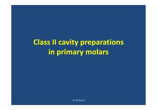
Class II cavity preparations in primary molars p y
Class II cavity preparations in primary molars p y
Class II cavity preparations in primary molars p y

Create successful ePaper yourself
Turn your PDF publications into a flip-book with our unique Google optimized e-Paper software.
<strong>Class</strong> <strong>II</strong> <strong>cavity</strong> <strong>preparations</strong><br />
<strong>in</strong> <strong>primary</strong> <strong>molars</strong><br />
Dr. Aly Sharaf
• Until eruption of the first permanent molar,<br />
the second <strong>primary</strong> molar is <strong>in</strong> contact only<br />
on its mesial surface which has an <strong>in</strong>cidence<br />
of caries ten times greater than that of the<br />
distal surface of the same tooth.<br />
• After the tooth’s distal contact is established<br />
little difference <strong>in</strong> susceptibility rema<strong>in</strong><br />
between the two surfaces.<br />
Dr. Aly Sharaf
• The susceptibility of caries on distal of the<br />
first <strong>primary</strong> molar is similar to mesial<br />
surface of the 2 nd <strong>primary</strong> molar. However,<br />
the mesial surface of 1 st <strong>primary</strong> molar is less<br />
caries prone. This is due to firm contact<br />
between first and second <strong>primary</strong> molar<br />
whereas a space is frequently present<br />
between the first molar and can<strong>in</strong>e.<br />
Dr. Aly Sharaf
• Caries on proximal surface most<br />
often beg<strong>in</strong>s just below the<br />
contact area and <strong>in</strong>itially spreads<br />
lt lateral lthen g<strong>in</strong>givally.<br />
i Dr. Aly Sharaf
• Radiograph exam<strong>in</strong>ation is particularly<br />
important to detect proximal caries. One<br />
study showed that as many as 75% of the<br />
proximal lesions affects the <strong>primary</strong> <strong>molars</strong><br />
would have gone undetected without the aid<br />
of radiographs.<br />
Dr. Aly Sharaf
Armamentarium<br />
• Mirror<br />
• Explorer<br />
• Enamel Hatchet 13‐14<br />
• Contra‐angle<br />
• No. 330 bur<br />
Dr. Aly Sharaf
Check the occlusion:<br />
Use articulat<strong>in</strong>g ribbon and tap the jaw of the<br />
manik<strong>in</strong>s. Remember the location of the<br />
marks and use them as an aid <strong>in</strong> carv<strong>in</strong>g the<br />
restoration.<br />
Dr. Aly Sharaf
<strong>Class</strong> <strong>II</strong> Amalgam OM #65<br />
Preparation<br />
Dr. Aly Sharaf
Establish Depth<br />
1. Use a #330 pear sharp bur.<br />
2. Start preparation by penetrat<strong>in</strong>g the occlusal<br />
surface from the central fossa and extend<br />
toward the mesial and term<strong>in</strong>ate <strong>in</strong> the<br />
mesial lesion.<br />
3. Use the periodontal probe or (330) to check<br />
the depth of preparation.<br />
Dr. Aly Sharaf
Criteria<br />
Depth<br />
1. Plastic tooth 1.25‐1.5 mm from the<br />
cavosurface. Measured at the mesial ilid ridge<br />
area.<br />
2.Natural tooth penetrate 0.25mm <strong>in</strong>to dent<strong>in</strong>.<br />
Dr. Aly Sharaf
Width<br />
• At the Isthmus area, about 1/3 to 1/4 the<br />
distance between cusp tip<br />
Note: Isthmus width on this second <strong>primary</strong><br />
molar is slightly greater than on the first<br />
<strong>primary</strong> molar.<br />
Dr. Aly Sharaf
Extended <strong>in</strong>to the Grooves<br />
1. Et Extend d<strong>in</strong>to grooves us<strong>in</strong>g #330 bur and the high h speed<br />
handpiece.<br />
2. Ma<strong>in</strong>ta<strong>in</strong> the depth as you extend <strong>in</strong>to the grooves.<br />
3. Tilt the bur toward the oblique ridge as you approach it to<br />
avoid underm<strong>in</strong>e the ridge.<br />
4. As you approach the oblique ridge create a dovetail il(do<br />
not underm<strong>in</strong>e the oblique ridge).<br />
5. Extend the buccal groove approximately equal to the<br />
width of the bur and create a dovetail.<br />
6. Extend mesially and create dovetail leav<strong>in</strong>g only th<strong>in</strong> wall<br />
of enamel so as to protect tthe adjacent tooth thfrom<br />
abrasion.<br />
7. Use #330 bur or slow speed f<strong>in</strong>ish and smooth the walls.<br />
Dr. Aly Sharaf
Criteria<br />
1. 1.25 – 1.5 mm depth must be ma<strong>in</strong>ta<strong>in</strong>ed<br />
i throughout.<br />
2. Buccal and l<strong>in</strong>gual walls should converge slightly<br />
<strong>in</strong> occlusal direction.<br />
3. Distal wall should be vertical <strong>in</strong> m<strong>in</strong>imal. In<br />
maximum model it diverge slightly.<br />
4. The oblique ridge must not be underm<strong>in</strong>ed.<br />
5. Only th<strong>in</strong> wall of enamel should be left mesially.<br />
6. All l<strong>in</strong>e angles should be rounded.<br />
Dr. Aly Sharaf
Prepare the Proximal Box<br />
1. Place #330 bur <strong>in</strong>side id the <strong>cavity</strong> aga<strong>in</strong>st tthe<br />
wall of tooth structure and move the bur <strong>in</strong> a<br />
g<strong>in</strong>gival direction, move the bur bucco‐l<strong>in</strong>gually<br />
with a pendulum motion. This will provide the<br />
slight convergence of the buccal and l<strong>in</strong>gual<br />
walls toward the occlusal (broader at g<strong>in</strong>gival<br />
than occlusal surface).<br />
2. Extend proximal box g<strong>in</strong>givally to break contact<br />
with the adjacent tooth so that tip of an<br />
explorer can be passed through.<br />
Dr. Aly Sharaf
1. Extend buccal and l<strong>in</strong>gual l walls of the<br />
proximal box to self‐cleans<strong>in</strong>g area. This can<br />
be determ<strong>in</strong>ed by the clearance of an<br />
explorer tip.<br />
2. If necessary use a Hatchet to plan the l<strong>in</strong>gual<br />
and buccal walls of the proximal box<br />
3. The axial wall follow the contour of tooth<br />
(mesial surface of the tooth).<br />
Dr. Aly Sharaf
Criteria<br />
1. The proper depth of proximal lbox is approximately<br />
2.5 –3 mm depend<strong>in</strong>g on the clearance.<br />
2. Proximal lbox wall extend so as explorer tip can pass<br />
through the embrasure.<br />
3. The proximal box should be wider g<strong>in</strong>givally than<br />
occlusally for retention of the restoration.<br />
4. Width of the g<strong>in</strong>gival seat is approximately 1 mm.<br />
5. Proximal box extends g<strong>in</strong>givally so that the tip of an<br />
explorer can pass through. The depth of the proximal<br />
box from the pulpo‐axial l<strong>in</strong>e angle to the g<strong>in</strong>gival seat<br />
approximately 1.25‐1.50 mm.<br />
Dr. Aly Sharaf
6. Rounded daxio‐pulpal ll<strong>in</strong>e angles, as are all l<strong>in</strong>es.<br />
7. Sharp cavosurface angle <strong>in</strong> the buccal l<strong>in</strong>gual<br />
walls/ a n<strong>in</strong>ety degree is desirable.<br />
8. There is a slight extension <strong>in</strong>to the fissured<br />
mesio‐buccal groove (sometimes familiarly<br />
referred to as a dovetail).<br />
9. The axial wall of the proximal box is at the same<br />
depth as the pulpal wall 1.0 to 1.25 mm.<br />
10. The g<strong>in</strong>gival wall on the m<strong>in</strong>imum model is just<br />
at the level of the g<strong>in</strong>giva: on the maximum, it is<br />
0.5 mm below the g<strong>in</strong>giva.<br />
g<br />
Dr. Aly Sharaf
Occluso‐distal # 74<br />
Extend <strong>in</strong>to grooves<br />
Term<strong>in</strong>ate your dovetail at central pit.<br />
The transverse ridge which is not underm<strong>in</strong>ed<br />
is lfi left <strong>in</strong>tact. The mesial ilwall at the<br />
transverse ridge is slightly undercut on the<br />
m<strong>in</strong>imum model or vertical on the maximum.<br />
This isthmus is 1.25 to 1.5 mm wide.<br />
Dr. Aly Sharaf
Prepare the proximal box<br />
• As mentioned before the pulpal and axial<br />
depth are 1.0 to 1.25 mm. G<strong>in</strong>gival wall is level<br />
with ihthe g<strong>in</strong>giva i <strong>in</strong> m<strong>in</strong>imal i model. dl0.5 mm<br />
below g<strong>in</strong>giva on the maximum model.<br />
Dr. Aly Sharaf
Two class <strong>II</strong> “ back‐to‐back” b k” cavities ii can be prepared as<br />
essentially one procedure rather than separately.<br />
Prepare the occlusal portion of each tooth then form<br />
the proximal boxes vertically simultaneously by mov<strong>in</strong>g<br />
the burs back and forth between the 2 proximal<br />
surfaces establish<strong>in</strong>g first the buccal walls then the<br />
l<strong>in</strong>gual or (vice verse).<br />
It must be remembered that the axial wall on a first<br />
<strong>primary</strong> molar will be slightly deep than on the<br />
adjacent second <strong>primary</strong> molar. Etblihb Establish both g<strong>in</strong>gival<br />
i walls by perpendicularly the bur to round them.<br />
Dr. Aly Sharaf
Dr. Aly Sharaf
Dr. Aly Sharaf