TECHNIQUES AND OUTCOMES - Redalyc
TECHNIQUES AND OUTCOMES - Redalyc
TECHNIQUES AND OUTCOMES - Redalyc

You also want an ePaper? Increase the reach of your titles
YUMPU automatically turns print PDFs into web optimized ePapers that Google loves.
322<br />
P. M. Lewandowski, S. Leslie, I. S. Gill, and M. M. Desai.<br />
fasciotomy is partially closed, the R-port reinserted and<br />
pneumoperitoneum re-established. Once hemostasis<br />
has been confirmed, the R-port is removed and the<br />
fascia and skin closed. Since the Quadport has a 15<br />
mm inlet, the Endocatch bag can be inserted after<br />
hilar dissection.<br />
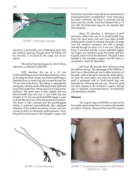
FIGURE 5. Articulating instruments.<br />
extraction is technically more challenging given that<br />
the umbilical opening, through which the kidney will<br />
be removed, is occupied by the single port access<br />
device.<br />
We outline three techniques by which kidney<br />
extraction is achieved in LESS-DN:<br />
Gill5 describes the use of a 15 mm<br />
EndoCatch® bag to assist with kidney extraction. Prior<br />
to dividing the renal vessels, the EndoCatch® bag is<br />
detached from its metal ring and inserted through the<br />
12 mm inlet of the R-port. The kidney is subsequently<br />
entrapped, with the mouth of the bag loosely tightened<br />
around the renal hilum, taking care not to compromise<br />
perfusion. The renal artery is then clipped with two<br />
Hem-O-Lock® clips and one 11 mm metal clip and<br />
divided. A 12 mm vascular EndoGIA® stapler is used<br />
to divide the renal vein at the interaortocaval location.<br />
The R-port is then removed and the pre-entrapped<br />
kidney is extracted trans-umbilically after adequate<br />
extension of the midline fasciotomy incision and skin<br />
incision to allow for atraumatic graft extraction. Once<br />
the graft has been given to the transplant surgeon, the<br />
Desai (7) describes a technique of graft<br />
extraction without the use of an EndoCatch® bag.<br />
Once the renal artery and vein have been divided<br />
and the graft is ready for retrieval, the graft is brought<br />
near the umbilical extraction site with a grasper<br />
inserted through an extra 3 or 5 mm port. Once the<br />
R-port is removed and the incision extended slightly,<br />
two fingers are inserted to grasp the kidney and then<br />
to gently extract it from the abdomen. The graft is then<br />
handed to the transplant surgeon and the R-port is<br />
reinserted to check for hemostasis.<br />
Del Pizzo (8) describe their technique using<br />
the GelPoint device. The advantage of the GelPoint is<br />
that it has a detachable gel cap to allow extraction of<br />
the graft, without having to remove the whole device.<br />
So once the renal artery and vein are divided, the<br />
graft is entrapped with an EndoCatch® bag and<br />
extracted by removing the gel cap. Once the graft<br />
has been passed to the transplant surgeon, the gel<br />
cap is replaced, pneumoperitoneum re-established<br />
and hemostasis checked.<br />
Outcomes<br />
The surgical steps of LESS-DN in each of the<br />
five studies demonstrate that it is a technically feasible<br />
procedure. However, there are three important aspects<br />
FIGURE 6. Curved Instruments.<br />
FIGURE 7. Postoperative photograph of a LESS donor’s<br />
abdomen at 1 week.