Discitis And Vertebral Osteomyelitis
Discitis And Vertebral Osteomyelitis
Discitis And Vertebral Osteomyelitis

Create successful ePaper yourself
Turn your PDF publications into a flip-book with our unique Google optimized e-Paper software.
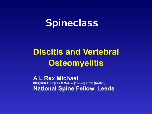
Spineclass<br />
<strong>Discitis</strong> and <strong>Vertebral</strong><br />
<strong>Osteomyelitis</strong><br />
A L Rex Michael<br />
DNB(T&O), FRCS(Ed.), M.Med.Sc. (Trauma) ,FRCS (Tr&Orth)<br />
National Spine Fellow, Leeds
Acknowledgements<br />
• Mr. P. A. Millner consultant Orthopaedic Spinal<br />
Surgeon Leeds Teaching Hospitals NHS Trust
Objectives<br />
• Epidemiology<br />
• Pathology<br />
• Clinical features<br />
• Management<br />
• Prognosis
Epidemiology<br />
• 2 - 4% all cases of “osteomyelitis”<br />
• rare: 1 in 250,000/yr but rising incidence<br />
• post-op discitis = 2-3%<br />
• pre-antibiotic mortality = 70%<br />
• delayed diagnosis common (weeks/months)
Risk factors<br />
• Age: peak incidence 7th decade<br />
• concurrent illness/infection<br />
– diabetes<br />
– immunosuppressed<br />
– steroid therapy<br />
– UTI (46%)<br />
• invasive procedures
Spine levels<br />
• spondylodiscitis / facet disease<br />
– lumbar (59%)<br />
– thoracic (33%)<br />
– cervical (8%)<br />
• epidural abscess<br />
– cervical (90%)<br />
– thoracic (33%)<br />
– lumbar (24%)
Pathology (1)<br />
• Organisms<br />
– S aureus >50% cases<br />
– after UTI = gram-negatives<br />
– Opportunistic / TB<br />
• Route of spread<br />
– direct extension<br />
– post-operative<br />
– haematogenous
Pathology (2)<br />
• <strong>Vertebral</strong> metaphyses (end plate region)<br />
= end-arteriole blood supply (filter)<br />
→ Septic emboli (? facet disease)<br />
• Direct spread from implantation<br />
→ secondary spread to discs, paraspinal tissues<br />
and spaces
Clinical features<br />
• pain and focal tenderness 90%<br />
• fever 61%<br />
• root symptoms/signs 60%<br />
• cord symptoms/signs 30%<br />
Also: deformity, muscle spasms, meningism,<br />
unexplained septicaemia
Diagnosis<br />
• Lab tests<br />
↑ white cell count 40-60%<br />
↑ ESR / PV / CRP 80-90%<br />
positive BC 20-25%<br />
Imaging<br />
Biopsy
Imaging studies (1)<br />
• Plain films<br />
– vertebral metaphyseal blurring (osteolysis)<br />
– loss of disc height<br />
– endplate blurring<br />
– subchondral reactive bone formation<br />
– bone destruction (and deformity)<br />
– soft-tissue shadows e.g.psoas abscess
Pyogenic spinal infections
pyogenic<br />
spondylodiscitis
pyogenic<br />
spondylodiscitis<br />
bad disc = good news
Imaging studies (2)<br />
• CT scans (with contrast)<br />
– delineate bony margins / involvement<br />
– soft-tissue invasion<br />
– poor for outlining neural elements<br />
– risk of spread if combined with myelography,<br />
but can obtain CSF<br />
– 3D/MPR useful for pre-op planning
Pyogenic spinal infections
Imaging studies (3)<br />
• Radionuclide studies<br />
– detect earlier than plain films<br />
– high sensitivity, ? specificity<br />
e.g. gallium + Tc = 95% accurate<br />
– little structural information<br />
– false negatives in neutropenics (gallium)<br />
– false negatives in bone infarction
Imaging studies (4)<br />
• MRI (with gadolinium) = modality of choice<br />
– T1 = ↓ signal in vertebral body and disc<br />
– T2 = ↑ signal in vertebral body and disc<br />
– (+ gad) = vascularised vs. pus (and in post-op)<br />
> 95% accuracy
Pyogenic spinal infections
pyogenic discitis/osteomyelitis<br />
bad disc = good news
Biopsy<br />
• microbiology and histology<br />
– infection vs. malignancy<br />
• closed needle biopsy (guided)<br />
– 68 - 86% accuracy (false negative 30%)<br />
• open biopsy<br />
– > 80% accurate (false negative 14%)<br />
• special lab techniques (DNA PCR, etc)
Treatment goals<br />
• Establish diagnosis<br />
• Protect / restore neurological function<br />
• Pain relief<br />
• Clear infection and prevent recurrence<br />
• Maintain / restore spine stability
Treatment (1)<br />
• antibiotics<br />
– sensitivities<br />
– adequate dose (iv then oral)<br />
– ensure MBC reached<br />
– adequate duration (> 6 weeks)<br />
– monitor response (clinical/ indices/ imaging)<br />
– toxicity profile and monitoring
Treatment (2)<br />
• immobilisation<br />
– bed rest<br />
– moulded orthoses (low thoracic / lumbar)<br />
– halo-vest or orthosis (cervical / high thoracic)<br />
• surgical intervention
Treatment (3)<br />
• Indications for surgical Rx<br />
– unable to obtain diagnosis by closed means<br />
– significant abscess formation<br />
– failed medical Rx (antibiotics / immobilisation)<br />
including intolerance<br />
– bone destruction with deformity / instability<br />
– progressive neurological deficit
Treatment (4)<br />
• Principles of surgical Rx<br />
– excise necrotic tissue back to healthy margins<br />
– decompress spinal canal where necessary<br />
– tissue samples for microbiology / histology<br />
– reconstruction / stabilisation of defect<br />
approach and technique dictated by spinal level<br />
and region to be debrided
Outcome (1)<br />
• Successful non-op Rx predicted by<br />
– age < 60 years<br />
– immune competence<br />
– S aureus infection<br />
↓ ESR (or other inflammatory index)<br />
(in absence of indication for surgical Rx)
Outcome (2)<br />
• Surgical Rx outcome<br />
– less chronic back pain overall (cf non-op)<br />
– related to pre-op neurological status<br />
(quad < paraplegic < paraparesis)<br />
– epidural abscess < granulation tissue
Summary<br />
• Rare, rising incidence<br />
• Delayed diagnosis common<br />
• Mainly elderly or ill<br />
• Specific diagnostic methods<br />
• Treatment options