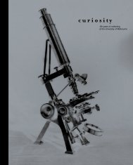
COLLECTIONS - University of Melbourne
COLLECTIONS - University of Melbourne
COLLECTIONS - University of Melbourne

You also want an ePaper? Increase the reach of your titles
YUMPU automatically turns print PDFs into web optimized ePapers that Google loves.
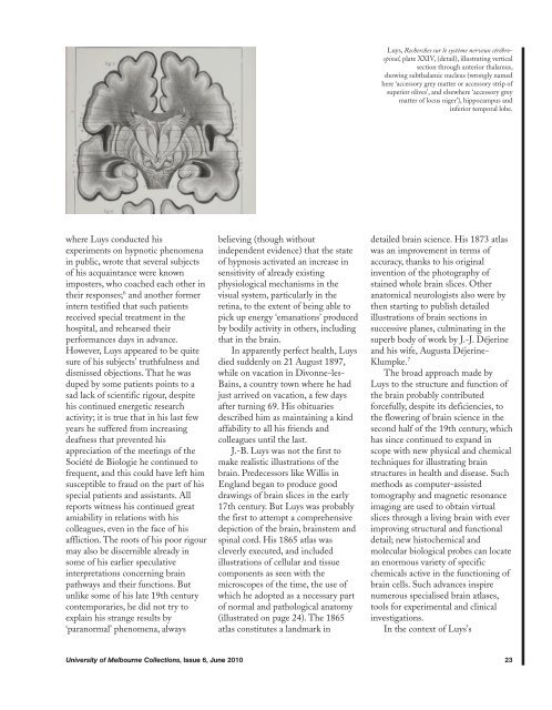
Luys, Recherches sur le système nerveux cérébrospinal,<br />
plate XXIV, (detail), illustrating vertical<br />
section through anterior thalamus,<br />
showing subthalamic nucleus (wrongly named<br />
here ‘accessory grey matter or accessory strip <strong>of</strong><br />
superior olives’, and elsewhere ‘accessory grey<br />
matter <strong>of</strong> locus niger’), hippocampus and<br />
inferior temporal lobe.<br />
where Luys conducted his<br />
experiments on hypnotic phenomena<br />
in public, wrote that several subjects<br />
<strong>of</strong> his acquaintance were known<br />
imposters, who coached each other in<br />
their responses; 6 and another former<br />
intern testified that such patients<br />
received special treatment in the<br />
hospital, and rehearsed their<br />
performances days in advance.<br />
However, Luys appeared to be quite<br />
sure <strong>of</strong> his subjects’ truthfulness and<br />
dismissed objections. That he was<br />
duped by some patients points to a<br />
sad lack <strong>of</strong> scientific rigour, despite<br />
his continued energetic research<br />
activity; it is true that in his last few<br />
years he suffered from increasing<br />
deafness that prevented his<br />
appreciation <strong>of</strong> the meetings <strong>of</strong> the<br />
Société de Biologie he continued to<br />
frequent, and this could have left him<br />
susceptible to fraud on the part <strong>of</strong> his<br />
special patients and assistants. All<br />
reports witness his continued great<br />
amiability in relations with his<br />
colleagues, even in the face <strong>of</strong> his<br />
affliction. The roots <strong>of</strong> his poor rigour<br />
may also be discernible already in<br />
some <strong>of</strong> his earlier speculative<br />
interpretations concerning brain<br />
pathways and their functions. But<br />
unlike some <strong>of</strong> his late 19th century<br />
contemporaries, he did not try to<br />
explain his strange results by<br />
‘paranormal’ phenomena, always<br />
believing (though without<br />
independent evidence) that the state<br />
<strong>of</strong> hypnosis activated an increase in<br />
sensitivity <strong>of</strong> already existing<br />
physiological mechanisms in the<br />
visual system, particularly in the<br />
retina, to the extent <strong>of</strong> being able to<br />
pick up energy ‘emanations’ produced<br />
by bodily activity in others, including<br />
that in the brain.<br />
In apparently perfect health, Luys<br />
died suddenly on 21 August 1897,<br />
while on vacation in Divonne-les-<br />
Bains, a country town where he had<br />
just arrived on vacation, a few days<br />
after turning 69. His obituaries<br />
described him as maintaining a kind<br />
affability to all his friends and<br />
colleagues until the last.<br />
J.-B. Luys was not the first to<br />
make realistic illustrations <strong>of</strong> the<br />
brain. Predecessors like Willis in<br />
England began to produce good<br />
drawings <strong>of</strong> brain slices in the early<br />
17th century. But Luys was probably<br />
the first to attempt a comprehensive<br />
depiction <strong>of</strong> the brain, brainstem and<br />
spinal cord. His 1865 atlas was<br />
cleverly executed, and included<br />
illustrations <strong>of</strong> cellular and tissue<br />
components as seen with the<br />
microscopes <strong>of</strong> the time, the use <strong>of</strong><br />
which he adopted as a necessary part<br />
<strong>of</strong> normal and pathological anatomy<br />
(illustrated on page 24). The 1865<br />
atlas constitutes a landmark in<br />
detailed brain science. His 1873 atlas<br />
was an improvement in terms <strong>of</strong><br />
accuracy, thanks to his original<br />
invention <strong>of</strong> the photography <strong>of</strong><br />
stained whole brain slices. Other<br />
anatomical neurologists also were by<br />
then starting to publish detailed<br />
illustrations <strong>of</strong> brain sections in<br />
successive planes, culminating in the<br />
superb body <strong>of</strong> work by J.-J. Déjerine<br />
and his wife, Augusta Déjerine-<br />
Klumpke. 7<br />
The broad approach made by<br />
Luys to the structure and function <strong>of</strong><br />
the brain probably contributed<br />
forcefully, despite its deficiencies, to<br />
the flowering <strong>of</strong> brain science in the<br />
second half <strong>of</strong> the 19th century, which<br />
has since continued to expand in<br />
scope with new physical and chemical<br />
techniques for illustrating brain<br />
structures in health and disease. Such<br />
methods as computer-assisted<br />
tomography and magnetic resonance<br />
imaging are used to obtain virtual<br />
slices through a living brain with ever<br />
improving structural and functional<br />
detail; new histochemical and<br />
molecular biological probes can locate<br />
an enormous variety <strong>of</strong> specific<br />
chemicals active in the functioning <strong>of</strong><br />
brain cells. Such advances inspire<br />
numerous specialised brain atlases,<br />
tools for experimental and clinical<br />
investigations.<br />
In the context <strong>of</strong> Luys’s<br />
<strong>University</strong> <strong>of</strong> <strong>Melbourne</strong> Collections, Issue 6, June 2010 23