Pharmaceutical Industry and Green Chemistry
Pharmaceutical Industry and Green Chemistry
Pharmaceutical Industry and Green Chemistry

Create successful ePaper yourself
Turn your PDF publications into a flip-book with our unique Google optimized e-Paper software.
oxygen <strong>and</strong> carbon dioxide is approximately 50 to 55<br />
mL/100 g/min. As blood perfusion is progressively<br />
reduced, oxygen extraction from hemoglobin, which<br />
is indicated by arteriovenous difference in oxygen,<br />
increases without clinical manifestation. When<br />
blood perfusion reaches 25 to 30 mL/100 g/min,<br />
electroencephalographic (EEG) abnormalities<br />
<strong>and</strong> consciousness alterations may occur. As blood<br />
perfusion falls further below 20 mL/100 g/min<br />
approximately, EEG becomes isoelectric <strong>and</strong> neurons<br />
increasingly switch to anaerobic metabolism, with<br />
concomitant increased production of lactate <strong>and</strong><br />
hydrogen ions. Once perfusion reaches 10 to 12<br />
mL/100 g/min, neurotransmission is lost, sodiumpotassium<br />
pumps fail 7 <strong>and</strong> cytotoxic edema ensues 8 .<br />
In the absence of cerebral hypothermia, perfusion of<br />
less than 6 to 10 mL/100 g/min triggers tissue death<br />
cascade mediated by calcium <strong>and</strong> glutamate 9 .<br />
Hypocapnia, on the other h<strong>and</strong>, is a state of<br />
reduced carbon dioxide, Even when marked,<br />
hypocapnia is normally well tolerated, often with few<br />
apparent effects. Transient induction of hypocapnia<br />
can lead to lifesaving physiological changes in patients<br />
with severe intracranial hypertension or neonatal<br />
pulmonary-artery hypertension, but hypocapnia of<br />
longer duration in critically ill patients may have a<br />
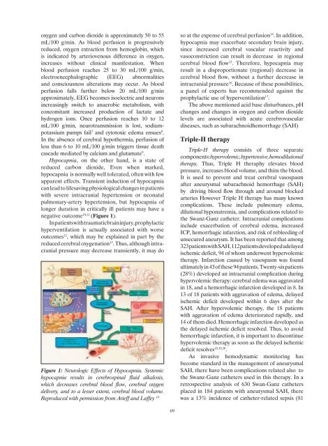
negative outcome 10,11 (Figure 1).<br />
In patients with traumatic brain injury, prophylactic<br />
hyperventilation is actually associated with worse<br />
outcomes 12 , which may be explained in part by the<br />
reduced cerebral oxygenation 13 . Thus, although intracranial<br />
pressure may decrease transiently, it may do<br />
Figure 1: Neurologic Effects of Hypocapnia. Systemic<br />
hypocapnia results in cerebrospinal fluid alkalosis,<br />
which decreases cerebral blood flow, cerebral oxygen<br />
delivery, <strong>and</strong> to a lesser extent, cerebral blood volume.<br />
Reproduced with permission from Arieff <strong>and</strong> Laffey 14<br />
so at the expense of cerebral perfusion 14 . In addition,<br />
hypocapnia may exacerbate secondary brain injury,<br />
since increased cerebral vascular reactivity <strong>and</strong><br />
vasoconstriction can result in decrease in regional<br />
cerebral blood flow 15 . Therefore, hypocapnia may<br />
result in a disproportionate (regional) decrease in<br />
cerebral blood flow, without a further decrease in<br />
intracranial pressure 16 . Because of these possibilities,<br />
a panel of experts has recommended against the<br />
prophylactic use of hyperventilation 17 .<br />
The above mentioned acid base disturbances, pH<br />
changes <strong>and</strong> changes in oxygen <strong>and</strong> carbon dioxide<br />
levels are associated with acute cerebrovascular<br />
diseases, such as subarachnoidhemorrhage (SAH)<br />
Triple-H therapy<br />
Triple-H therapy consists of three separate<br />
components: hypervolemic, hypertensive, hemodilutional<br />
therapy, Thus, Triple H theraphy elevates blood<br />
pressure, increases blood volume, <strong>and</strong> thins the blood.<br />
It is used to prevent <strong>and</strong> treat cerebral vasospasm<br />
after aneurysmal subarachnoid hemorrhage (SAH)<br />
by driving blood flow through <strong>and</strong> around blocked<br />
arteries However Triple H therapy has many known<br />
complications. These include pulmonary edema,<br />
dilutional hyponatremia, <strong>and</strong> complications related to<br />
the Swanz-Ganz catheter. Intracranial complications<br />
include exacerbation of cerebral edema, increased<br />
ICP, hemorrhagic infarction, <strong>and</strong> risk of rebleeding of<br />
unsecured aneurysm. It has been reported that among<br />
323 patients with SAH, 112 patients developed adelayed<br />
ischemic deficit, 94 of whom underwent hypervolemic<br />
therapy. Infarction caused by vasospasm was found<br />
ultimately in 43 of these 94 patients. Twenty-six patients<br />
(28%) developed an intracranial complication during<br />
hypervolemic therapy: cerebral edema was aggravated<br />
in 18, <strong>and</strong> a hemorrhagic infarction developed in 8. In<br />
13 of 18 patients with aggravation of edema, delayed<br />
ischemic deficit developed within 6 days after the<br />
SAH. After hypervolemic therapy, the 18 patients<br />
with aggravation of edema deteriorated rapidly, <strong>and</strong><br />
14 of them died. Hemorrhagic infarction developed as<br />
the delayed ischemic deficit resolved. Thus, to avoid<br />
hemorrhagic infarction, it is important to discontinue<br />
hypervolemic therapy as soon as the delayed ischemic<br />
deficit resolves 18,19,20 .<br />
As invasive hemodynamic monitoring has<br />
become st<strong>and</strong>ard in the management of aneurysmal<br />
SAH, there have been complications related also to<br />
the Swanz-Ganz catheters used in this therapy. In a<br />
retrospective analysis of 630 Swan-Ganz catheters<br />
placed in 184 patients with aneurysmal SAH, there<br />
was a 13% incidence of catheter-related sepsis (81<br />
69