Integration of Topological Constraints in Medical ... - Florent Ségonne
Integration of Topological Constraints in Medical ... - Florent Ségonne
Integration of Topological Constraints in Medical ... - Florent Ségonne

You also want an ePaper? Increase the reach of your titles
YUMPU automatically turns print PDFs into web optimized ePapers that Google loves.
2 F. Ségonne and B. Fischl<br />
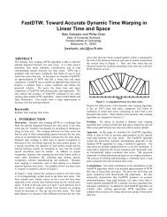
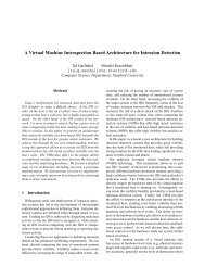
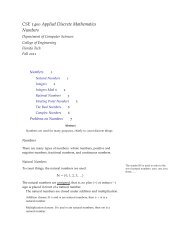
FIGURE 1. a) Subcortical structures have a spherical topology. For <strong>in</strong>stance, the<br />
shape <strong>of</strong> the hippocampus can be cont<strong>in</strong>uously deformed onto a sphere. b) The<br />
human cerebral cortex is a highly folded ribbon <strong>of</strong> gray matter that lies <strong>in</strong>side<br />
the cerebrosp<strong>in</strong>al fluid (the red <strong>in</strong>terface) and outside the white matter <strong>of</strong> the<br />
bra<strong>in</strong> (the green <strong>in</strong>terface). When the midl<strong>in</strong>e connections between the left and<br />
right hemisphere are artificially closed, these two surfaces have the topology <strong>of</strong> a<br />
sphere. c) Due to the partial volume effect, subject motion, etc..., it becomes hard<br />
to dist<strong>in</strong>guish opposite banks <strong>of</strong> a the gray matter. d) Segmentation algorithms<br />
that do not constra<strong>in</strong> the topology <strong>of</strong>ten produce cortical segmentations with<br />
several topological defects (i.e. handles, cavities, disconnected components). e) A<br />
close-up <strong>of</strong> a topologically-<strong>in</strong>correct cortical surface representation.<br />
teristic spatial pattern relative to one another (e.g. <strong>in</strong> the human bra<strong>in</strong>, the<br />
amygdala is anterior and superior to the hippocampus). The set <strong>of</strong> <strong>in</strong>dividual<br />
topological properties and specific relationships between anatomical<br />
structures determ<strong>in</strong>e the global topology <strong>of</strong> a region <strong>of</strong> <strong>in</strong>terest.<br />
Although many cl<strong>in</strong>ical and research applications require accurate segmentations<br />
that respect the true anatomy <strong>of</strong> the targeted structures, only<br />
a few techniques have been proposed to achieve accurate and topologicallycorrect<br />
segmentations.<br />
1.2 Motivation<br />
Many neurodegenerative disorders, psychiatric disorders, and healthy ag<strong>in</strong>g<br />
are frequently associated with structural changes <strong>in</strong> the bra<strong>in</strong>. These<br />
changes, which can cause alterations <strong>in</strong> the imag<strong>in</strong>g properties <strong>of</strong> the bra<strong>in</strong><br />
tissue, as well as <strong>in</strong> morphometric properties <strong>of</strong> bra<strong>in</strong> structures, can be<br />
captured and detected by sophisticated segmentation techniques. Certa<strong>in</strong><br />
cl<strong>in</strong>ical and research applications depend crucially on the accuracy and correctness<br />
<strong>of</strong> the representations (visualization [11, 12, 56], spherical coord<strong>in</strong>ate<br />
system and surface-based atlases [12, 16, 17, 18, 22, 50, 56], shape analysis<br />
[16, 20, 31, 43, 53, 54], surface-based process<strong>in</strong>g <strong>of</strong> functional data [12],<br />
and <strong>in</strong>ter-subject registration [22, 51, 55], among others). Small geometric<br />
errors <strong>in</strong> a segmentation can easily change the apparent connectivity <strong>of</strong> the<br />
segmented structure, pos<strong>in</strong>g a problem to studies that aim at analyz<strong>in</strong>g<br />
Locally, its <strong>in</strong>tr<strong>in</strong>sic “unfolded” structure is that <strong>of</strong> a 2D sheet, several millimeters thick.<br />
In the absence <strong>of</strong> pathology and assum<strong>in</strong>g that the midl<strong>in</strong>e hemispheric connections are<br />
artificially closed, each cortical hemisphere can be considered as a simply-connected 2D<br />
sheet <strong>of</strong> neurons that carries the simple topology <strong>of</strong> a sphere - see Fig. 1-b
![{ public static void main (String[] args) { System.out.println (](https://img.yumpu.com/49719541/1/190x143/-public-static-void-main-string-args-systemoutprintln-hello-.jpg?quality=85)