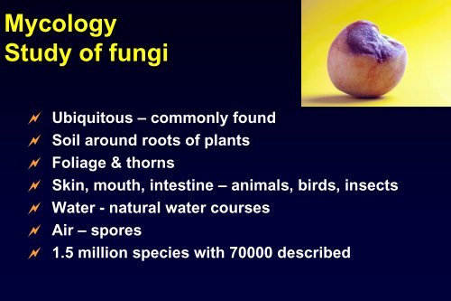
- Page 1: Mycology for Beginners Louise Hafne
- Page 7 and 8: Human infections 8-10 dermatophyte
- Page 9 and 10: Fungi Domain Bacteria ) contains th
- Page 11 and 12: Taxonomy - „Classical‟ Based o
- Page 13 and 14: Structure - Yeast Unicellular Rep
- Page 15 and 16: Hyphae - non-septate Coenocytic T
- Page 17 and 18: Reproduction Asexual - mitosis - a
- Page 19 and 20: Mycosis - fungal disease Superfici
- Page 21 and 22: Other disease Toxicity - mycotoxin
- Page 23 and 24: Pathogenicity Accidental exposure
- Page 25 and 26: Pathogenesis of indoor fungi diseas
- Page 27 and 28: Laboratory Mould form dimorphic fu
- Page 29 and 30: Images from Kaminski‟s Digital Im
- Page 31 and 32: Mycology • Fungal microscopy & cu
- Page 33 and 34: Why? • Appearance of lesion decep
- Page 35 and 36: Immune Response • Immunocompromis
- Page 37 and 38: Significance • How do you know?
- Page 39 and 40: The Gram Stain • Gram colour is n
- Page 41 and 42: Yeasts • Candida Oval cells narro
- Page 43 and 44: Body Fluids • Large volume fluids
- Page 45 and 46: Acknowledgements Microbiology Scien
- Page 47 and 48: Dermatophyte Identification Louise
- Page 49 and 50: Martha E Kern Medical Mycology
- Page 51 and 52: Geographical distribution T. rubrum
- Page 53 and 54:
Clinical disease Tinea barbae T. ru
- Page 55 and 56:
Culture Surface colour - white, buf
- Page 57 and 58:
Microscopy - culture Macroconidia c
- Page 59 and 60:
Trichophyton rubrum Onychomycosis G
- Page 61 and 62:
Epidermophyton floccosum Tinea crur
- Page 63 and 64:
Trichophyton mentagrophytes Patient
- Page 65 and 66:
Microsporum canis Patient: 7yr old
- Page 67 and 68:
Images from Kaminski‟s Digital Im
- Page 69 and 70:
Yeasts ‣ Unicellular fungal organ
- Page 71 and 72:
Candidiasis ‣ Candida albicans -
- Page 73 and 74:
Candida - culture ‣ Clinical spec
- Page 75 and 76:
Identification - CAC ‣ ChromAgar
- Page 77 and 78:
Cryptococcosis ‣ Cryptococcus neo
- Page 79 and 80:
A B C D Worldwide Subtropical Subtr
- Page 81 and 82:
Cryptococcus - microscopy ‣ Spher
- Page 83 and 84:
Trichosporon sp. (beigelii) ‣ T.
- Page 85 and 86:
Geotrichum sp. ‣ Rare infections
- Page 87 and 88:
Yeast identification ‣ Positive g
- Page 90 and 91:
Biofilm production by clinical isol
- Page 92 and 93:
Other Yeast-like isolates ‣ Sporo
- Page 94 and 95:
Rare Yeast-like - travellers ‣ Bl
- Page 96 and 97:
Wot R U? Louise Hafner Ann McPhee A
- Page 98 and 99:
Mould Identification
- Page 100 and 101:
Opportunistic fungi ‣ Cosmopolita
- Page 102 and 103:
Zygomycetes Rhizopus oryzae
- Page 104 and 105:
Hyphomycetes - identification ‣Co
- Page 106 and 107:
Aspergillosis ‣ Paranasal sinusit
- Page 108 and 109:
Pseudallescheriasis Scedosporium in
- Page 110 and 111:
Scedosporium aurantiacum ‣ New sp
- Page 113 and 114:
Phialidic conidia production
- Page 115 and 116:
Culture
- Page 117 and 118:
Paecilomyces lilacinus ‣ Growth r
- Page 119 and 120:
Hyphomycetes ‣ Phaeohyphomycetes
- Page 121 and 122:
‘Jackson 5’… ABC Easy As 123
- Page 123 and 124:
ABCDE of Mycotic keratitis, Paranas
- Page 125 and 126:
Others - Coelomycetes ostiole • C
- Page 127 and 128:
Coelomycetes ‣ Plant pathogens
- Page 129 and 130:
Disease ‣ Sporotrichosis Sporothr
- Page 131 and 132:
Dematiaceous fungi Clinicopathologi
- Page 133 and 134:
Exophiala jeanselmei complex ‣ Ve
- Page 135 and 136:
References Atlas of Clinical Fungi
- Page 137:
Graduation