Ramsay Hunt Syndrome with Hemiparesis and ... - Vol.22 No.1
Ramsay Hunt Syndrome with Hemiparesis and ... - Vol.22 No.1
Ramsay Hunt Syndrome with Hemiparesis and ... - Vol.22 No.1
Create successful ePaper yourself
Turn your PDF publications into a flip-book with our unique Google optimized e-Paper software.
279<br />
The coexistence of RHS <strong>and</strong> VZV encephalitis is<br />
rare <strong>and</strong> Kin et al. (6) found only 8 cases, including one<br />
case of their own, in English <strong>and</strong> Japanese literature (6) .<br />
When the data were examined more closely, we found<br />
that these patients could be divided into two groups<br />
according to the presence of disturbed consciousness<br />
(encephalitis) <strong>and</strong> pyramidal sign (stroke-like), similar to<br />
the classification of Booss <strong>and</strong> Esiri (10) . Since our cases<br />
showed hemiparesis <strong>with</strong>out disturbance of consciousness,<br />
we compare our cases <strong>with</strong> those previously reported<br />
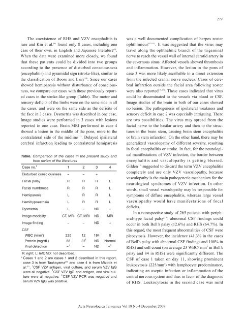
cases in the stroke-like group (Table). The motor <strong>and</strong><br />
sensory deficits of the limbs were on the same side in all<br />
the cases, <strong>and</strong> were on the same side as the deficits of<br />
the face in 3 cases. Dysmetria was described in one case.<br />
Image studies were performed in 3 cases <strong>with</strong> lesions<br />
reported in one case. Brain MRI performed in case 4<br />
showed a lesion in the middle of the pons, more to the<br />
contralateral side of the midline (11) . Delayed ipsilateral<br />
cerebral infarction leading to contralateral hemiparesis<br />
Table. Comparison of the cases in the present study <strong>and</strong><br />
from review of the literatures<br />
Case no.* 1 2 3 4<br />
Disturbed consciousness – – +<br />
Facial palsy R R R L<br />
Facial numbness R R R L<br />
<strong>Hemiparesis</strong> L R R L<br />
Hemihypoesthsia L R R L<br />
Dysmetria L – ND –<br />
Image modality CT, MRI CT, MRI ND MRI<br />
Image finding – – ND +<br />
CSF<br />
WBC (/mm 3 ) 225 12 184 0<br />
Protein (mg/dL) 88 37 ND Normal<br />
Viral detection – † – ND – <br />
R: right; L: left; ND: not described.<br />
* Cases 1 <strong>and</strong> 2 are cases 1 <strong>and</strong> 2 described in this report,<br />
case 3 is from Taukayama (20)<br />
<strong>and</strong> case 4 is from Mizock et<br />
al. (11) . † CSF VZV antigen, viral culture, <strong>and</strong> serum VZV IgG<br />
were all negative. CSF VZV IgG <strong>and</strong> antigen, <strong>and</strong> viral culture<br />
were all negative. CSF VZV PCR was negative <strong>and</strong><br />
serum VZV IgG was positive.<br />
was a well documented complication of herpes zoster<br />
ophthlmicus (12-15) . It was suggested that the virus may<br />
travel along the ophthalmic branch of the trigeminal<br />
nerve to reach the vessel wall of internal carotid artery in<br />
the cavernous sinus. Affected vessels showed thrombosis<br />
<strong>and</strong> inflammation. However, the lesion in the pons of<br />
case 3 was more likely ascribable to a direct extension<br />
from the infected cranial nerve nucleus. Cases of cerebral<br />
infarction outside the facial area following zoster<br />
were also reported (16,17) . These cases indicated that virus<br />
could be disseminated to the vessels via blood or CSF.<br />
Image studies of the brain in both of our cases showed<br />
no lesion. The pathogenesis of ipsilateral weakness <strong>and</strong><br />
sensory deficit in case 2 was especially intriguing. There<br />
are two possibilities. The virus may spread from the<br />
facial nerve to the basilar artery <strong>and</strong> then to the structures<br />
in the brain stem, causing brain stem encephalitis<br />
or brain stem infarction. On the other h<strong>and</strong>, there may be<br />
generalized vasculopathy of different severity, resulting<br />
in focal encephalitis or stroke. In fact, for the neurological<br />
manifestations of VZV infection, the border between<br />
encephalitis <strong>and</strong> vasculopathy is getting blurred.<br />
Gilden (18) suggested to discard the term VZV encephalitis<br />
completely <strong>and</strong> use only VZV vasculopathy, because<br />
vasculopathy is the main pathogenetic mechanism for the<br />
neurological syndromes of VZV infection. In other<br />
words, small vessel vasculopathy may be responsible for<br />
symptoms of diffuse encephalitis, whereas large vessel<br />
vasculopathy would have manifestations of focal<br />
deficits.<br />
In a retrospective study of 265 patients <strong>with</strong> peripheral-type<br />
facial palsy (19) , abnormal CSF findings could<br />
occur in both Bell’s palsy (12.6%) <strong>and</strong> RHS (64.7%). In<br />
this regard, the most frequent abnormalities of CSF were<br />
pleocytosis. However, the incidence (41.3% in the cases<br />
of Bell’s palsy <strong>with</strong> abnormal CSF findings <strong>and</strong> 100% in<br />
RHS) <strong>and</strong> cell count (on average 23 WBC/ mm 3 in Bell’s<br />
palsy <strong>and</strong> 84 in RHS) were significantly different. The<br />
CSF of case 1 taken on day 11, showing prominent<br />
leukocytosis (225/mm 3 ) <strong>with</strong> lymphocyte predominance,<br />
indicating an aseptic infection or inflammation of the<br />
central nervous system <strong>and</strong> thus in favor of the diagnosis<br />
of RHS. Leukocytosis in the second case was mild<br />
Acta Neurologica Taiwanica Vol 18 No 4 December 2009


