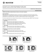
Spectrum® Mammary Implants - Mentor
Spectrum® Mammary Implants - Mentor
Spectrum® Mammary Implants - Mentor

Create successful ePaper yourself
Turn your PDF publications into a flip-book with our unique Google optimized e-Paper software.
0211024 Intl Spec Surg Guide 6/6/06 2:31 PM Page 3<br />
The following information<br />
is a compilation of<br />
research on the <strong>Mentor</strong><br />
Spectrum ® <strong>Mammary</strong><br />
<strong>Implants</strong> from multiple<br />
sources including<br />
the developer of<br />
the Spectrum<br />
Expander/Implant.<br />
This monograph is<br />
designed to provide<br />
the surgeon using the<br />
Spectrum implants with<br />
the most current vital<br />
technique information to<br />
THE BREAST SURGERY MEASUREMENT TOOL<br />
The Pre-Operative Evaluation Sheet is available in pads of 25 sheets.<br />
This form is designed to facilitate the surgical plan.<br />
Sample of form<br />
LEGEND<br />
FH = Family History<br />
CA = Cancer<br />
PA = Periareolar<br />
TA = Transaxillary<br />
AD = Areolar Diameter<br />
Sn-N = Sternal Notch-Nipple<br />
N-IMF = Nipple-<br />
Inframammary Fold<br />
SURGICAL TECHNIQUE<br />
Augmentation<br />
The key points for good results with the Spectrum include:<br />
• liberal muscle release<br />
• pocket dissection with blunt dissection and intraoperative expansion<br />
• minimal initial lateral dissection<br />
• proper implant positioning at the inframammary fold and adequate release of<br />
muscle and fascia inferiorly<br />
Reconstruction<br />
The key points for good results with the Spectrum include:<br />
• muscle release under direct fiber optic vision<br />
• use of a coagulation cautery and an extension suction bovie<br />
• complete submuscular pocket dissection beneath the pectoralis major and<br />
serratus anterior<br />
• pectoralis muscle release medially and inferiorly<br />
• fixation of the IM Fold<br />
Implant Selection<br />
Augmentation<br />
The width of the selected implant should be close to or equal to<br />
the breast base. For augmentation, use a Spectrum placed 90% submuscular.<br />
For revision augmentation, it is recommended that you select a Spectrum<br />
placed submuscular.<br />
optimize results.<br />
Reconstruction<br />
The Spectrum can be used for both immediate and delayed reconstruction.<br />
It can be used as part of a two-stage reconstruction or as the long-term<br />
implant in a single-stage procedure.<br />
Incision Planning<br />
Augmentation<br />
The standard three incisions (IMF, periareolar, and transaxillary) can be used<br />
with the Spectrum implants.<br />
Corporate Headquarters<br />
Santa Barbara, CA 93111 USA<br />
www.mentorcorp.com<br />
Customer Service<br />
Tel: +1 805 879 6000<br />
Manufacturer<br />
<strong>Mentor</strong><br />
3041 Skyway Circle North<br />
Irving, TX 75038 USA<br />
PRE-OPERATIVE CONSULTATION<br />
Consideration should be given to taking the following measurements during<br />
the pre-operative consultation. These measurements will allow for optimal<br />
pre-operative planning for the Spectrum expander/implant.<br />
1. Nipple to IMF (taken on stretch). Allows pre-operative planning of<br />
new inframammary fold level.<br />
2. Breast base width. The implant base width should be equal to the breast<br />
base width measured from the para-sternal region at the pectoralis major<br />
origin to the lateral border of the breast. This will reduce the chances of<br />
implant palpability as well as other complications.<br />
3. Desired Volume. Implant volume can be determined by placing an<br />
implant in the patient’s bra. The base diameter and desired volume are<br />
both taken into consideration when assessing implant size.*<br />
Injection Dome Placement<br />
1. It is important to have a sufficient soft tissue tunnel between the<br />
implant and the injection dome.<br />
2. Place the dome close to the incision so that the dome can be<br />
removed through the original incision. The dome should be<br />
secured in a snug tunnel or sutured to prevent rotation.<br />
3. Use a 23-gauge butterfly to fill, and allow to flush prior to filling.<br />
4. Once volume adjustments are completed, remove the injection<br />
dome under local anesthetic. Make a small incision close to<br />
the dome. Grasp beyond the connector and remove the<br />
tube before taking out the injection dome to avoid disruption<br />
of the connector.<br />
Injection dome placement sites<br />
MENTOR<br />
* Postoperative adjustability allows for compensation if there is a discrepancy<br />
between base diameter and desired volume.<br />
The technique described in this guide is the opinion of Hilton Becker, M.D.