Diagnostic Ultrasound of the Ankle and Foot
Diagnostic Ultrasound of the Ankle and Foot
Diagnostic Ultrasound of the Ankle and Foot

You also want an ePaper? Increase the reach of your titles
YUMPU automatically turns print PDFs into web optimized ePapers that Google loves.
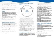
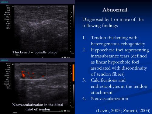
Abnormal<br />
Diagnosed by 1 or more <strong>of</strong> <strong>the</strong><br />
following findings<br />
Thickened – ‘Spindle Shape’<br />
Neovascularization in <strong>the</strong> distal<br />
third <strong>of</strong> tendon<br />
1. Tendon thickening with<br />
heterogeneous echogenicity<br />
2. Hypoechoic foci representing<br />
intrasubstance tears (defined<br />
as linear hypoechoic foci<br />
associated with discontinuity<br />
<strong>of</strong> tendon fibres)<br />
3. Calcifications <strong>and</strong><br />
en<strong>the</strong>siophytes at <strong>the</strong> tendon<br />
attachment<br />
4. Neovascularization<br />
(Levin, 2005; Zanetti, 2003)